. 2023 Aug;620(7976):1025-1030.
doi: 10.1038/s41586-023-06370-4. Epub 2023 Aug 2.

Africa-specific human genetic variation near CHD1L associates with HIV-1 load

Paul J McLaren #  1   2 Immacolata Porreca #  3 Gennaro Iaconis #  4 Hoi Ping Mok #  4 Subhankar Mukhopadhyay #  5 Emre Karakoc #  3 Sara Cristinelli  6 Cristina Pomilla  3 István Bartha  7   8 Christian W Thorball  7   8   9 Riley H Tough  10   11 Paolo Angelino  8 Cher S Kiar  5 Tommy Carstensen  3   4 Segun Fatumo  12   13 Tarryn Porter  3 Isobel Jarvis  4 William C Skarnes  14 Andrew Bassett  3 Marianne K DeGorter  15 Mohana Prasad Sathya Moorthy  15   16 Jeffrey F Tuff  10 Eun-Young Kim  17 Miriam Walter  17 Lacy M Simons  17 Arman Bashirova  18   19 Susan Buchbinder  20 Mary Carrington  18   19   21 Andrea Cossarizza  22 Andrea De Luca  23   24 James J Goedert  25 David B Goldstein  26 David W Haas  27 Joshua T Herbeck  28 Eric O Johnson  29 Pontiano Kaleebu  30   31 William Kilembe  32 Gregory D Kirk  33 Neeltje A Kootstra  34 Alex H Kral  35 Olivier Lambotte  36   37 Ma Luo  11   38 Simon Mallal  27   39 Javier Martinez-Picado  40   41   42   43 Laurence Meyer  44   45 José M Miro  43   46 Pravi Moodley  47 Ayesha A Motala  48 James I Mullins  49 Kireem Nam  17 Niels Obel  50 Fraser Pirie  48 Francis A Plummer  11 Guido Poli  51   52 Matthew A Price  53   54 Andri Rauch  55 Ioannis Theodorou  56 Alexandra Trkola  57 Bruce D Walker  21   58 Cheryl A Winkler  59 Jean-François Zagury  60 Stephen B Montgomery  15   16 Angela Ciuffi  6 Judd F Hultquist  17 Steven M Wolinsky  17 Gordon Dougan  3   4 Andrew M L Lever  4   61 Deepti Gurdasani  62   63 Harriet Groom  4 Manjinder S Sandhu  64   65   66 Jacques Fellay  67   68   69
Affiliations

Africa-specific human genetic variation near CHD1L associates with HIV-1 load

Paul J McLaren et al. Nature. 2023 Aug.

Erratum in

  • Author Correction: Africa-specific human genetic variation near CHD1L associates with HIV-1 load.
    McLaren PJ, Porreca I, Iaconis G, Mok HP, Mukhopadhyay S, Karakoc E, Cristinelli S, Pomilla C, Bartha I, Thorball CW, Tough RH, Angelino P, Kiar CS, Carstensen T, Fatumo S, Porter T, Jarvis I, Skarnes WC, Bassett A, DeGorter MK, Sathya Moorthy MP, Tuff JF, Kim EY, Walter M, Simons LM, Bashirova A, Buchbinder S, Carrington M, Cossarizza A, De Luca A, Goedert JJ, Goldstein DB, Haas DW, Herbeck JT, Johnson EO, Kaleebu P, Kilembe W, Kirk GD, Kootstra NA, Kral AH, Lambotte O, Luo M, Mallal S, Martinez-Picado J, Meyer L, Miro JM, Moodley P, Motala AA, Mullins JI, Nam K, Obel N, Pirie F, Plummer FA, Poli G, Price MA, Rauch A, Theodorou I, Trkola A, Walker BD, Winkler CA, Zagury JF, Montgomery SB, Ciuffi A, Hultquist JF, Wolinsky SM, Dougan G, Lever AML, Gurdasani D, Groom H, Sandhu MS, Fellay J. McLaren PJ, et al. Nature. 2023 Sep;621(7979):E42. doi: 10.1038/s41586-023-06591-7. Nature. 2023. PMID: 37670157 No abstract available.

Abstract

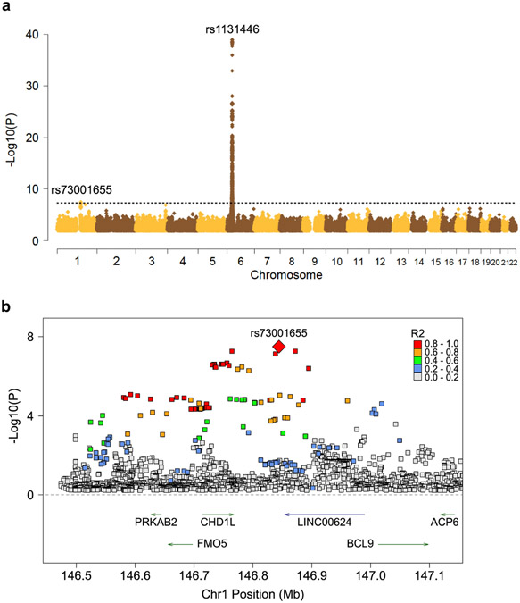
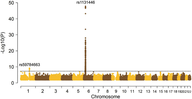
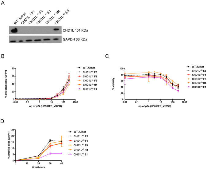
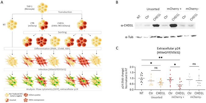
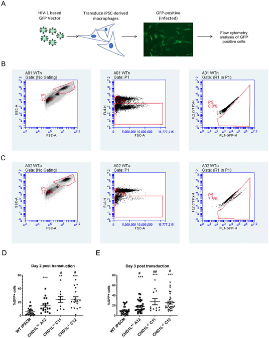
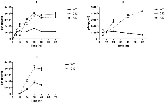
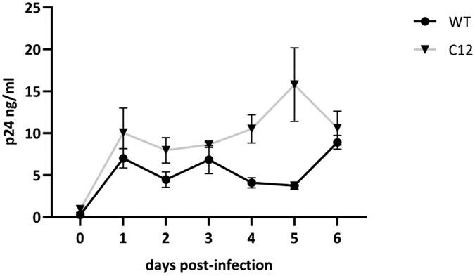
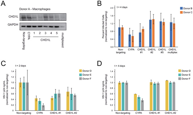
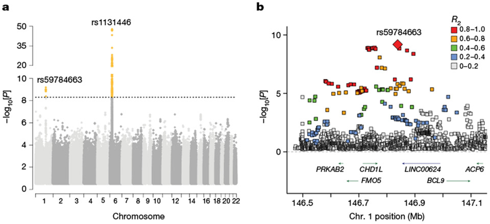
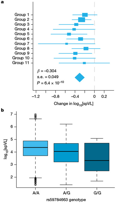
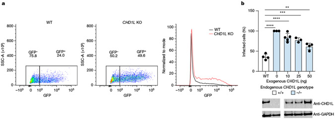
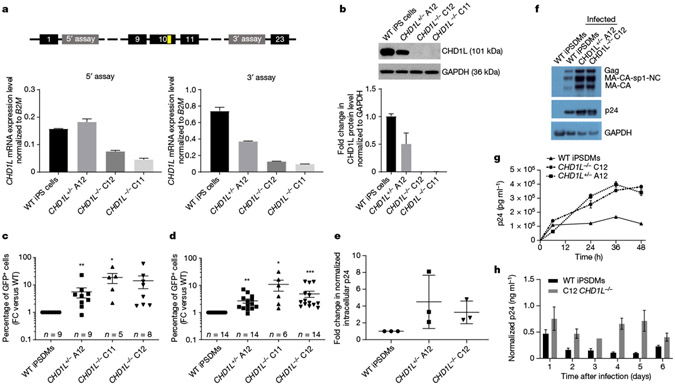
HIV-1 remains a global health crisis1, highlighting the need to identify new targets for therapies. Here, given the disproportionate HIV-1 burden and marked human genome diversity in Africa2, we assessed the genetic determinants of control of set-point viral load in 3,879 people of African ancestries living with HIV-1 participating in the international collaboration for the genomics of HIV3. We identify a previously undescribed association signal on chromosome 1 where the peak variant associates with an approximately 0.3 log10-transformed copies per ml lower set-point viral load per minor allele copy and is specific to populations of African descent. The top associated variant is intergenic and lies between a long intergenic non-coding RNA (LINC00624) and the coding gene CHD1L, which encodes a helicase that is involved in DNA repair4. Infection assays in iPS cell-derived macrophages and other immortalized cell lines showed increased HIV-1 replication in CHD1L-knockdown and CHD1L-knockout cells. We provide evidence from population genetic studies that Africa-specific genetic variation near CHD1L associates with HIV replication in vivo. Although experimental studies suggest that CHD1L is able to limit HIV infection in some cell types in vitro, further investigation is required to understand the mechanisms underlying our observations, including any potential indirect effects of CHD1L on HIV spread in vivo that our cell-based assays cannot recapitulate.


Figures

Extended Data Fig. 1 ∣
Extended Data Fig. 1 ∣. A discovery genome-wide association analysis identifies a potentially novel locus associated with HIV spVL in individuals with African ancestries.
a, Genome-wide association results of the impact of common polymorphisms on HIV-1 spVL in the discovery set of 2,682 individuals of African ancestry. Genetic variants (yellow/brown diamonds) are plotted by chromosome position (GRCh37, x-axis) and statistical significance (y-axis). The dashed line indicates the screening threshold for significance (P < 5 × 10⁻⁸). Variants in two genomic regions, the HLA region on chromosome 6 and a novel chromosome 1 locus, are significantly associated with spVL. The top associated variant per region is listed above the association peak. b, Association results across the newly identified chromosome 1 region in the discovery sample of 2,682 individuals of African ancestry. Variants (boxes and diamond) are plotted by position (GRCh37) and −log10(P). The top associated variant, rs73001655 (P = 3.2 × 10⁻⁸) is represented by the red diamond. Association was calculated per group using linear regression and meta-analysed across groups. Additional variants are coloured by their correlation to rs73001655 calculated from the African subset of the 1000 Genomes Project reference phase 3 sample. Arrows below the dashed line indicate the location and direction of transcription of protein-coding genes (green) and non-coding RNA (blue).
Extended Data Fig. 2 ∣
Extended Data Fig. 2 ∣. Genome-wide association results of the impact of common polymorphisms on HIV-1 spVL in the combined set of 3,879 individuals with African ancestries.
Genetic variants (yellow/brown triangles) are plotted by chromosome position (GRCh37, x-axis) and statistical significance (−log10(P), y-axis). The dashed line indicates the threshold for genome-wide significance in samples with African ancestries (P < 5 × 10⁻⁹). Variants in two regions are significantly associated with spVL. The top associated variant per region is listed above the association peak.
Extended Data Fig. 3 ∣
Extended Data Fig. 3 ∣. Characterization and infection assays in Jurkat CHD1L mono and biallelic knockout mutants.
a, Western blot for CHD1L shows reduced (E5) and ablated (F1, F5, E1, H4) CHD1L expression, consistent with the respective genotypes. Levels of GAPDH are shown as loading control. b,c, The percentage of GFP positive cells (b) and viable cells (c) in CHD1L knockout clones was evaluated by flow cytometry at 48 h post-infection with different concentrations of NL4-3-deltaEnv-GFP/VSV-G (0-300 ng of p24). d, The percentage of GFP positive cells was evaluated at different time points (24, 36, 48 h) post-transduction with 300ng of p24 NL4-3-deltaEnv-GFP/VSV-G virus.
Extended Data Fig. 4 ∣
Extended Data Fig. 4 ∣. Impact of CHD1L overexpression on HIV replication in THP-1 differentiated cells.
a, Experimental design. THP-1 were transduced with lentiviral particles encoding, either CHD1L IRES mcherry (CHD1L), or mCherry alone as a control (CTR), or left untreated (NT). Successfully transduced cells were sorted by FACS. The resulting sorted monocyte populations were differentiated into macrophages during 48 h in presence of 25 nM PMA and let recover for 24 h additional hours. Differentiated cell lines were infected with the single-round amphotropic HIVeGFP/VSV.G virus. b, Western blot confirming CHD1L overexpression in THP-1 cells transduced with CHD1L-encoding vector. c, Extracellular p24 was measured by ELISA at day 3 post-infection (n = 4). Results are normalized to the NT sample at day 3, mean and individual values of at least two experiments in triplicate are plotted. Multiple comparison One-way ANOVA showed statistical significance between CTR and CHD1L overexpressing cells (p < 0.005).
Extended Data Fig. 5 ∣
Extended Data Fig. 5 ∣. Infection of iPSC-derived macrophages (iPSDMs) with the HIV-1 vector, NL4-3-deltaEnv-GFP/VSV-G.
a, Experimental design: VSV-G pseudotyped HIV-1 vector was used to infect iPSDMs. Viral activity was assessed by GFP expression through flow cytometry analysis. b,c, Gating strategy for uninfected (b) and infected (c) WT cells of a single experiment. Live cells were selected by light scattering exclusion of debris (left panels) and dead cells exclusion by DRAQ-7 staining (middle panels). To circumvent autofluorescence, GFP-positivity was controlled through FL1/FL2 comparison (right panels). d,e, Raw infection data for WT and CHD1L knockout iPSDMs. Data refer to Fig. 4c and d of the main text. Data from individual wells of each experiment are reported as raw percentage of GFP positive cells. *, ** and *** represent statistically significant differences (p ≤ 0.05, 0.01 and 0.001, respectively) between WT and mutant clones using Wilcoxon matched-pairs signed rank test. #,## represent statistically significant differences (p ≤ 0.05 and 0.01, respectively) between the CHD1L+/− A12 clone and the CHD1L−/− C12 and C11 clones using Wilcoxon matched-pairs signed rank test.
Extended Data Fig. 6 ∣
Extended Data Fig. 6 ∣. Viral Gag particle release from WT and CHD1L knockout macrophages.
Viral Gag particle release was measured by p24 ELISA assay on the culture supernatants at different time points post-transduction. The three graphs show independent biological replicates. A12 cells were not available for all time points. Data are reported as the average and standard deviation of duplicate p24 ELISA readings. In each independent replicate, C12 was significantly different from WT as determined by repeated measures ANOVA (1: F (6, 12) = 188.8, P < 0.0001, 2: F (5, 10) = 503.6, P < 0.0001, 3: F (5, 10) = 81.58, P < 0.0001).
Extended Data Fig. 7 ∣
Extended Data Fig. 7 ∣. p24 release from CHD1L KO cells infected with replication competent HIV.BE_GIN.
Raw supernatant p24 values corresponding to Fig. 4h in the main text.
Extended Data Fig. 8 ∣
Extended Data Fig. 8 ∣. Assessing the impact of CHD1L knock-out in primary monocyte-derived macrophages on HIV infection.
a, CHD1L was efficiently knocked out in primary MDMs by 3 of 5 crRNP constructs and a combined, multiplexed pool. b, Percent infected cells 4 days post-challenge as measured by flow cytometry showed an increase in three of the four CHD1L knockout pools compared to the non-targeting control, but these differences were not statistically significant. c,d, p24 levels in the culture supernatants as measured by ELISA were lower in CHD1L knockout cell pools 2 days post-infection (c), but recovered to the level of the non-targeting control by 4 days post-infection (d).
Fig. 1 ∣
Fig. 1 ∣. GWAS analysis identifies a locus on chromosome 1 that is associated with HIV spVL in individuals with African ancestries.
a, Genome-wide association results of the impact of common polymorphisms on HIV-1 spVL in the combined set of 3,879 individuals of African ancestries. Genetic variants were tested for association with spVL per group using linear regression and evidence was combined across groups using inverse-variance weighted meta-analysis in a fixed-effects framework. Genetic variants (grey and yellow diamonds) are plotted by chromosome position (GRCh37, x axis) and statistical significance (−log10[P], y axis). The dashed line indicates the threshold for genome-wide significance in populations of African ancestries (P < 5 × 10⁻⁹). Variants in two regions are significantly associated with spVL (yellow diamonds). These include the HLA region on chromosome 6 and a locus on chromosome 1. The top associated variant per region is listed above the association peak. To improve visualization of the chromosome 1 region, the y axis was split. Full genome-wide results are presented in Extended Data Fig. 2. b, Association results across the newly identified chromosome 1 region. Variants (boxes and diamond) are plotted by position (GRCh37) and −log10[P]. The most strongly associated variant, rs59784663 (P = 6.4 × 10⁻¹⁰), is represented by a red diamond. Additional variants are coloured by their correlation with rs59784663 calculated from the African subset of the 1000 Genomes Project reference phase 3 sample. The arrows below the dashed line indicate the location and direction of transcription of protein-coding genes (green) and non-coding RNA (blue).
Fig. 2 ∣
Fig. 2 ∣. The viral-load-decreasing effect of rs59784663(G).
a, The per group effect of rs59784663(G) on HIV-1 spVL. The boxes indicate the per-group effect estimate (β from linear regression) with s.e. (whiskers). The summary effect (diamond) and P value are the result of meta-analysis across groups. Results from the meta-analysis are shown; the β, s.e. and P value are given at the bottom left. The relationship between group number, cohort/clinical centre and ancestry is presented in Supplementary Table 1. b, The additive allelic effect of the rs59784663 genotype on spVL is summarized across all individuals.
Fig. 3 ∣
Fig. 3 ∣. CHD1L expression decreases single-round HIV infection in U2OS cells.
a, Representative flow cytometry plots of WT and CHD1L-KO U2OS cells infected with HIV NL4.3-deltaEnv-GFP/VSV-G. After 48 h, CHD1L-WT U2OS cells (left) showed an approximately twofold reduction in number of GFP-positive cells compared with CHD1L-KO cells (middle, overlaid on the right). b, U2OS CHD1L-KO cells were transfected with increasing concentrations (0, 10, 25 or 50 ng) of CHD1L expression plasmid. Then, 24 h after transfection, WT U2OS or transfected KO cells were infected with HIV NL4.3-deltaEnv-GFP/VSV-G and assessed for GFP positivity after 48 h. A representative immunoblot for CHD1L and GAPDH is shown below. n ≥ 4. Statistical analysis was performed using unpaired t-tests; ****P ≤ 0.0001, ***P ≤ 0.0005, **P ≤ 0.005. The full blot is provided in Supplementary Fig. 8.
Fig. 4 ∣
Fig. 4 ∣. KO of CHD1L increases HIV-1 infection in human-iPS cell-derived macrophages.
a, Quantitative PCR (qPCR) validation of the level of full-length CHD1L mRNA after induction of frameshift deletions. The diagram shows the position of the 5′ and 3′ TaqMan assays used to confirm exon 10 KO (puromycin resistance cassette in yellow). The bar plots show the relative expression of CHD1L normalized to B2M expression for WT and heterozygous and homozygous KO clones. b, Immunoblot analysis of CHD1L and GAPDH protein in WT and KO clones. The blot is representative of three replicates (all data are shown in Supplementary Fig. 10). The fold changes in normalized CHD1L levels are given below. n = 3. Data are mean ± s.d. c,d, The percentage of GFP-positive cells in HIV-1-GFP-transduced WT and CHD1L-KO clones evaluated 2 days (c) and 3 days (d) after transduction, normalized to the WT. Each point represents an independent experiment, conducted in duplicate. Statistical analysis was performed using Wilcoxon matched-pairs signed-rank tests, comparing between the WT and mutant clones; *P ≤ 0.05, **P ≤ 0.01, ***P ≤ 0.001. e, Intracellular viral capsid p24 protein measured by enzyme-linked immunosorbent assay (ELISA) 2 days after transduction. n = 3. f, Western blot analyses of intracellular viral capsid p24 2 days after transduction. iPSDMs, iPS cell-derived macrophages. g, Time-dependent extracellular viral Gag particle release measured by p24 ELISA. Data are mean ± s.d. of duplicate readings and are representative of three independent experiments (all data are shown in Extended Data Fig. 6). h, p24 production after infection of CHD1L-WT and CHD1L-KO cells with HIV.BE_GIN (BaL Env and GFP-IRES-nef) replication-competent virus. The graphs show p24 (ng ml−1) normalized to the GFP-positive cell proportion for 2–4 replicates (except for C12 at day 3, for which n = 1). Data are mean ± s.e.m. A significance value of P ≤ 0.005 was determined for normalized WT/C12 comparisons using a paired t-test. Raw p24 values are shown in Extended Data Fig. 7.

Similar articles

Cited by

References

    1. UNAIDS Data 2021 (UNAIDS, 2021); https://www.unaids.org/en/resources/documents/2021/2021_unaids_data.
    1. Gurdasani D, Barroso I, Zeggini E & Sandhu MS Genomics of disease risk in globally diverse populations. Nat. Rev. Genet 20, 520–535 (2019). - PubMed
    1. McLaren PJ et al. Association study of common genetic variants and HIV-1 acquisition in 6,300 infected cases and 7,200 controls. PLoS Pathog. 9, e1003515 (2013). - PMC - PubMed
    1. Ahel D. et al. Poly(ADP-ribose)-dependent regulation of DNA repair by the chromatin remodeling enzyme ALC1. Science 325, 1240–1243 (2009). - PMC - PubMed
    1. Prevention Gap Report (UNAIDS, 2016).