. 2023 Sep;17(9):865-873.
doi: 10.1111/crj.13672. Epub 2023 Aug 2.

Imaging and pathological characteristics, treatment, and prognosis of pulmonary sequestration-A retrospective study of 13 cases

Affiliations

Imaging and pathological characteristics, treatment, and prognosis of pulmonary sequestration-A retrospective study of 13 cases

Xiangjin Liu et al. Clin Respir J. 2023 Sep.

Abstract

Objective: This study aimed to summarize and analyze the characteristics of pulmonary sequestration to improve our understanding of this disease.

Methods: Between January 2019 and April 2023, the clinical data of 13 patients with pulmonary sequestration underwent surgical treatment at the First Affiliated Hospital of Gannan Medical University.

Results: The male-to-female ratio was 4:9, the age was 0.5 to 60 years, and the average age was 38 ± 19 years. There were 10 and 3 cases of intralobar and extralobar pulmonary sequestration, respectively. Chest enhanced computed tomography (CT) and three-dimensional vascular reconstruction showed that the abnormal blood vessels were derived from the descending thoracic aorta in nine cases and from other blood vessels in four cases. Three patients underwent thoracoscopic lobectomy, two underwent thoracoscopic segmentectomy, and eight underwent thoracoscopic wedge resection. All the patients successfully completed the surgery and were discharged postoperatively.

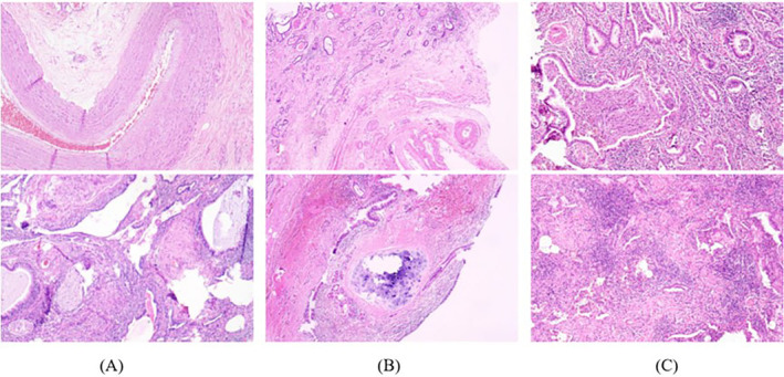
Conclusions: Some patients with pulmonary sequestration exhibit no obvious symptoms. Patients with clinical symptoms are easily confused for pneumonia, bronchial cysts, lung abscesses, and lung tumors; therefore, patients with pulmonary sequestration are prone to missed diagnosis and misdiagnosis. Currently, enhanced chest CT combined with three-dimensional vascular reconstruction can accurately show the course, branches, and relationship with the mass of the feeding artery. Routine pathological examination is helpful to further clarify the diagnosis of pulmonary sequestration. Minimally invasive thoracoscopic surgery is the preferred treatment for patients with pulmonary sequestration. Surgical resection is safe and feasible, and satisfactory results are typically obtained.

Keywords: imaging characteristics; pathological characteristics; prognosis; pulmonary sequestration; treatment.


Conflict of interest statement

The authors declare that the research was conducted in the absence of any commercial or financial relationships that could be construed as a potential conflict of interest.

Figures

FIGURE 1
FIGURE 1
Chest CT images of patients with pulmonary sequestration (mediastinal window + pulmonary window). (A) The lumpy solid mass in the lower lobe of the right lung has uniform density and clear boundaries. (B) An irregular mixed‐density cystic shadow can be observed in the lower lobe of the left lung, with septation and scattered small patchy calcifications at the edge. (C) Patchy, dense shadows can be observed in the posterior basal segment of the left lower lobe, with unclear boundaries and reduced density of the adjacent lung parenchyma. (D) The lower lobe of the left lung shows a patchy increase in density and the boundary is slightly blurred, showing ground glass‐like changes.
FIGURE 2
FIGURE 2
The thick branches of the thoracic aorta with a diameter of 15 mm entered the lesion. Chest enhanced CT combined with three‐dimensional vascular reconstruction shows that the posterior basal segment of the left lower lobe has patchy high‐density and unclear boundaries, and the descending thoracic aorta has a large branch with a diameter of 15 mm into the lesion.
FIGURE 3
FIGURE 3
The thoracic aorta sent out three branch arteries into the lesion. Enhanced chest CT combined with three‐dimensional vascular reconstruction shows a mass‐like density shadow in the lower lobe of the left lung, uneven enhancement, and a strip shadow around it. Three branch arteries from the thoracic aorta enter the lesion.
FIGURE 4
FIGURE 4
Preoperative chest CT and chest X‐ray 2 months after operation in patients with pulmonary sequestration complicated with pectus excavatum. (A) Preoperative chest CT tomography showing that the middle segment of the sternum and the corresponding bilateral costal cartilage are symmetrically depressed, with a range of approximately 13 × 14 cm, and the deepest depression was approximately 2.0 cm. A soft tissue density shadow can be observed in the lower lobe of the right lung, and pleural thickening can be observed between the two lung lobes. (B) Two months after surgery, the patient returned to the hospital for review of chest radiographs in the front and side positions. The orthopedic plate was well fixed, thoracic depression significantly improved, and the left lower lobe changed after the operation.
FIGURE 5
FIGURE 5
Pathological morphology of pulmonary sequestration lesions under microscope (hematoxylin and eosin stain, ×100).

References

    1. Pryce DM. Lower accessory pulmonary artery with intralobar sequestration of lung; a report of seven cases. J Pathol Bacteriol. 1946;58(3):457‐467. doi:10.1002/path.1700580316 - DOI - PubMed
    1. Gabelloni M, Faggioni L, Accogli S, Aringhieri G, Neri E. Pulmonary sequestration: what the radiologist should know. Clin Imaging. 2021;73:61‐72. doi:10.1016/j.clinimag.2020.11.040 - DOI - PubMed
    1. Onorati I, Diso D, Anzidei M, De Giacomo T, Anile M. Right intralobar sequestration with arterial supply from left gastric artery. Thorax. 2015;70(9):912. doi:10.1136/thoraxjnl-2015-206817 - DOI - PubMed
    1. Boyle N, Waters DK, McCann J, Butler M, Dodd JD, McCarthy C. Pulmonary embolus and hemoptysis revealing rare Intralobar sequestration. Am J Respir Crit Care Med. 2023;207(3):e4‐e5. doi:10.1164/rccm.202206-1196IM - DOI - PubMed
    1. Kestenholz PB, Schneiter D, Hillinger S, Lardinois D, Weder W. Thoracoscopic treatment of pulmonary sequestration. Eur J Cardiothorac Surg. 2006;29(5):815‐818. doi:10.1016/j.ejcts.2006.02.018 - DOI - PubMed