Review
. 2023 Aug 1:4:0043.
doi: 10.34133/cbsystems.0043. eCollection 2023.

Bioprinting Methods for Fabricating In Vitro Tubular Blood Vessel Models

Affiliations
Review

Bioprinting Methods for Fabricating In Vitro Tubular Blood Vessel Models

Seon-Jin Kim et al. Cyborg Bionic Syst. .

Abstract

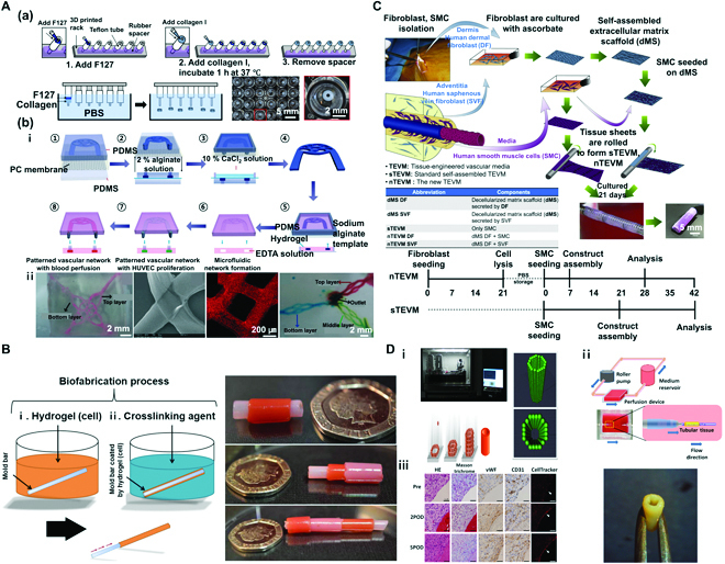
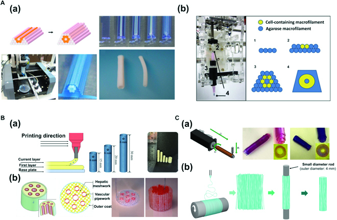
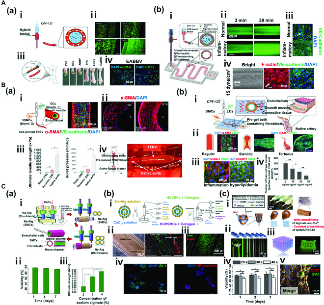
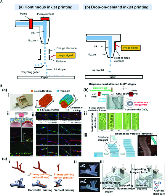
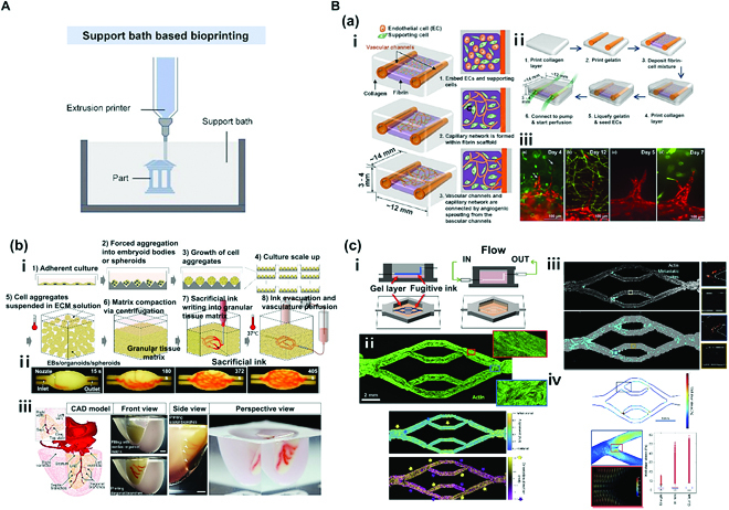
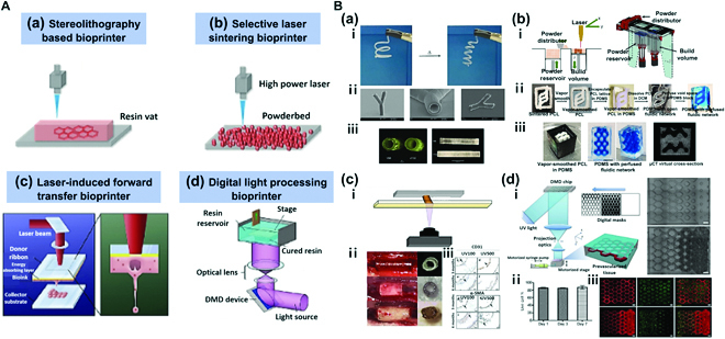
Dysfunctional blood vessels are implicated in various diseases, including cardiovascular diseases, neurodegenerative diseases, and cancer. Several studies have attempted to prevent and treat vascular diseases and understand interactions between these diseases and blood vessels across different organs and tissues. Initial studies were conducted using 2-dimensional (2D) in vitro and animal models. However, these models have difficulties in mimicking the 3D microenvironment in human, simulating kinetics related to cell activities, and replicating human pathophysiology; in addition, 3D models involve remarkably high costs. Thus, in vitro bioengineered models (BMs) have recently gained attention. BMs created through biofabrication based on tissue engineering and regenerative medicine are breakthrough models that can overcome limitations of 2D and animal models. They can also simulate the natural microenvironment in a patient- and target-specific manner. In this review, we will introduce 3D bioprinting methods for fabricating bioengineered blood vessel models, which can serve as the basis for treating and preventing various vascular diseases. Additionally, we will describe possible advancements from tubular to vascular models. Last, we will discuss specific applications, limitations, and future perspectives of fabricated BMs.


Figures

Fig. 1.
Fig. 1.
(A) Hydrogel casting method. (a) Method 1: Fabrication of freestanding cell-laden hydrogel blood vessel rings using a casting mold. Reproduced with permission [41]. Copyright 2022, The Authors. (b) Method 2: Fabrication of interconnected 3D vascular network construct using a sacrificial template and a casting mold. (i) Schematic diagram of the fabrication process. (ii) An image of a 3D vascular network construct in gelatin hydrogel, an image of a perfusion test using red microspheres, and an image of 3-layer interconnected vascular networks. Reproduced with permission [42]. Copyright 2014, The Royal Society of Chemistry. (B) Micro-dip coating method. Schematic diagram of tubular structure biofabrication process via dip coating. Reproduced with permission [43]. Copyright 2017, The Authors. (C) Extracellular sheet matrix rolling assembly method. Schematic diagram of biofabrication process of a tubular structure via sheet rolling, and timeline required to produce vascular constructs for comparing nTEVM (the new TEVM) versus sTEVM (standard self-assembled TEVM). Reproduced with permission [44]. Copyright 2012, Elsevier. (D) Tubular structures fabricated using the Kenzan method. (i) The system of lamination of MCS (Bio-3D printer) involves skewering of MCSs into needle array according to a 3D structure predesigned using a computer system. (ii) Schematic illustration of a bioreactor system and scaffold-free vascular graft generated from MCSs. (iii) Graft post-implantation is patented and remodeled. Reproduced with permission [45]. Copyright 2015, The Authors.
Fig. 2.
Fig. 2.
(A) Microextrusion bioprinting: (a) Fabrication of scaffold-free vascular tissue using agarose rods and cellular cylinders. Reproduced with permission [46]. Copyright 2023, Elsevier. (b) A Fab@Home printing system and printing protocol for building a cellular tubular structure by depositing hydrogel macrofilaments in additive manufacturing. Reproduced with permission [47]. Copyright 2010, Elsevier. (B) Concentric ring bioprinting. (a) Method 1: Direct concentric ring 3D printing using gelatin (Gt)-Alg-montmorillonite (MMT) bioinks. Images of the concentric ring printing method, 3D model, and 3D printed Gt-Alg-MMT hydrogel vascular scaffold. Reproduced with permission [48]. Copyright 2022, Elsevier. (b) Method 2: Modeling of liver tissue construct with a vascular network using direct concentric 3D printing method with gelatin/Alg/chitosan (GAC) hydrogel. Reproduced with permission [49]. Copyright 2009, SAGE Publications. (C) Rod bioprinting. (a) Formation of vascular structure and multimaterial vascular structure. Schematic image of a rod printer and rotating rods of various diameters. The relationship between thickness of the vascular structure and rod diameter is also shown. Printing hydrogel, multilayer vessel structure. Reproduced with permission [50]. Copyright 2021, The Authors. (b) Fabrication of artificial poly (lactic acid) (PLA) blood vessels using electrospinning and 3D rod printing. Reproduced with permission [51]. Copyright 2021, The Authors, licensed under a Creative Commons Attribution 3.0 International License.
Fig. 3.
Fig. 3.
(A) Coaxial nozzle bioprinting (monolayer). (a) Tissue-engineered bio-blood vessels developed using coaxial nozzle cell printing for ischemic disease. (i) Schematic diagram of coaxial nozzle and materials. (ii) Live/dead assay for printed EPC (endothelial progenitor cell)-laden BBV during 7 days. Cells were stained with calcein AM (live, green) and ethidium homodimer I (dead, red). (iii) Representative images of 6 groups (PBS, BBV, ABBV, EPC, EBBV, and EABBV). (iv) Immunostaining results of EPC/APMS-laden BBV. The 6 groups were stained with anti-CD31 antibody and anti-α-SMA antibody after 28 days. BBV, bio-blood vessel; ABBV, atorvastatin-loaded poly (lactic-co-glycolic) acid microsphere (APMS)-laden BBV; EBBV, EPC-laden BBV; EABBV, EPC/APMS-laden BBV. Reproduced with permission [52]. Copyright 2017, The Authors, licensed under a Creative Commons Attribution 4.0 International License. (b) Freestanding, perfusable, and functional in vitro vascular models developed using coaxial nozzle cell printing for mimicking native endothelium pathophysiology. (i) Schematic diagram of coaxial nozzle cell printing process. (ii) Permeability test for a normal vascular model (VM) and an inflammatory model. (iii) Immunostaining results of normal and inflammatory VM indicating disruptions of the endothelial barrier in the inflammatory model. (iv) Response to shear stress in VM. Reproduced with permission [53]. Copyright 2018, WILEY-VCH Verlag GmbH & Co. KGaA, Weinheim. (B) Coaxial nozzle bioprinting (multilayer). (a) Tissue-engineered vascular grafts containing an endothelial layer and a muscle layer using triple-coaxial cell printing technology. (i) Schematic diagram of triple-coaxial nozzle, materials, and tissue-engineered blood vessels (TEBVs), and immunostaining results of TEBV. (ii) Culturing of TEBV under static conditions (left) and dynamic conditions (right). (iii) Mechanical property test (UTS and BP). (iv) Abdominal aorta graft of TEBV in the rat model. Reproduced with permission [54]. Copyright 2019, AIP Publishing. (b) Fabrication of atherosclerotic in vitro model containing endothelium and smooth muscles. (i) Schematic diagram of the cell printing process, native artery, and materials. (ii) Triple-layer arterial constructs (regular, stenotic, and tortuous). (iii) Endothelial dysfunction response of the arterial construct [inflammation: tumor necrosis factor-α (TNF-α), hyperlipidemia: low-density lipoprotein (LDL)]. (iv) Evaluation of the effect of atorvastatin on an arterial construct (turbulent flow). Reproduced with permission [55]. Copyright 2020, Wiley-VCH GmbH. (C) Vessel-like structure with multilevel fluidic channels. (a) Fabrication process of Na-Alg cell-laden hydrogel vessel-like structure containing endothelium and smooth muscles using coaxial nozzle and rod printing methods. (i) Schematic of the fabrication process. (ii) Live/dead assay (live, green; dead, red) of vessel-like structure. (iii) Test of mechanical properties (UTS) of the construct. Reproduced with permission [56]. Copyright 2017, American Chemical Society. (b) Fabrication of multicellular vessel structure using 4 flow channels. (i) Schematic diagram of the 4-channel coaxial nozzle. (ii) Images showing the distribution of cells via cell-laden hydrogel perfusion. (iii) Live/dead assay (live, green; dead, red). (iv) Immunostaining image showing expression of ZO1 tight junction and VE-cadherin proteins in HUVECs and HUVSMCs. Reproduced with permission [57]. Copyright 2018, Elsevier. (c) Direct 3D bioprinting of perfusable vascular constructs. (i) Schematic diagram of various coaxial nozzle and materials. (ii) 3D-printed vascular constructs with various sizes. (iii) Perfusable 10 layers of a 3D bioprinted construct. (iv) Live/dead assay (live, green; dead, red) of vascular constructs. (v) Immunostaining images showing expression of CD31 and SMA. Reproduced with permission [58]. Copyright 2016, Elsevier.
Fig. 4.
Fig. 4.
(A) Simplified representation of 2 different mechanisms of inkjet printing. (a) Continuous inkjet printing (CIJ) and (b) drop-on-demand printing (DOD). Reproduced with permission [60]. Copyright 2022, The Authors. (B) DOD inkjet bioprinting: (a) Fabrication of multilayered vessel using DOD inkjet bioprinting. (i) The cooling system of the bioprinter for optimal gelation timing (left) and detailed structure and shape of the printed tubular structure. (ii) Schematic diagram of the cross-section of the channel with a single layer of endothelial cells and fluorescence micrographs of vascular-like channels in the cross-section. (iii) Fluorescence micrographs of the endothelium in sections with qualitative and quantitative assessment of collagen IV expression [CD31: green; VE-cadherin and collagen IV: red; 4′,6-diamidino-2-phenylindole (DAPI): blue]. Reproduced with permission [62]. Copyright 2018, The Authors. (b) Fabrication of bifurcated vascular tree using DOD inkjet bioprinting. (i) Basic tubular structure of a vascular network. (ii) Horizontal and vertical bifurcations. (iii) CaCl2 solution provides a supporting buoyant force for spanning and overhang regions in horizontal printing and overhang regions in vertical printing. Schematic diagram of the gelation process is shown. Reproduced with permission [65]. Copyright 2012, Wiley Periodicals Inc. (c) Fabrication of zigzag-shaped overhang structure using DOD inkjet printing in CaCl2 bath. (i) Schematic of the proposed platform-assisted 3D inkjet bioprinting system. (ii) Gel lines printed using a 120-mm dispense head. (iii) Zig-zag tube printing process and a printed zigzag tube sample. Reproduced with permission [66]. Copyright 2014, Wiley Periodicals Inc.
Fig. 5.
Fig. 5.
(A) Schematic illustration of support bath-based bioprinting. Reproduced with permission [67]. Copyright 2022, The Authors. (B) Support bath bioprinting: (a) Fabrication of tubular tissue by applying droplet bioprinter to printing. (i) Schematic of growth and maturation process of multiscale vascular system fabricated using 3D bioprinting technology. (ii) Channel construction and fibrin deposition procedure performed using a 3D bioprinter. (iii) Different types of angiogenic sprouts. Reproduced with permission [69]. Copyright 2014, Biomedical Engineering Society. (b) Fabrication of perfusable vascular network using organ building blocks (OBBs), organoids, as a suspension. (i) Step-by-step illustration of the fabrication process. (ii) An image sequence showing embedded 3D printing of a branched, hierarchical vascular network within a compacted EB-based tissue matrix connected to inlet and outlet tubes (scale bar = 10 mm). (iii) A PDMS mold (1:2 scale) was formed using 3D computed tomography data. A left anterior descending artery, together with diagonal and septal branches, was embedded into a septal–anterior wall wedge of the cardiac tissue matrix (scale bar = 5 mm). Reproduced with permission [70]. Copyright 2019, The Authors. (c) Fabrication of perfusable vascular network by applying microextrusion bioprinter to suspension-based printing. (i) Schematic representation of vascular bed printing using sacrificial ink. (ii) Cytoskeletal morphology of microvascular endothelial cells and alignment of cytoskeleton in perfused engineered beds. The preferential alignment follows flow direction. (iii) Full endothelialized vascular bed with identified CTC (circulating tumor cell) sites (cyan). Full acellular vascular beds exhibited a higher CTC burden than endothelialized beds (4% versus 2% of vessel area). (iv) WSS (wall shear stress) and laminar flow during CTC perfusion. WSS profile from vessel sidewalls at center Z slices gathered from simulation data was generated with parameters matching those used for CTC perfusion experiments. Reproduced with permission [71]. Copyright 2020, The Authors.
Fig. 6.
Fig. 6.
(A) Simplified representation of 4 different categories of laser-assisted bioprinting. (a) Working principle of stereolithography-based bioprinter. (b) Working principle of selective laser sintering bioprinter. Reproduced with permission [72]. Copyright 2016, WILEY-VCH Verlag GmbH & Co. KGaA, Weinheim. (c) Working principle of laser-induced forward transfer bioprinter. Reproduced with permission [74]. Copyright 2019, Elsevier. (d) Working principle of digital light processing bioprinter. Reproduced with permission [75]. Copyright 2020, The Authors. (B) Laser-assisted bioprinting. (a) Fabrication of cylindrical structures using stereolithography and multiphoton polymerization (MPP). (i) Shape memory effect. (ii) Scanning electron micrographs of a branched tubular structure generated by 2-photon polymerization of PTHF-PA 1. (iii) 3D tubes prepared from polytetrahydrofuranether-diacrylate (PTHF-DA) 1 with 0.5% Irgacure-184 and 10% additional crosslinker trimethylolpropane triacrylate. Reproduced with permission [79]. Copyright 2012, The Authors. (b) Fabrication of a perfusable network with a complex structure using OpenSLS bioprinting. (i) Custom open-source selective laser sintering (OpenSLS) hardware. (ii) Process sequence for creating a fluidic network using a sacrificial PCL structure. (iii) Sacrificial templating of a reduced diamond lattice model resulted in formation of a complex, interconnected fluidic network in PDMS, and perfusion with blue dye. Reproduced with permission [80]. Copyright 2016, The Authors. (c) Fabrication of patient-specific implantable vascular grafts using digital light processing (DLP) bioprinting method. (i) Fabrication method using DLP-mediated printing. (ii) Evolution of 3D printed graft over 6 months in vivo. (iii) At 3 and 6 months, an endothelial monolayer was observed despite detachment of the endothelial cell layer during a histological preparation process. Reproduced with permission [82]. Copyright 2015, WILEY-VCH Verlag GmbH & Co. KGaA, Weinheim. (d) Fabrication of vascularized tissues with regular patterns using DLP bioprinting. (i) Schematic diagram of the bioprinting platform (left) with bioprinted acellular construct and cellular construct (right). (ii) Results of cell viability assay of bioprinted tissue constructs encapsulated with HUVECs demonstrating over 85% cell viability. (iii) Fluorescent images demonstrating bioprinting of heterogeneous cell-laden tissue constructs with uniform and gradient channel width (scale bar = 250 μm). Reproduced with permission [84]. Copyright 2017, Elsevier.

References

    1. Naito H, Iba T, Takakura N. Mechanisms of new blood-vessel formation and proliferative heterogeneity of endothelial cells. Int Immunol. 2020;32(5):295–305. - PubMed
    1. Pugsley MK, Tabrizchi R. The vascular system: An overview of structure and function. J Pharmacol Toxicol Methods. 2000;44(2):333–340. - PubMed
    1. Ryu H, Oh S, Lee HJ, Lee JY, Lee HK, Jeon NL. Engineering a blood vessel network module for body-on-a-chip applications. J Lab Autom. 2015;20(3):296–301. - PubMed
    1. Şahin B, İlgün G. Risk factors of deaths related to cardiovascular diseases in World Health Organization (WHO) member countries. Health Soc Care Community. 2022;30(1):73–80. - PubMed
    1. Quyyumi AA. Endothelial function in health and disease: New insights into the genesis of cardiovascular disease. Am J Med. 1998;105(1A):32S–39S. - PubMed

LinkOut - more resources