. 2023 Jul 18:14:1215893.
doi: 10.3389/fimmu.2023.1215893. eCollection 2023.

Characterization of the humoral and cellular immunity induced by a recombinant BCG vaccine for the respiratory syncytial virus in healthy adults

Affiliations

Characterization of the humoral and cellular immunity induced by a recombinant BCG vaccine for the respiratory syncytial virus in healthy adults

Gaspar A Pacheco et al. Front Immunol. .

Abstract

Introduction: The human respiratory syncytial virus (hRSV) is responsible for most respiratory tract infections in infants. Even though currently there are no approved hRSV vaccines for newborns or infants, several candidates are being developed. rBCG-N-hRSV is a vaccine candidate previously shown to be safe in a phase I clinical trial in adults (clinicaltrials.gov identifier #NCT03213405). Here, secondary immunogenicity analyses were performed on these samples.

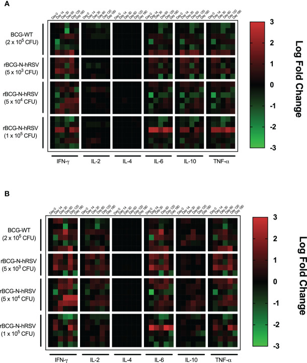
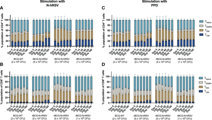
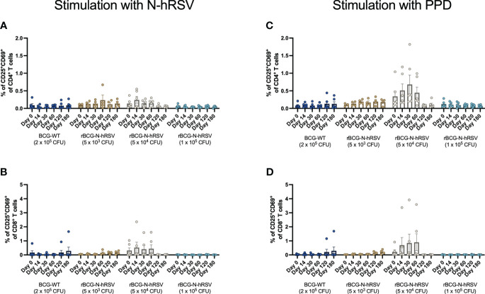
Methods: PBMCs isolated from immunized volunteers were stimulated with hRSV or mycobacterial antigens to evaluate cytokines and cytotoxic T cell-derived molecules and the expansion of memory T cell subsets. Complement C1q binding and IgG subclass composition of serum antibodies were assessed.

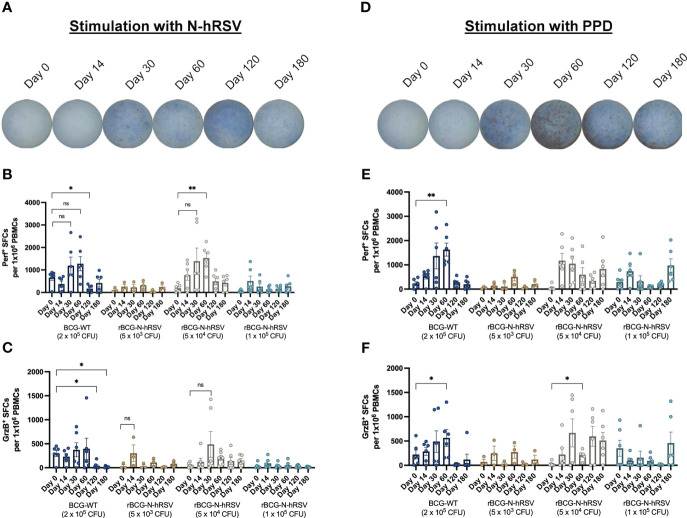
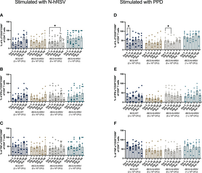
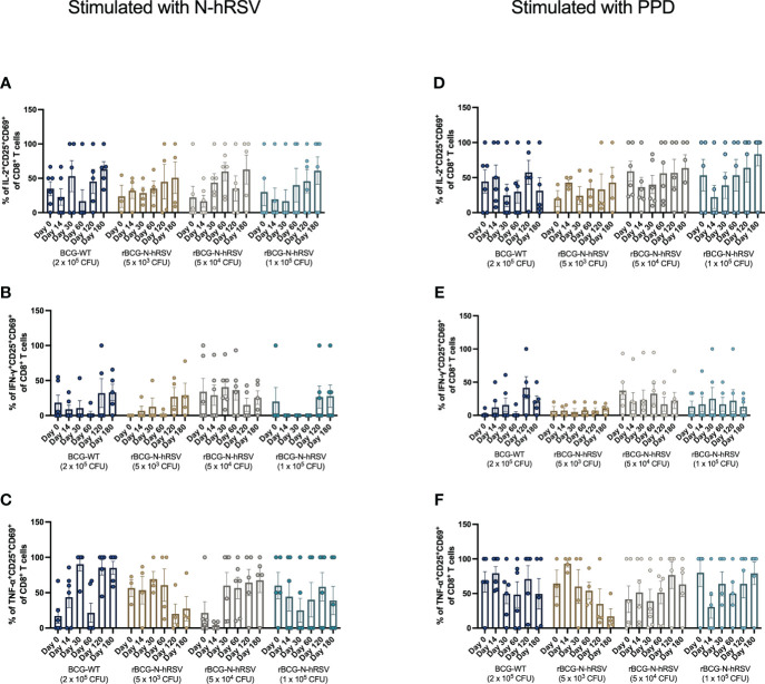
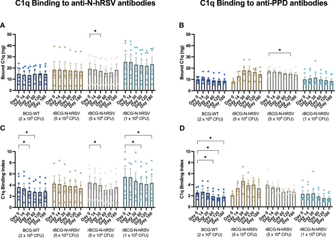
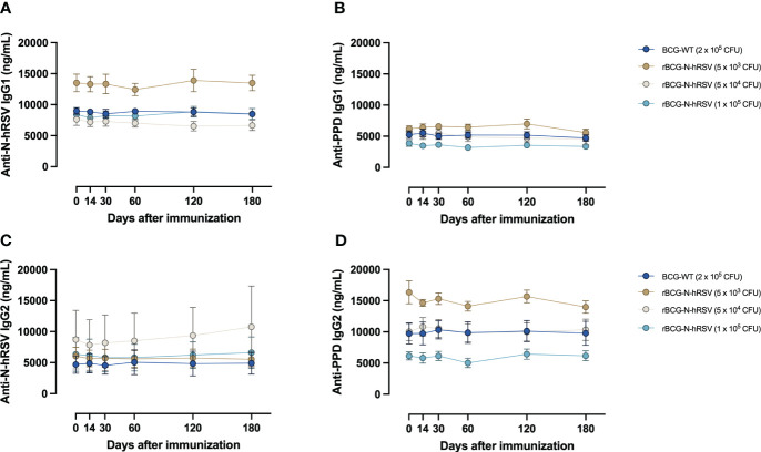
Results: Compared to levels detected prior to vaccination, perforin-, granzyme B-, and IFN-γ-producing PBMCs responding to stimulus increased after immunization, along with their effector memory response. N-hRSV- and mycobacterial-specific antibodies from rBCG-N-hRSV-immunized subjects bound C1q.

Conclusion: Immunization with rBCG-N-hRSV induces cellular and humoral immune responses, supporting that rBCG-N-hRSV is immunogenic and safe in healthy individuals.

Clinical trial registration: https://classic.clinicaltrials.gov/ct2/show/, identifier NCT03213405.

Keywords: BCG; cellular response; clinical trial; hRSV; vaccine.


Conflict of interest statement

PG, SB, and AK hold a patent for the rBCG-N-hRSV vaccine with Pontificia Universidad Católica de Chile PCT/US2008/076682. The remaining authors declare that the research was conducted in the absence of any commercial or financial relationships that could be construed as a potential conflict of interest.

Figures

Figure 1
Figure 1
PBMCs from subjects immunized with 5x10⁴ CFU of rBCG-N-hRSV show enhanced Perf and GrzB production upon stimulation of with N-hRSV or PPD. Representative images of wells in which PBMCs from subjects immunized with 5x10⁴ CFU of rBCG-N-hRSV were stimulated with either (A) N-hRSV or (B) PPD. Images are representative of subjects immunized with 5x10⁴ CFU of rBCG-N-hRSV. Blue spots are Perf+ spot-forming cells (SFCs), and red spots are GrzB+ SFCs. (B, E) Perf+ SFCs and (C, F) GrzB+ SFCs were counted after PBMCs were stimulated for 48 h with either 1.25 µg/mL of N-hRSV (B, C) or 750 IU/mL of PPD (E, F). Stimulation of PBMCs with 5 µg/mL ConA or 0.5% sterile PBS were used as positive and negative stimulation controls, respectively (data not shown). The concentration of the stimulus was selected according to the purpose of the evaluation to be performed. Data for subjects potentially exposed to hRSV during the post-immunization period, as suggested by hRSV-specific serological assays, were excluded. Bars represent the mean value of SFCs, and error bars represent the SEM. A two-way ANOVA for repeated measures with post-hoc Dunnett's test corrected for multiple comparisons against Day 0 was performed for data analysis. ns= not significant, * = P<0.05, ** = P<0.01.
Figure 2
Figure 2
PBMCs from subjects immunized with 5x10³ or 5x10⁴ CFU of rBCG-N-hRSV produce IFN-γ upon stimulation with N-hRSV and PPD. Heatmap of log10 fold change of the concentration of the cytokines IFN-γ, IL-2, IL-4, IL-6, IL-10, and TNF-α in supernatants of PBMC cultures stimulated with (A) 1.25 µg/mL N-hRSV or with (B) 750 IU/mL PPD respect to day 0. Each block of columns represents a particular cytokine, labeled below. Individual columns represent time points after immunization, specified above. Each block of rows represents one specific cohort of immunized study subjects labeled left. Individual rows represent individual subjects. A color scale is depicted on the right. Data for subjects potentially exposed to hRSV during the post-immunization period, as suggested by hRSV-specific serological assays, were excluded.
Figure 3
Figure 3
Expansion of T cell memory subsets after an immunization with a single dose of rBCG-N-hRSV. T cell subsets defined by the expression of CD62L and CD45RA were evaluated after stimulation of PBMCs with (A, B) N-hRSV or (C, D) PPD by flow cytometry. Stimulation of PBMCs with 5 µg/mL ConA or 0.5% sterile PBS were used as positive and negative stimulation controls, respectively (data not shown). TNaïve, TEMRA, TEM, and TCM subsets are shown for (A, C) CD4+ T cells and (B, D) CD8+ T cells. Data for subjects potentially exposed to hRSV during the post-immunization period, as suggested by hRSV-specific serological assays, were excluded. Bars indicate means, while error bars represent SEM. A two-way ANOVA for repeated measures with post-hoc Dunnett's test corrected for multiple comparisons against Day 0 was performed for data analysis.
Figure 4
Figure 4
Activation of T cells in subjects immunized with rBCG-N-hRSV. Activation of T cells was assessed by flow cytometry. PBMCs were stimulated with either (A, B) N-hRSV or (C, D) PPD. Percentages of CD25+CD69+ (A, C) CD4+ T cell or (B, D) CD8+ T cell populations are shown. Stimulation of PBMCs with 5 µg/mL ConA or 0.5% sterile PBS were used as positive and negative stimulation controls, respectively (data not shown). Data for subjects potentially exposed to hRSV during the post-immunization period, as suggested by hRSV-specific serological assays, were excluded. Bars indicate means, error bars represent SEM. A two-way ANOVA for repeated measures with post-hoc Dunnett's test corrected for multiple comparisons against Day 0 was performed for data analysis.
Figure 5
Figure 5
Cytokine production by activated CD4+ T cells from rBCG-N-hRSV immunized subjects. The profile of the activated CD4+ T cells was assessed by flow cytometry. PBMCs were stimulated with either (A–C) N-hRSV or (D–F) PPD. Percentages of IL-2+CD25+CD69+ in (A, D), IFN-γ+CD25+CD69+ (B, E), or TNF-α+CD25+CD69+ (C, F) CD4+ T cell populations are shown. Stimulation of PBMCs with 5 µg/mL ConA or 0.5% sterile PBS were used as positive and negative stimulation controls, respectively (data not shown). Data for subjects potentially exposed to hRSV during the post-immunization period, as suggested by hRSV-specific serological assays, were excluded. Bars indicate means, while error bars represent SEM. A two-way ANOVA for repeated measures with post-hoc Dunnett's test corrected for multiple comparisons against Day 0 was performed for data analysis. * = P<0.05.
Figure 6
Figure 6
Cytokine production by activated CD8+ T cells from rBCG-N-hRSV immunized subjects. The profile of activated CD8+ T cells was assessed by flow cytometry. PBMCs were stimulated with either (A–C) N-hRSV or (D–F) PPD. Percentages of IL-2+CD25+CD69+ in (A, D), IFN-γ+CD25+CD69+ (B, E), or TNF-α+CD25+CD69+ (C, F) CD8+ T cell populations are shown. Stimulation of PBMCs with 5 µg/mL ConA or 0.5% sterile PBS were used as positive and negative stimulation controls, respectively (data not shown). Data for subjects potentially exposed to hRSV during the post-immunization period, as suggested by hRSV-specific serological assays, were excluded. Bars indicate means, while error bars represent SEM. A two-way ANOVA for repeated measures with post-hoc Dunnett's test corrected for multiple comparisons against Day 0 was performed for data analysis.
Figure 7
Figure 7
N-hRSV- and anti-PPD-specific serum antibodies from rBCG-N-hRSV immunized subjects show reduced binding of C1q. Total C1q binding is shown for (A) total anti-N-hRSV antibodies and (B) total anti-PPD antibodies. A C1q Binding Index was calculated by dividing total bound C1q by the log10 of total IgG against (C) N-hRSV or (D) PPD. Data for subjects potentially exposed to hRSV during the post-immunization period, as suggested by hRSV-specific serological assays, were excluded. Bars indicate means, while error bars represent SEM. A two-way ANOVA for repeated measures with post-hoc Dunnett's test corrected for multiple comparisons against Day 0 was performed for data analysis. * = P<0.05.
Figure 8
Figure 8
Immunization with rBCG-N-hRSV does not induce differential secretion of anti-N-hRSV or anti-PPD IgG1 and IgG2 antibodies. (A, B) IgG1 and (C, D) IgG2 antibody titers were measured using ELISA. Depicted are (A, C) anti-N-hRSV and (B, D) anti-PPD antibody concentrations over time. Data for subjects potentially exposed to hRSV during the post-immunization period, as suggested by hRSV-specific serological assays, were excluded. Dots represent mean titers, while error bars represent SEM. A two-way ANOVA for repeated measures with post-hoc Dunnett's test corrected for multiple comparisons against Day 0 was performed for analysis of data.

Similar articles

Cited by

References

    1. Nair H, Nokes DJ, Gessner BD, Dherani M, Madhi SA, Singleton RJ, et al. . Global burden of acute lower respiratory infections due to respiratory syncytial virus in young children: a systematic review and meta-analysis. Lancet (2010) 375:1545–55. doi: 10.1016/S0140-6736(10)60206-1 - DOI - PMC - PubMed
    1. Bont L, Checchia PA, Fauroux B, Figueras-Aloy J, Manzoni P, Paes B, et al. . Defining the epidemiology and burden of severe respiratory syncytial virus infection among infants and children in Western countries. Infect Dis Ther (2016) 3:271–98. doi: 10.1007/s40121-016-0123-0 - DOI - PMC - PubMed
    1. Tregoning JS, Schwarze J. Respiratory viral infections in infants: causes, clinical symptoms, virology, and immunology. Clin Microbiol Rev (2010) 23:74–98. doi: 10.1128/CMR.00032-09 - DOI - PMC - PubMed
    1. Espinoza JA, Bohmwald K, Cespedes PF, Gomez RS, Riquelme SA, Cortes CM, et al. . Impaired learning resulting from respiratory syncytial virus infection. Proc Natl Acad Sci U.S.A. (2013) 110:9112–7. doi: 10.1073/pnas.1217508110 - DOI - PMC - PubMed
    1. Peña M, Jara C, Flores JC, Hoyos-Bachiloglu R, Iturriaga C, Medina M, et al. . Severe respiratory disease caused by human respiratory syncytial virus impairs language learning during early infancy. Sci Rep (2020) 10(1):22356. doi: 10.1038/s41598-020-79140-1 - DOI - PMC - PubMed

Publication types

Associated data