. 2023 Jun 26;12(7):e1233-e1240.
doi: 10.1016/j.eats.2023.03.014. eCollection 2023 Jul.

Modified Arthroscopic Latissimus Dorsi Transfer at the Infraspinatus Footprint With Anterior Extracortical Fixation

Affiliations

Modified Arthroscopic Latissimus Dorsi Transfer at the Infraspinatus Footprint With Anterior Extracortical Fixation

Gonzalo de Cabo et al. Arthrosc Tech. .

Abstract

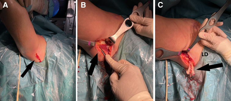
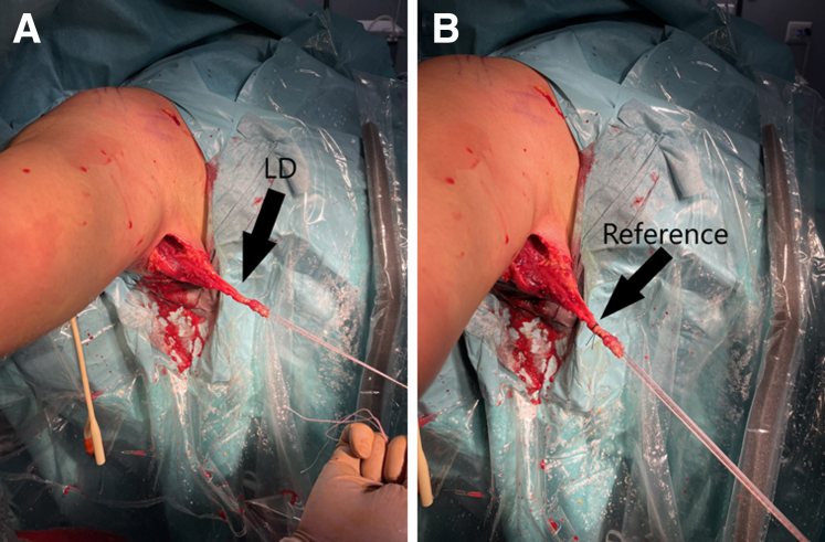
Massive irreparable cuff tears may represent as many as 20% to 40% of total cases of operated rotator cuff tears and can be a challenging clinical problem. Many treatment options have been proposed for their treatment. Among these options, latissimus dorsi tendon transfer can be considered a good alternative, especially in young patients before they develop glenohumeral arthritic changes. This technique aims at rebalancing the shoulder with a functioning subscapularis muscle and restoring both active external rotation and elevation with the aid of a properly functioning deltoid muscle. The modified arthroscopic latissimus dorsi transfer at the infraspinatus footprint with anterior extracortical fixation rebalances the pair of forces acting on the shoulder, stabilizing it in the transverse plane, minimizing the risk of latissimus dorsi transferred rupture and associated complications.


Figures

Fig 1
Fig 1
The surgical procedure is performed with the patient under general and locoregional anesthesia (interscalene block). The patient is placed in the beach-chair position with the arm parallel to the body. Right shoulder, no traction is used.
Fig 2
Fig 2
After subacromial bursectomy and resection of the remaining rotator interval using a shaver, we clean the entire rotator interval and expose the base of the coracoid to avoid postoperative pain and stiffness. The patient is placed in the beach-chair position with the arm parallel to the body. Right shoulder, no traction is used. Black arrow: supraspinatus remnants (C, coracoid; G, glenoid; HH, humeral head; St, Subscapularis tendon.).
Fig 3
Fig 3
(A) Latissimus dorsi (LD) dissection and detachment should be started from laterally to medially following the upper and lower borders. (B) This allows detachment of the LD tendon, achieving the optimal length while preserving the insertion of the teres major (TM). (C) Once the tendon of the LD is detached, it should be released from its adhesions to the surrounding structures: the TM posteriorly and the triceps inferiorly. The patient is placed in the beach-chair position with the arm parallel to the body. Right shoulder, no traction is used.
Fig 4
Fig 4
(A-B) We place a Foley catheter in the posterior area that we had previously prepared, after dissecting the infraspinatus fascia without injuring the axillary nerve. (C) The Foley catheter is inflated and left for a later step. (D) We will use it for the subsequent passage of the Latissimus dorsi to the posterior region. The patient is placed in the beach-chair position with the arm parallel to the body. Right shoulder, no traction is used.
Fig 5
Fig 5
(A) A 4- to 6-cm straight vertical incision is performed in the middle of the posterior half and distally to the axillary fold. (B). The subcutaneous tissue is divided until the “white tissue” of the LD is found and followed anteriorly to the LD border. (C) Then, dissection is continued superiorly until the previously arthroscopically released tendon is visible. If the tendon has been properly liberated during arthroscopy, it will “pop out” easily without the need for dissection at its humeral insertion. The patient is placed in the beach-chair position with the arm parallel to the body. Right shoulder, no traction is used. (LD, latissimus dorsi.)
Fig 6
Fig 6
(A) The insertional part of the LD is removed and tubulated using 2 high-strength sutures. (B) A mark is placed at 3 to 3.5 cm with a No. 0 Sofsilk, which we will use as a reference when we bring the LD up to the infraspinatus insertional area, to know how much tendon we have tunneled into the humeral head. The patient is placed in the beach-chair position with the arm parallel to the body. Right shoulder, no traction is used. (LD, Latissimus dorsi.)
Fig 7
Fig 7
(A) The triceps fascia is bluntly opened through the mini-open approach until the Foley catheter, which was previously left swollen, is palpated. It is deflated and pulled out through the axillar mini-open approach. (B) The sutures are tied to the Foley catheter and pulled proximally between the triceps and deltoid (care must be taken not to injure the axillary nerve). The patient is placed in the beach-chair position with the arm parallel to the body. Right shoulder, no traction is used.
Fig 8
Fig 8
(A) Arthroscopic preparation of the infraspinatus footprint on the humeral head, “just in front of subscapularis” is performed. (B) Using anterior cruciate ligament reconstruction instruments (VersiTomic; Stryker, Kalamazoo, MI), we pass a Beath pin from posterior to anterior and then drill 20- to 25-mm deep with an 8-mm thick drill bit. The drill bit is used to create a blind tunnel. The humeral cortex is drilled to a thickness of 8 mm and a depth of 20 to 25 mm. The patient is placed in the beach-chair position with the arm parallel to the body. Right shoulder, no traction is used.
Fig 9
Fig 9
(A,B) The high-strength sutures prepared in the LD are passed through the beath pin. Recovering them through the anterior cortex of the humerus. Using the ProCinch plate (ProCinch; Stryker, Kalamazoo, MI), we pass the sutures through it and place it on the anterior cortex of the humerus. We then tie the knot. The patient is placed in the beach-chair position with the arm parallel to the body. Right shoulder, no traction is used. (HH, humeral head; If, infraspinatus footprint; LD, latissimus dorsi.)
Fig 10
Fig 10
(A-C) With the aim of leaving the subscapularis insertion totally mirrored to the area where we have put in LD (infraspinatus footprint). We use a double suture-loaded Iconix anchor (Stryker, Kalamazoo, MI) on the anterior aspect of the humeral head and ascend the subscapularis tendon (ST). The patient is placed in the beach-chair position with the arm parallel to the body. Right shoulder, no traction is used. (HH, humeral head; LD, latissimus dorsi.)
Fig 11
Fig 11
In this way, we manage to lower the humeral head, so that the deltoid is the one that raises the arm, compensating for the absence of the supraspinatus. The patient is placed in the beach-chair position with the arm parallel to the body. Right shoulder, no traction is used. (G, glenoid; HH, humeral head.)

References

    1. Kany J., Grimberg J., Amaravathi R.S., Sekaran P., Scorpie D., Werthel J.D. Arthroscopically-assisted latissimus dorsi transfer for irreparable rotator cuff insufficiency: Modes of failure and clinical correlation. Arthroscopy. 2018;34:1139–1150. - PubMed
    1. Lädermann A., Denard P.J., Collin P. Massive rotator cuff tears: Definition and treatment. Int Orthop. 2015;39:2403–2414. - PubMed
    1. Parsons I.M., Apreleva M., Fu F.H., Woo S.L. The effect of rotator cuff tears on reaction forces at the glenohumeral joint. J Orthop Res. 2002;20:439–446. - PubMed
    1. Cutbush K., Peter N.A., Hirpara K. All-arthroscopic latissimus dorsi transfer. Arthrosc Tech. 2016;5:e607–e613. - PMC - PubMed
    1. Mulieri P., Dunning P., Klein S., Pupello D., Frankle M. Reverse shoulder arthroplasty for the treatment of irreparable rotator cuff tear without glenohumeral arthritis. J Bone Joint Surg Am. 2010;92:2544–2556. - PubMed

LinkOut - more resources