. 2023 Jul 18:14:1205162.
doi: 10.3389/fphys.2023.1205162. eCollection 2023.

Sex differences in pituitary corticotroph excitability

Affiliations

Sex differences in pituitary corticotroph excitability

Peter J Duncan et al. Front Physiol. .

Abstract

Stress-related illness represents a major burden on health and society. Sex differences in stress-related disorders are well documented, with women having twice the lifetime rate of depression compared to men and most anxiety disorders. Anterior pituitary corticotrophs are central components of the hypothalamic-pituitary-adrenal (HPA) axis, receiving input from hypothalamic neuropeptides corticotrophin-releasing hormone (CRH) and arginine vasopressin (AVP), while regulating glucocorticoid output from the adrenal cortex. The dynamic control of electrical excitability by CRH/AVP and glucocorticoids is critical for corticotroph function; however, whether corticotrophs contribute to sexually differential responses of the HPA axis, which might underlie differences in stress-related disorders, is very poorly understood. Using perforated patch clamp electrophysiology in corticotrophs from mice expressing green fluorescent protein under the control of the Pomc promoter, we characterized basal and secretagogue-evoked excitability. Both male and female corticotrophs show predominantly single-spike action potentials under basal conditions; however, males predominantly display spikes with small-amplitude (<20 mV) afterhyperpolarizations (B-type), whereas females displayed a mixture of B-type spikes and spikes with a large-amplitude (>25 mV) afterhyperpolarization (A-type). In response to CRH, or CRH/AVP, male cells almost exclusively transition to a predominantly pseudo-plateau bursting, whereas only female B-type cells display bursting in response to CRH±AVP. Treatment of male or female corticotrophs with 1 nM estradiol (E2) for 24-72 h has no effect on the proportion of cells with A- or B-type spikes in either sex. However, E2 results in the cessation of CRH-induced bursting in both male and female corticotrophs, which can be partially reversed by adding a BK current using a dynamic clamp. RNA-seq analysis of purified corticotrophs reveals extensive differential gene expression at the transcriptional level, including more than 71 mRNAs encoding ion channel subunits. Interestingly, there is a two-fold lower level (p < 0.01) of BK channel pore-forming subunit (Kcnma1) expression in females compared to males, which may partially explain the decrease in CRH-induced bursting. This study identified sex differences at the level of the anterior pituitary corticotroph ion channel landscape and control of both spontaneous and CRH-evoked excitability. Determining the mechanisms of sex differences of corticotroph and HPA activity at the cellular level could be an important step for better understanding, diagnosing, and treating stress-related disorders.

Keywords: BK channel; HPA axis; RNA-seq; corticotroph; electrophysiology; sex differences.


Conflict of interest statement

The authors declare that the research was conducted in the absence of any commercial or financial relationships that could be construed as a potential conflict of interest.

Figures

FIGURE 1
FIGURE 1
Male and female corticotrophs show different proportions of spontaneous A- and B-type action potentials. Representative traces from (A) A-type male cells (10%), (B) B-type male cells (86%), (C) A-type female cells (40%), and (D) B-type female cells (57%). (E–H) For each group, individual spikes at higher temporal resolution are shown below their respective traces. A-type spikes are characterized by a sharp upstroke, followed by a large AHP, while B-type spikes show a slower time to peak and a smaller AHP.
FIGURE 2
FIGURE 2
The distinct properties of A- and B-type action potentials do not vary between male and female corticotrophs. Spontaneous activity of male (blue) and female (red) corticotrophs was classified as A-type (n = 3 and n = 12 cells, respectively) or B-type (n = 25 and n = 17 cells, respectively). The properties of 10 consecutive spikes were averaged for each cell. There was no significant difference in (A) membrane potential, (B) spike frequency, or (C) spike amplitude between A- and B-type spikes for either male or female cells. (D) Spike width was significantly longer in B-type cells for both males and females. (E) Time to peak was significantly longer in only female B-type cells than that in A-type. Cells were categorized based on the AHP amplitude. A-type spikes have a large AHP (typically >25 mV), and B-type spikes have smaller AHP (typically <20 mV). (F) AHP amplitude was significantly larger in A-type cells for both male and female corticotrophs. **p < 0.01 linear regression model (lm) with Tukey’s post hoc test.
FIGURE 3
FIGURE 3
CRH and CRH/AVP evoke bursting in a lower proportion of female versus male corticotrophs. Secretagogue-evoked activity was measured after stimulating corticotrophs for 3 minutes with 0.2 nM CRH (n = 10 male/female cells), 2 nM AVP (n = 9 male and n = 10 female cells), or a combined stimulus of CRH and AVP (n = 10 male/female cells). Stimulation of male cells (blue) with CRH resulted in (A) an increase in single-spike frequency in 10% of cells and (B) a transition to bursting in 90% of cells. In contrast, CRH stimulation of female cells (red) resulted in (C) an an increase in single-spike frequency in 40% of cells and (D) a transition to bursting in 60% of cells. Comparing secretagogue-evoked activity revealed no sexual differences in (E) membrane potential for all secretagogue stimulations. (F) Event frequency was significantly higher in only female cells stimulated with CRH/AVP. (G) Event duration and (H) burst factor were significantly higher in male corticotrophs stimulated with CRH or CRH/AVP than those in the respective female cells. *p < 0.05 and ** p < 0.01 linear regression model (lm), with Tukey’s post hoc test.
FIGURE 4
FIGURE 4
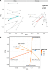
PCA of CRH-induced activity demonstrates bursting dominates in males, whilst either bursting or increased spiking occurs in females. A total of 12 parameters of corticotrophs were measured (cell capacitance, membrane potential, event frequency, spike frequency, burst frequency, event duration, spike duration, burst duration, burst factor, active time, event amplitude, and days in culture) to determine the main contributions to basal and CRH-evoked corticotroph excitability. Variables with high correlation (≥0.8) were removed (active time, burst factor, event frequency, and event duration) as they are predicted to measure the same underlying (but “latent”) aspect of a collection of variables. (A) Under basal conditions (white circles), male and female cells occupy a similar space on the PCA plot. The trajectory of each individual cell can be observed in response to CRH (black dots, connected to basal by a line). Male cells tend to move toward the negative PC1 direction. A proportion of female cells follow a similar trajectory to the male cells, but another group moves toward negative values of PC2. (B) Loading plots, showing the electrophysiological features used to calculate PCA. This shows that a decrease in PC1 corresponds to increased bursting behavior (burst duration and burst frequency) and a decrease in PC2 to an increased spike frequency (spike frequency and membrane potential).
FIGURE 5
FIGURE 5
CRH induces bursting in B-type cells and increased spike frequency in A-type cells. Female corticotrophs stimulated with CRH were categorized as either A type (n = 4 cells, yellow) or B type (n = 6 cells, orange). (A) Female corticotrophs with A-type spikes failed to transition to bursting when adding a BK conductance using dynamic clamp. (B) Under the same conditions, B-type cells could be transitioned to bursting. CRH significantly increased (C) event duration and (D) burst factor in B-type but not A-type cells. Bursting behavior could be enhanced in B-type cells with the addition of BK current, while the dynamic clamp failed to induce bursting in A-type cells. The parameters were gBK = 1 nS; vf = −10 mV; sf = 2 mV; and τBK = 2 ms, but slight variations were made in some cells depending on their intrinsic properties. ** p < 0.01 linear regression model (lm), with Tukey’s post hoc test.
FIGURE 6
FIGURE 6
Estradiol (E2) prevents CRH-induced bursting in male corticotrophs. Male corticotrophs (n = 7 cells, green) were cultured with 1 nM E2 at the time of plating, and electrophysiological recordings were obtained 24–72 h post-treatment. (A) E2-treated corticotrophs were spontaneously active. (B) Stimulation with CRH (0.2 nM for 3 min) resulted in an increase in single-spike frequency but not bursting in all cells tested (7/7 cells). (C) Addition of BK current induced bursting in 4/5 cells tested. CRH stimulation of E2-treated male cells resulted in (D) a significant increase in event frequency but not (E) event duration or (F) burst factor. Addition of the BK conductance significantly increased both event duration and burst factor. The parameters were gBK = 1 nS; vf = −10 mV; sf = 2 mV; and τBK = 2 ms, but slight variations were made in some cells depending on their intrinsic properties. **p < 0.01 mixed-effect linear model (lme), with Tukey’s post hoc test.
FIGURE 7
FIGURE 7
Estradiol (E2) prevents CRH-induced bursting in female corticotrophs. Female corticotrophs (n = 13 cells, purple) were cultured with 1 nM E2 at the time of plating, and electrophysiological recordings were obtained in the subsequent 24–72 h. (A) E2-treated corticotrophs were spontaneously active. (B) Stimulation with CRH (0.2 nM for 3 min) resulted in an increase in single-spike frequency in 10/13 cells, while CRH-induced bursting was observed in 3/13 cells. (C) Addition of BK current induced bursting in 7/11 cells tested. CRH stimulation of E2-treated female cells resulted in (D) a significant increase in event frequency but not (E) event duration or (F) burst factor. Addition of BK conductance significantly increased both event duration and burst factor. The parameters were gBK = 1 nS; vf = −10 mV; sf = 2 mV; and τBK = 2 ms, but slight variations were made in some cells depending on their intrinsic properties. **p < 0.01 mixed-effect linear model (lme), with Tukey’s post hoc test.
FIGURE 8
FIGURE 8
Sexual differences at the corticotroph ion channel gene transcription level. Corticotrophs from male and female mice were FACS-sorted and the transcriptome analyzed by bulk RNA-seq. (A) MA plots reveal a total of 2,480 differentially expressed genes (p < 0.05, red dots) between male and female corticotrophs. Genes above the zero line are enriched in female cells (1,332), while genes below the zero line are enriched in males (1,148). Ion channel genes were filtered from the dataset using GO:0005216 (ion channel activity). (B) A total of 71 ion channels were differentially expressed (p < 0.05), with 44 enriched in males and 27 enriched in females. (C) Heatmap showing differentially expressed ion channels in male (blue) and female (red) corticotrophs. Relative expression across each row (z-score) is shown as per the color code to the right of the panel. Columns indicate each sample comprising cells isolated from three pituitaries.

Similar articles

Cited by

References

    1. Aguilera G. (2011). HPA axis responsiveness to stress: Implications for healthy aging. Exp. Gerontol. 46, 90–95. 10.1016/j.exger.2010.08.023 - DOI - PMC - PubMed
    1. Andonegui-Elguera S., Silva-Roman G., Pena-Martinez E., Taniguchi-Ponciano K., Vela-Patino S., Remba-Shapiro I., et al. (2022). The genomic landscape of corticotroph tumors: From silent adenomas to ACTH-secreting carcinomas. Int. J. Mol. Sci. 23, 4861. 10.3390/ijms23094861 - DOI - PMC - PubMed
    1. Atkinson H. C., Waddell B. J. (1997). Circadian variation in basal plasma corticosterone and adrenocorticotropin in the rat: Sexual dimorphism and changes across the estrous cycle. Endocrinology 138, 3842–3848. 10.1210/endo.138.9.5395 - DOI - PubMed
    1. Bale T. L., Epperson C. N. (2015). Sex differences and stress across the lifespan. Nat. Neurosci. 18, 1413–1420. 10.1038/nn.4112 - DOI - PMC - PubMed
    1. Bangasser D. A., Cuarenta A. (2021). Sex differences in anxiety and depression: Circuits and mechanisms. Nat. Rev. Neurosci. 22, 674–684. 10.1038/s41583-021-00513-0 - DOI - PubMed

LinkOut - more resources