. 2023 Oct 3;25(10):1895-1909.
doi: 10.1093/neuonc/noad124.

Mapping pediatric brain tumors to their origins in the developing cerebellum

Affiliations

Mapping pediatric brain tumors to their origins in the developing cerebellum

Konstantin Okonechnikov et al. Neuro Oncol. .

Erratum in

Abstract

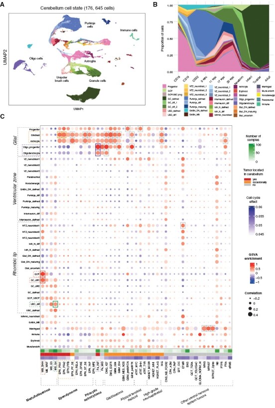
Background: Distinguishing the cellular origins of childhood brain tumors is key for understanding tumor initiation and identifying lineage-restricted, tumor-specific therapeutic targets. Previous strategies to map the cell-of-origin typically involved comparing human tumors to murine embryonal tissues, which is potentially limited due to species-specific differences. The aim of this study was to unravel the cellular origins of the 3 most common pediatric brain tumors, ependymoma, pilocytic astrocytoma, and medulloblastoma, using a developing human cerebellar atlas.

Methods: We used a single-nucleus atlas of the normal developing human cerebellum consisting of 176 645 cells as a reference for an in-depth comparison to 4416 bulk and single-cell transcriptome tumor datasets, using gene set variation analysis, correlation, and single-cell matching techniques.

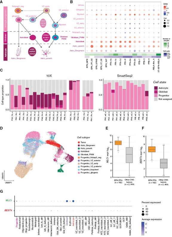
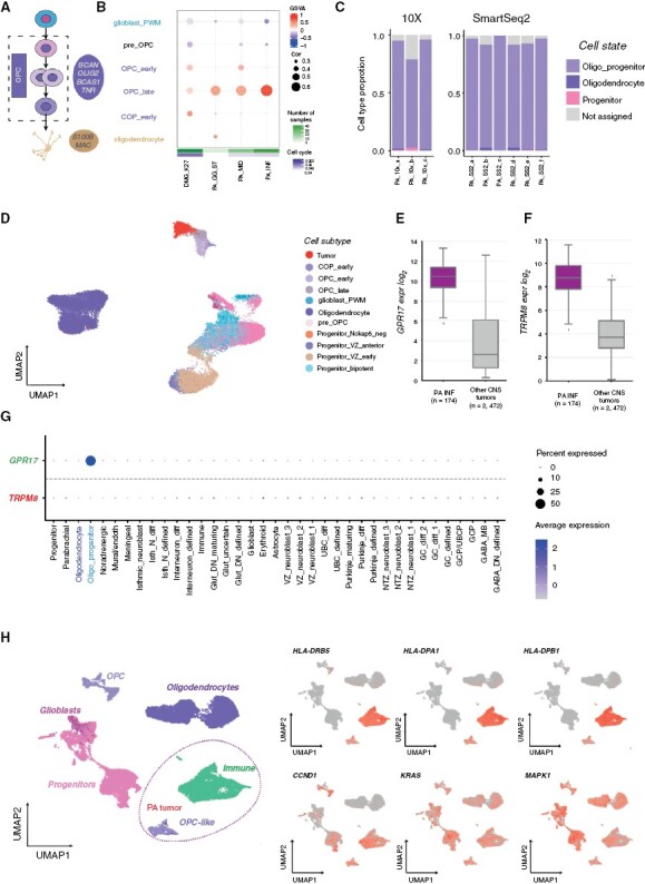
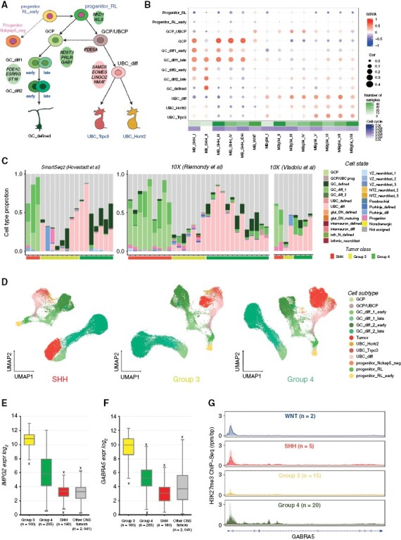
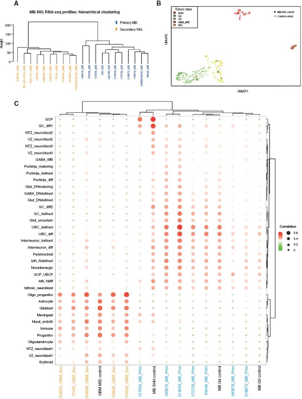
Results: We find that the astroglial cerebellar lineage is potentially the origin for posterior fossa ependymomas. We propose that infratentorial pilocytic astrocytomas originate from the oligodendrocyte lineage and MHC II genes are specifically enriched in these tumors. We confirm that SHH and Group 3/4 medulloblastomas originate from the granule cell and unipolar brush cell lineages. Radiation-induced gliomas stem from cerebellar glial lineages and demonstrate distinct origins from the primary medulloblastoma. We identify tumor genes that are expressed in the cerebellar lineage of origin, and genes that are tumor specific; both gene sets represent promising therapeutic targets for future study.

Conclusion: Based on our results, individual cells within a tumor may resemble different cell types along a restricted developmental lineage. Therefore, we suggest that tumors can arise from multiple cellular states along the cerebellar "lineage of origin."

Keywords: cerebellum; ependymoma; medulloblastoma; pilocytic astrocytoma; radiation-induced glioma; tumor origin.


Conflict of interest statement

The authors declare no competing financial interests.

Figures

Figure 1.
Figure 1.
Human single-nuclei developmental cerebellum cell atlas comparisons reveal putative lineages-of-origin of cerebellar tumors. (A) UMAP of human developmental cerebellum single-nuclei data used as a reference. Color codes represent cell state annotation and labels denote the main cell types. (B) Proportions of different states across developmental time. (C) Heatmap of comparisons of bulk central nervous system tumor transcriptome profiles (in columns) to cerebellar cell state (in rows), based on gene signature enrichment score (via Gene Set Variance Analysis (GSVA, indicated by color) and Pearson correlation score (indicated by size). Highest associations marked by rectangles, based on cutoff limits of min GSVA enrichment = 0.4 and min correlation = 0.4.
Figure 2.
Figure 2.
Ependymoma tumors arise from the astroglial lineage. (A) Astroglial cell lineage derived from early ventricular zone (VZ) progenitors. (B) Comparison of bulk ependymoma tumor gene expression profiles to cell subtypes of the astroglial lineage, based on GSVA enrichment and correlation measures. (C) Ependymoma single cells assigned to the closest normal cerebellum cell state; cells with <50% similar are classified as “not assigned.” (D) Projection of an individual single-cell tumor data (PFA1_10x_g, 10x v2) onto astroglial cell subtypes as visualized via UMAP. (E) Median expression boxplot of MLC1, a PFA-ependymoma enriched gene expressed within the astroglial lineage (limma adjusted P-value: 5.67E-31). (F) Median expression boxplot of BEST4, a PFA-ependymoma-specific gene (limma adjusted P-value: 1.75E−61). (G) Specificity of MLC1 and BEST4 gene expression across cerebellum cell states.
Figure 3.
Figure 3.
Pilocytic astrocytomas arise from the oligodendrocyte lineage. (A) Oligodendrocyte cerebellar lineage. (B) Comparison of bulk pilocytic astrocytoma gene expression profiles to cell subtypes from the oligodendrocyte lineage based on GSVA enrichment and correlation measures. (C) Pilocytic astrocytoma single-cell data SVM-based comparison to cerebellum cell state. (D) Projection of a single-cell tumor sample (ID: PA_10X_a, 10x v2) onto astroglia and oligodendrocyte cell subtypes as visualized via UMAP. (E) Median expression boxplot of GPR17, a pilocytic astrocytoma enriched gene expressed within the oligodendrocyte lineage (limma adjusted P-value: 1.25E−105). (F) Median expression boxplot of TRPM8, a pilocytic astrocytoma unique gene (limma adjusted P-value: 2.14E−72). (G) Specificity of GPR17 and TRPM8 gene expression across cerebellum cell states. (H) Comparison of a single-cell sample (ID: PA_10X_a, nontumorous immune cells included) with the oligodendrocyte lineage, and relative gene expression of MHC- and MAPK-associated genes.
Figure 4.
Figure 4.
Medulloblastomas arise from the granule cell and unipolar brush cell lineages. (A) Granule/unipolar brush cell (GC/UBC) lineage arising from rhombic lip progenitors. (B) Comparison of bulk medulloblastoma gene expression profiles to the GC/UBC lineage, based on GSVA enrichment and correlation measures. (C) Medulloblastoma single-cell data SVM-based comparison to cerebellum cell state. (D) 10X genomics medulloblastoma samples (IDs: SHH_10X_b, G3_10x_b, G4_10x_c) projection onto the GC/UBC lineage as visualized via UMAP. (E) Median expression boxplot of IMPG2, a medulloblastoma enriched gene expressed within the GC/UBC lineage (limma adjusted p-value: 5.72E−148). (F) Median expression boxplot GABRA5, a medulloblastoma-specific gene (limma adjusted P-value: 2.89E−77). (G) H3K27me3 ChIP-seq profiles (reads per million mapped reads per bp, rpm/bp) of GABRA5 loci in the four medulloblastoma groups.
Figure 5.
Figure 5.
Secondary radiation-induced gliomas (RIGs) originate de novo from the glial cell lineage and not from the original primary tumor. (A) Unsupervised hierarchical clustering of batch-effect adjusted RNA-seq profiles from primary medulloblastoma and secondary RIG tumors (based on top 500 HVGs). (B) UMAP visualization of medulloblastoma and glioblastomas. Primary medulloblastoma and corresponding secondary RIGs are marked with circles. (C) Comparison of gene expression profiles of primary medulloblastoma and secondary RIG tumors combined with corresponding FFPE bulk control to cerebellum cell state, based on correlation measures.

References

    1. Udaka YT, Packer RJ.. Pediatric brain tumors. Neurol Clin. 2018;36(3):533–556. - PubMed
    1. Chemaitilly W, Armstrong GT, Gajjar A, Hudson MM.. Hypothalamic-pituitary axis dysfunction in survivors of childhood CNS tumors: importance of systematic follow-up and early endocrine consultation. J Clin Oncol. 2016;34(36):4315–4319. - PubMed
    1. Jones DT, Banito A, Grünewald TG, et al. . Molecular characteristics and therapeutic vulnerabilities across paediatric solid tumours. Nat Rev Cancer. 2019;19(8):420–438. - PubMed
    1. Baslan T, Hicks J.. Unravelling biology and shifting paradigms in cancer with single-cell sequencing. Nat Rev Cancer. 2017;17(9):557–569. - PubMed
    1. Kaatsch P, Rickert CH, Kühl J, Schüz J, Michaelis J.. Population-based epidemiologic data on brain tumors in German children. Cancer: Interdisc Int J Am Cancer Soc. 2001;92(12):3155–3164. - PubMed

Publication types