. 2023 Nov;160(5):435-452.
doi: 10.1007/s00418-023-02224-2. Epub 2023 Aug 3.

Selective targeting of lectins and their macropinocytosis in urothelial tumours: translation from in vitro to ex vivo

Affiliations

Selective targeting of lectins and their macropinocytosis in urothelial tumours: translation from in vitro to ex vivo

Nataša Resnik et al. Histochem Cell Biol. 2023 Nov.

Abstract

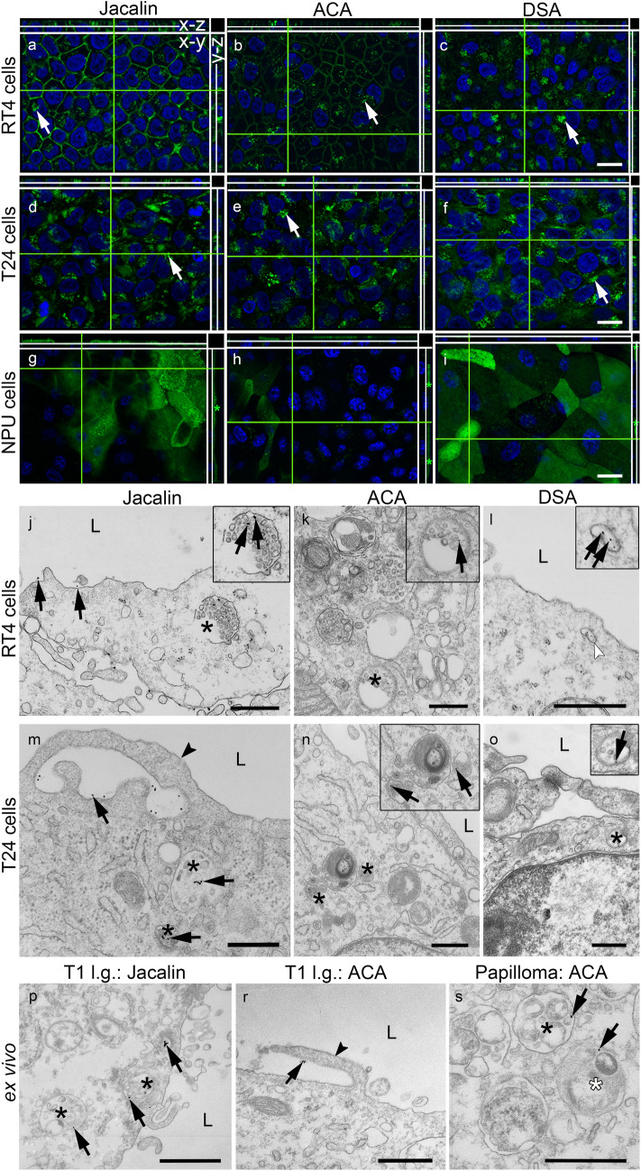
Urinary bladder cancer can be treated by intravesical application of therapeutic agents, but the specific targeting of cancer urothelial cells and the endocytotic pathways of the agents are not known. During carcinogenesis, the superficial urothelial cells exhibit changes in sugar residues on the apical plasma membranes. This can be exploited for selective targeting from the luminal side of the bladder. Here we show that the plant lectins Jacalin (from Artocarpus integrifolia), ACA (from Amaranthus caudatus) and DSA (from Datura stramonium) selectively bind to the apical plasma membrane of low- (RT4) and high-grade (T24) cancer urothelial cells in vitro and urothelial tumours ex vivo. The amount of lectin binding was significantly different between RT4 and T24 cells. Endocytosis of lectins was observed only in cancer urothelial cells and not in normal urothelial cells. Transmission electron microscopy analysis showed macropinosomes, endosome-like vesicles and multivesicular bodies filled with lectins in RT4 and T24 cells and also in cells of urothelial tumours ex vivo. Endocytosis of Jacalin and ACA in cancer cells was decreased in vitro after addition of inhibitor of macropinocytosis 5-(N-ethyl-N-isopropyl) amiloride (EIPA) and increased after stimulation of macropinocytosis with epidermal growth factor (EGF). Clathrin, caveolin and flotillin did not colocalise with lectins. These results confirm that the predominant mechanism of lectin endocytosis in cancer urothelial cells is macropinocytosis. Therefore, we propose that lectins in combination with conjugated therapeutic agents are promising tools for improved intravesical therapy by targeting cancer cells.

Keywords: Cancer urothelial cells; Endocytosis; Glycosylation; Lectins; Macropinocytosis; Urinary bladder tumours.


Conflict of interest statement

The authors declare no conflict of interest.

Figures

Fig. 1
Fig. 1
UP expression in urothelial normal and cancer cells in in vitro and ex vivo models. UP expression (red) is highest in NPU cells (a) and in normal tissue (e, k). UP expression is restricted to individual RT4 cells (b) and to individual superficial cells of Ta (f, ln) and T1 tissue samples (g, or). T24 cells do not express UPs (c). Immunoblotting of UPs (upper strong band corresponds to UPIIIa-47 kDa and weak lower band corresponds to UPIa-27 kDa) shows strong expression in NPU cells, very weak expression in RT4 cells and no expression in T24 cells (d). Staining with haematoxylin and eosin shows that the morphology of the urothelium is altered in Ta (i) and T1 (j) tumours compared to normal urothelium (h). Immuno-electron microscopy shows abundant UPs in the apical plasma membrane (arrows) and in discoidal vesicles (arrowheads) in superficial cells of normal urothelium (k). In the superficial cells of Ta (ln) and T1 (or) tumours, immunolabelling of UPs in the apical plasma membrane (arrows) and in round vesicles (arrowheads) is weaker. The insets in the left corners (lr) are 250% magnifications of marked region. L = lumen. Scale bar: ah 50 µm, ij 100 µm, kr 1 µm
Fig. 2
Fig. 2
Lectins bind selectively to superficial urothelial cells in normal and cancer biopsy samples ex vivo. The apical plasma membrane of normal urothelial cells (ac2) and cells in Ta tumours (df2) is labelled with Jacalin, ACA and DSA (green). The apical plasma membrane of urothelial cells in T1 tumours (gi2) is labelled with Jacalin (g) and is negative for ACA (h) and DSA (i). Differentiated superficial urothelial cells in normal urothelium (a1, b1, c1), Ta l.g. (d1, e1, f1) and T1 l.g. (g1, h1, i1) express UPs (red). In merged images colocalisation or no colocalisation between lectins and UPs is clearly observed: UP-positive cells with lectin labelling = colocalisation (a2, b2, c2, d2, f2, g2; yellow arrows); UP-negative cells without lectin labelling = no colocalisation (a2, d2, f2, h2, i2; white arrows); UP-positive cells without lectin labelling = no colocalisation (a2, b2, e2, f2, h2, i2; red arrows); UP-negative cells with lectin labelling = no colocalisation (b2, c2, d2, e2, f2, g2; green arrows). L = lumen; l.g. = low grade. Scale bar for all images: 50 µm
Fig. 3
Fig. 3
Selective lectin binding to urothelial normal and cancer cells in vitro. FITC-conjugated lectins Jacalin (a, b, c), ACA (d, e, f) and DSA (g, h, i) (green) bind to the plasma membrane of NPU, RT4 and T24 cells. The plasma membranes of all T24 and RT4 cells are uniformly labelled with Jacalin, ACA and DSA, whereas NPU cells have stronger (green arrowheads) and weaker (white arrowheads) labelling with Jacalin, ACA and DSA. The green lines on the x–y sections correspond to the white lines on the x–z and y–z views of the optical sections. NPU cells have a heterogeneous morphology shown on x–y sections of the apical parts of the cells. (j) Lectin binding to NPU, RT4 and T24 cells is calculated from absolute values of FITC fluorescence intensities. Shown are average values ± SE; *p < 0.05, **p < 0.001. Scale bar: 10 µm
Fig. 4
Fig. 4
Correlation of FITC conjugated lectins Jacalin (a), ACA (b) and DSA (c) binding (green) and UP labelling (red) in NPU cells. UP-positive cells with lectins (a, c; three asterisks), UP-negative cells without lectin (a, b, c; hash), UP-positive cells without lectins (b; two asterisks), UP-negative cells with lectins (a, b; one asterisk). Shown are x–y, x–z and y–z views of optical sections. Scale bar: 10 µm
Fig. 5
Fig. 5
Endocytosis of FITC-conjugated lectins in urothelial cells in vitro and ex vivo. Jacalin (a, d), ACA (b, e) and DSA (c, f) were found in the cytoplasm of T24 and RT4 cells (white arrows). It is important to note that in NPU cells Jacalin (g), ACA (h) and DSA (i) are distributed only in the apical plasma membrane (y–z, green asterisk) and that the cytoplasm is without lectins. Shown are x–y, x–z and y–z views of optical sections. In the TEM images (js), the colloidal gold-conjugated lectins are indicated by black arrows. Jacalin is surrounded by membrane ruffles (m; arrowhead), which are an ultrastructural feature of macropinocytosis. After endocytosis, Jacalin (j, m; arrows), ACA (k, n; arrows) and DSA (l, o; arrows) are observed in MVBs (j, k, m, n, o; asterisk) and endosome-like vesicles (l; white arrowhead). The framed images are enlargements of the MVBs and vesicles with lectins. In the tumour biopsy samples ex vivo, the lectins (arrows) bind to the apical plasma membrane (r) and are associated with the plasma membrane ruffles (r; arrowhead). In the cytoplasm, lectins are gathered in multivesicular bodies (MVBs; p, s; black asterisk) and amphisome (s; white asterisk). L = lumen; l.g. = low grade. Scale bar: ai 20 µm, js 500 nm
Fig. 6
Fig. 6
Macropinocytosis of the FITC-conjugated lectins Jacalin, ACA and DSA in cancer urothelial cells in vitro. (a, e, c, d, g, h) TRITC-conjugated dextran (3 kDa) (Dex 3 kDa, red), (b, f) TRITC-conjugated dextran (70 kDa) (Dex 70 kDa, red) and FITC-conjugated lectin (green) Jacalin (a, b, e, f), ACA (c, g) and DSA (d, h) colocalise in T24 and RT4 cells (yellow). Details of dextran and lectin colocalisation (yellow arrows) are shown enlarged in the white frames. (i) Quantification of lectin binding and internalisation in RT4 and T24 cells after treatment with EIPA (lectin + EIPA), EGF (lectin + EGF) and Dyngo (lectin + Dyngo) and without treatment (lectin). The intensities of the fluorescence were normalised to the endocytosis of the single lectin in cells without treatment (value 1). Shown are averages ± SE; *p < 0.05, **p < 0.001. (j) The distribution of TRITC-conjugated dextran (70 kDa) (red) and actin filaments (FITC-conjugated phalloidin; green) in T24 cells without EGF stimulation (control) and with EGF stimulation. In EGF stimulated cells, TRITC-conjugated dextran (70 kDa) (red) is surrounded by actin filaments (green) in the apical cytoplasm (white arrow and green frames in x–z and y–z views). EGF-stimulated cells have more macropinosomes than control (unstimulated) cells (red arrows). Scale bar: ah 20 µm, j 10 µm

References

    1. Allen RD, Schroeder CC, Fok AK. Intracellular binding of wheat-germ agglutinin by golgi complexes, phagosomes, and lysosomes of paramecium-multimicronucleatum. J Histochem Cytochem. 1989;37(2):195–202. doi: 10.1177/37.2.2911005. - DOI - PubMed
    1. Amin MB, Greene FL, Edge SB, Compton CC, Gershenwald JE, Brookland RK, Meyer L, Gress DM, Byrd DR, Winchester DP. The eighth edition AJCC cancer staging manual: continuing to build a bridge from a population-based to a more “personalized” approach to cancer staging. CA Cancer J Clin. 2017;67(2):93–99. doi: 10.3322/caac.21388. - DOI - PubMed
    1. Aragon-Ching JB, Werntz RP, Zietman AL, Steinberg GD. Multidisciplinary management of muscle-invasive bladder cancer: current challenges and future directions. Am Soc Clin Oncol Educ Book. 2018;38:307–318. doi: 10.1200/edbk_201227. - DOI - PubMed
    1. Azevedo R, Peixoto A, Gaiteiro C, Fernandes E, Neves M, Lima L, Santos LL, Ferreira JA. Over forty years of bladder cancer glycobiology: where do glycans stand facing precision oncology? Oncotarget. 2017;8(53):91734–91764. doi: 10.18632/oncotarget.19433. - DOI - PMC - PubMed
    1. Bergman S, Javadpour N. Cell-surface antigen A, B or O(H) as an indicator of malignant potential in stage a bladder carcinoma - preliminary-report. J Urology. 1978;119(1):49–51. doi: 10.1016/S0022-5347(17)57378-4. - DOI - PubMed