. 2023 Aug 4;381(6657):502-508.
doi: 10.1126/science.abq3178. Epub 2023 Aug 3.

Peptide YY: A Paneth cell antimicrobial peptide that maintains Candida gut commensalism

Affiliations

Peptide YY: A Paneth cell antimicrobial peptide that maintains Candida gut commensalism

Joseph F Pierre et al. Science. .

Abstract

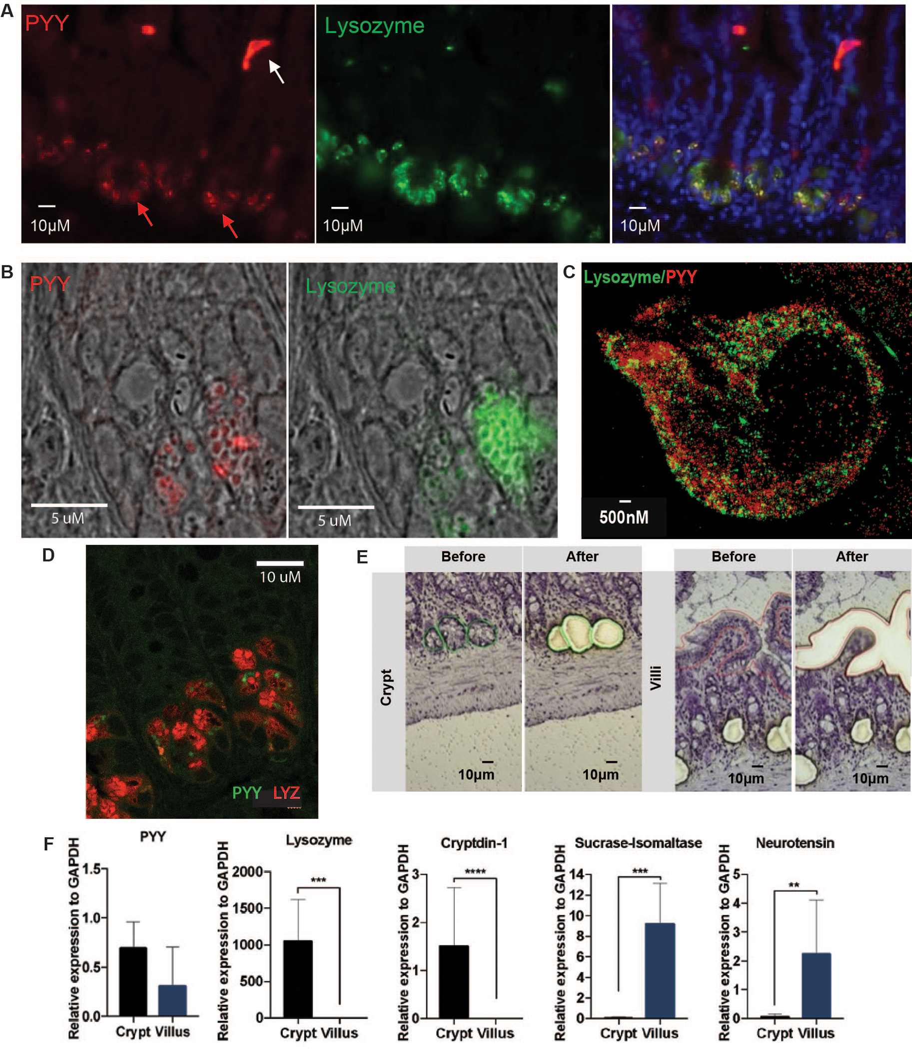
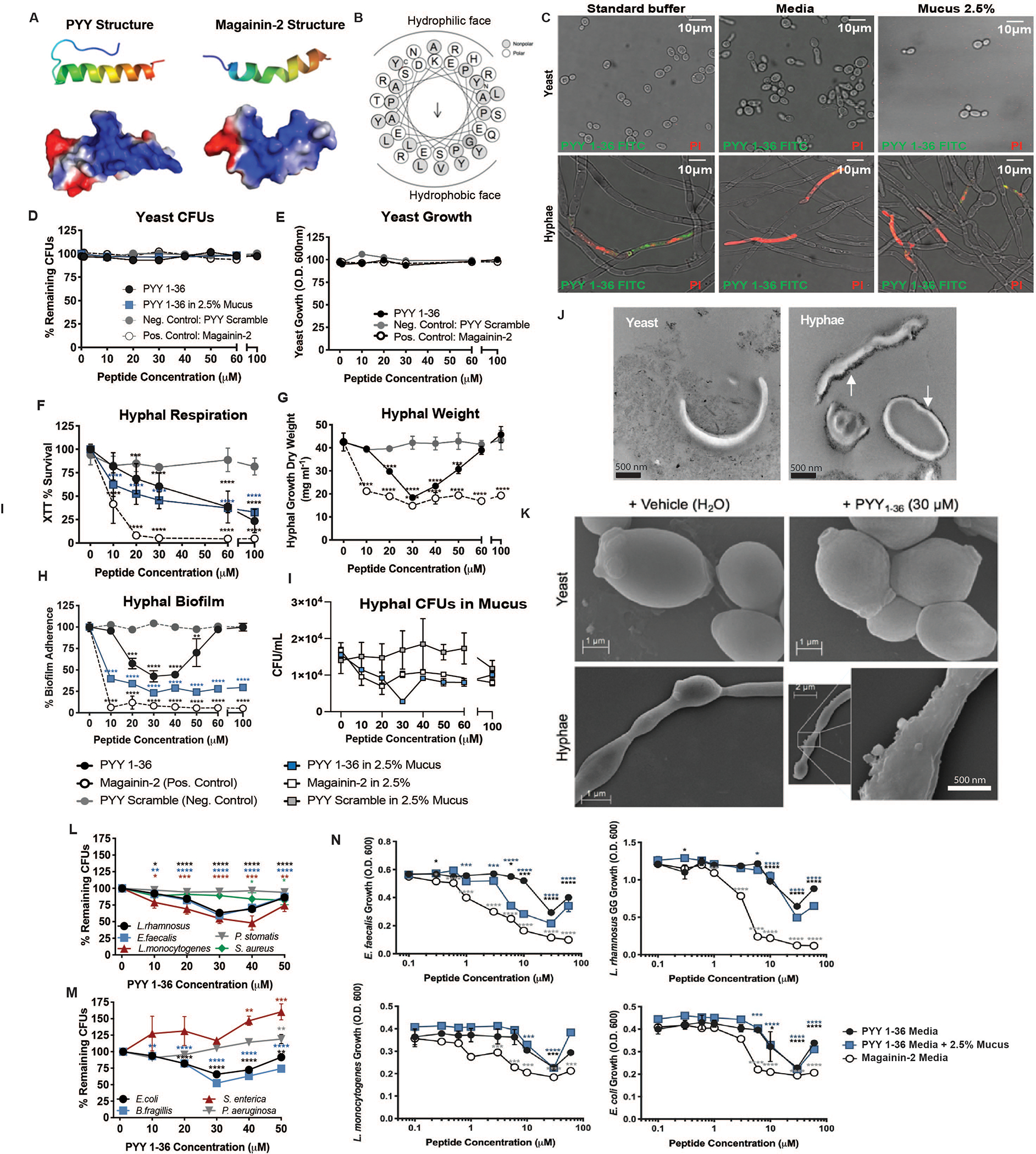
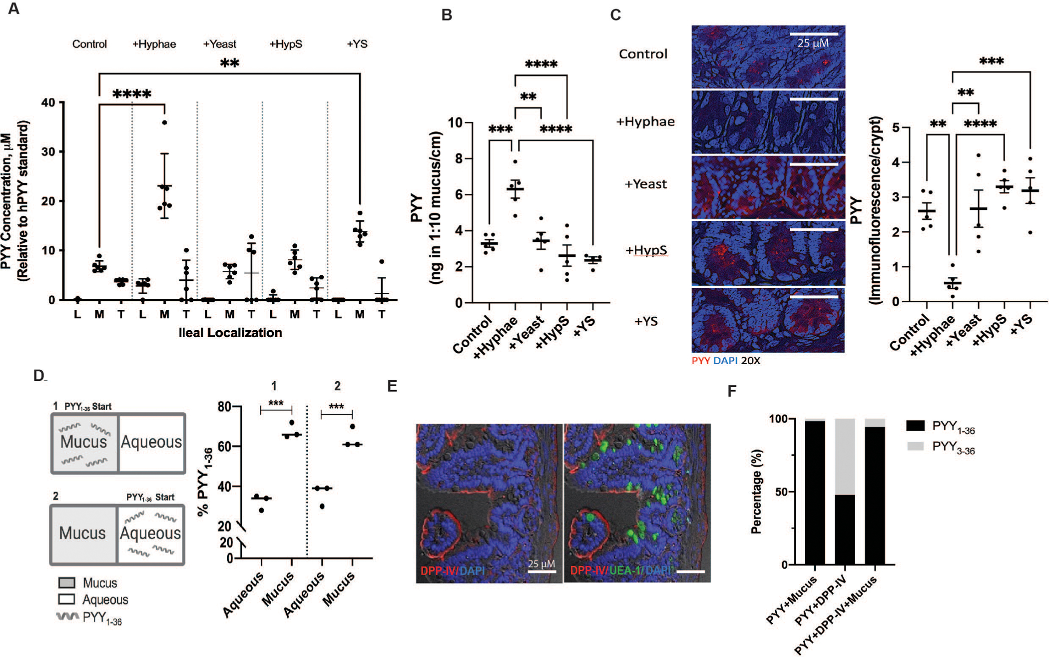
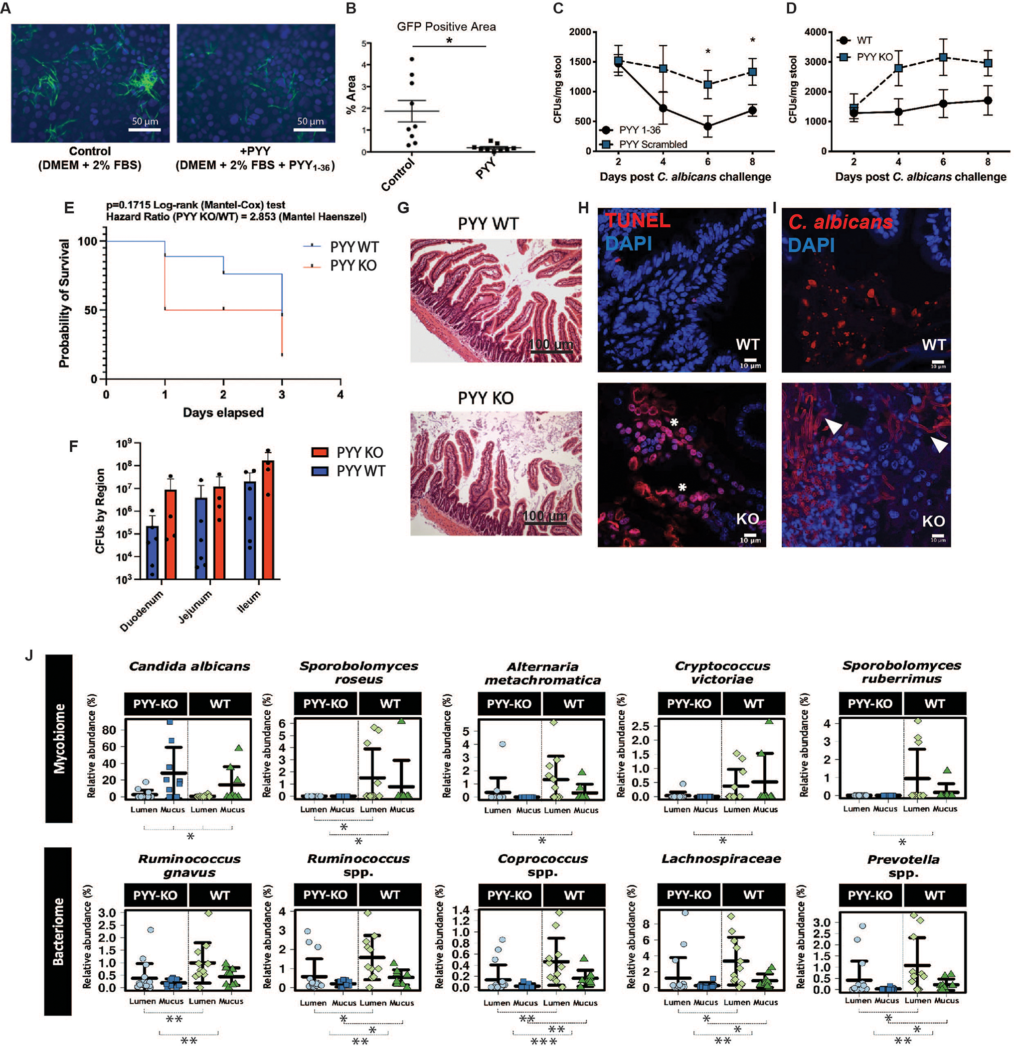
The mammalian gut secretes a family of multifunctional peptides that affect appetite, intestinal secretions, and motility whereas others regulate the microbiota. We have found that peptide YY (PYY1-36), but not endocrine PYY3-36, acts as an antimicrobial peptide (AMP) expressed by gut epithelial paneth cells (PC). PC-PYY is packaged into secretory granules and is secreted into and retained by surface mucus, which optimizes PC-PYY activity. Although PC-PYY shows some antibacterial activity, it displays selective antifungal activity against virulent Candida albicans hyphae-but not the yeast form. PC-PYY is a cationic molecule that interacts with the anionic surfaces of fungal hyphae to cause membrane disruption and transcriptional reprogramming that selects for the yeast phenotype. Hence, PC-PYY is an antifungal AMP that contributes to the maintenance of gut fungal commensalism.


Conflict of interest statement

Competing interests: EBC, JFP, and KGH are co-inventors of Patent PCT/US18/32997, Publication No 20200062823, “Compositions and methods for treating and/or preventing pathogenic fungal infection and for maintenance of microbiome commensalism”.

Figures

Fig. 1.
Fig. 1.. Peptide YY (PYY) localizes to ileal Paneth cells.
(A) PYY (red) detection in Paneth cells (PC, red arrow) and L-cells (white arrow) via anti-PYY antibody immunofluorescence (IF). PC PYY staining co-localized (right panel) with PC Lysozyme (LYZ, green; middle panel). (B) Confirmation of PC PYY localization via mRNA Fluorescent In Situ Hybridization (red, left panel) and counterstaining with anti-LYZ antibody (green, right panel). (C) High-resolution stimulated emission depletion IF microscopy of PC PYY and LYZ packaging in discrete secretory granules within the cytosol. (D) SP8 confocal microscopy of PYY (green) and LYZ (red) in ileum PCs. (E) Representative extractions from ileal crypts and villus epithelial cells via Laser-capture microdissection (LCM). (F) Relative expression of PYY and marker mRNA for crypt secretion compared with villus epithelium secretion estimated from LCM. Lysozyme and Cryptdin-1 are AMPs that are also secreted from PCs; Sucrase-isomaltase is released from the villus surface brush border; Neurotensin is a marker for enteroendocrine cells. Significance was determined using t-test (n=6, repeated twice, *p<0.05; **p<0.01; ***p<0.001; ****p<0.0001).
Fig. 2.
Fig. 2.. PYY displays antimicrobial activity towards Candida albicans hyphae and some bacteria.
(A) Ribbon diagram (top) and space-filling model indicating electrostatic surface charge (bottom; red negative, blue positive) for amphipathic alpha-helix structures of PYY1–36 and Magainin-2. (B) Amphipathic PYY1–36 helical wheel projection displaying surface localization of residues and calculated hydrophobic dipole moment (μH 0.208, arrow). (C) Propidium iodide (PI) staining of C. albicans yeast and hyphae following exposure to PYY1–36-FITC (green) in standard antimicrobial peptide assay buffer (AMP), preferred growth media for each form (YPD-yeast or RPMI-hyphae), or standard buffer + 2.5% w/v porcine mucus. C. albicans yeast (D) survival (Colony Forming Units, CFUs) and (E) growth (Optical density, O.D.) following exposure to PYY1–36 +/− mucus, PYY scramble, and Magainin-2 peptides. Pathogenic C. albicans hyphal (F) respiration by tetrazolium salt (XTT) assay (G) dry weight (H) biofilm adherence and (I) CFUs following exposure to PYY1–36 +/− mucus, PYY scramble, and Magainin-2 peptides. (J) Transmission electron microscopy for visualization of cationic surfaces (black probe accumulation) in C. albicans yeast and hyphae membranes. (K) Scanning electron microscopy of C. albicans yeast and hyphae following 2-hour exposure to vehicle (H2O) or PYY1–36. Dose-dependent killing of Gram-positive (L) and Gram-negative (M) bacteria induced by PYY1–36. CFUs remaining were normalized to 0 μM PYY. Significance was determined via ANOVA. (N) Impact of varying concentrations of PYY1–36 +/− mucus versus Magainin-2 on bacteria growth. O.D. values were normalized to wells containing 0 μM PYY. All assays were performed in duplicate three times. Significance was determined via ANOVA (*p<0.05; **p<0.01; ***p<0.001; ****p<0.0001).
Fig 3.
Fig 3.. Exposure to C. albicans hyphae but not yeast enhances PYY localization into ileal mucus.
(A) PYY1–36 quantification using liquid chromatography-electrospray ionization-mass spectrometry (LC-ESI-MS) in lumen (L), mucus (M), and tissue (T) of ex vivo ileal loops (n=6 /treatment) and in (B) mucus of in vivo ileal loops (n=5 /treatment) following stimulation with vehicle (Control), C. albicans hyphae (+Hyphae), C. albicans yeast (+Yeast), hyphae conditioned media (CM) supernatant (+HypS), or yeast CM supernatant (+YS). (C) Representative staining and quantification of PC PYY protein (red) following in vivo ileal loop stimulation with vehicle control, +Hyphae, +Yeast, +HypS, or +YS. Significance was measured using ANOVA and Dunnett’s Multiple Comparisons Test; **p<0.01, *** p<0.005, **** p<0.001. (D) PYY mucus vs. aqueous partitioning in vitro. PYY1–36 was added to either mucus (1) or aqueous (2) wells separated by a 10 kDa MWCO filter. Significance was measured using t-test; ***p<0.001. (E) Dipeptidyl peptidase IV (DDP-IV) immunofluorescence (IF) in murine ileum brush border. Lectin UEA-I-FITC (Ulex europaeus) counterstain = ileal mucus; DAPI = nuclei. (F) PYY1–36 and PYY3–36 exposure to DPP-IV +/− mucus to assess peptide degradation via LC-ESI-MS (n=3/treatment), expressed as a percent of total peptide. All experiments were done in triplicate with 3 replicate experiments.
Fig 4.
Fig 4.. PYY reduces gastrointestinal colonization of C. albicans in vitro and in vivo.
(A, B) C. albicans hyphae abundance (Hyphae-GFP) on confluent Caco2 cells exposed to vehicle (Control) or PYY1–36 (20μm) after 6 hours. (C) Fungal colony forming units (CFUs) in wild-type C57BL/6 mice (WT) +/− exogenously administered PYY1–36 or scrambled peptide via oral gavage 8 days post C. albicans (4×10⁶ CFU) challenge (n=8/group, *p<0.05 vs control). (D) C. albicans colonization (4×10⁶ CFUs) in PYY-KO relative to WT mice (n=8–9/timepoint/group). (E) 72-hour PYY-KO and WT mouse survival after gavage with C. albicans (2×10⁷ CFU; n=8/group, repeated twice with both males and females) under cefoxitin and clindamycin (2 WT were humanely euthanized). (F) CFUs recovered from in small intestinal regions of PYY-KO and WT animals gavaged with C. albicans (2×10⁷ CFU) (G) Intestinal mucosal gross morphology remained intact, while (H) epithelial cell apoptosis (red = TUNEL; white asterisk) and (I) virulent Candida morphology (hyphae; white arrows) was increased vs yeast in the mucosa of PYY-KO vs WT animals (36.3±12.1 vs. 11.77±3.8 %, p<0.04) following oral gavage challenge under clindamycin (2×10⁷ CFU/mouse; n=8/group; repeated twice with both males and females, red = Candida). (J) Differentially altered fungal (top panel, n=39 samples) and bacterial (bottom panel, n=46 samples) populations from small intestinal mucus and lumen of PYY-KO vs. WT animals without Candida challenge as determined via Wilcoxon rank test (between groups) and Kruskal-Wallis test (across groups) *: p<0.05, **: p<0.01, ***: p<0.001. To account for uneven sequencing depth, data were transformed into relative abundances based on total sum scaling.

Comment in

References

    1. Costello EK, Lauber CL, Hamady M, Fierer N, Gordon JI, Knight R, Bacterial community variation in human body habitats across space and time. Science. 326, 1694–1697 (2009). - PMC - PubMed
    1. Batterham RL, Cowley MA, Small CJ, Herzog H, Cohen MA, Dakin CL, Wren AM, Brynes AE, Low MJ, Ghatei MA, Cone RD, Bloom SR, Gut hormone PYY(3–36) physiologically inhibits food intake. Nature. 418, 650–4 (2002). - PubMed
    1. Witchley JN, Penumetcha P, Abonv N, Woolford C, Michell A, Noble SM, Candida albicans Morphogenesis Programs Control the Balance between Gut Commensalism and Invasive Infection. Cell Host Microbe. 25, 432–443 (2019). - PMC - PubMed
    1. Bevins CL, Salzman NH, Paneth cells, antimicrobial peptides and maintenance of intestinal homeostasis. Nat Rev Microbiol. 9, 356–68 (2011). - PubMed
    1. Clevers HC, Bevins CL, Paneth cells: Maestros of the small intestinal crypts. Annu Rev Physiol. 75 (2013), pp. 289–311. - PubMed