. 2023 Aug 3;14(8):494.
doi: 10.1038/s41419-023-06025-2.

Smurf1 and Smurf2 mediated polyubiquitination and degradation of RNF220 suppresses Shh-group medulloblastoma

Affiliations

Smurf1 and Smurf2 mediated polyubiquitination and degradation of RNF220 suppresses Shh-group medulloblastoma

Yuwei Li et al. Cell Death Dis. .

Abstract

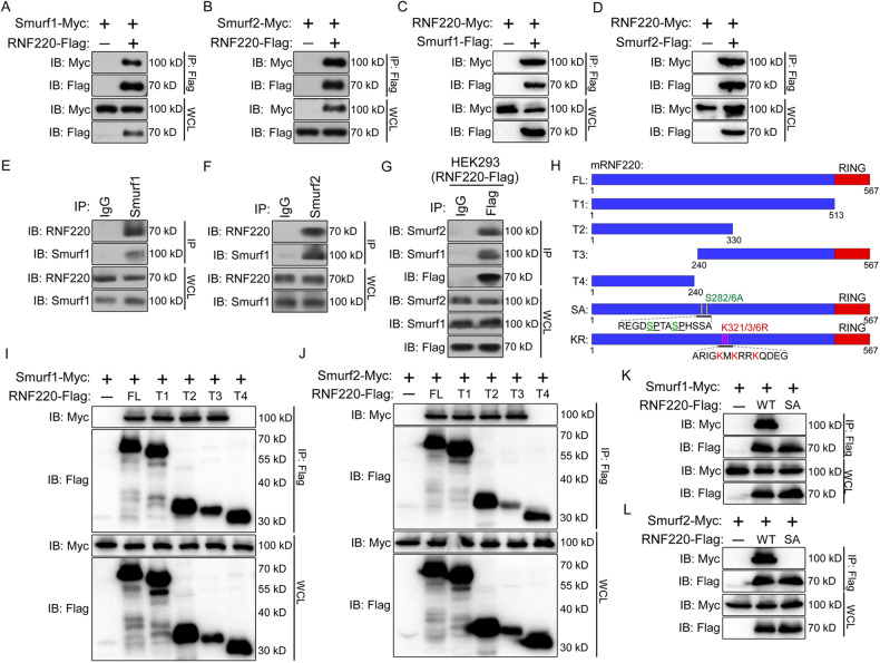
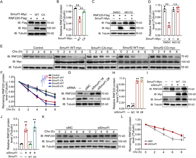
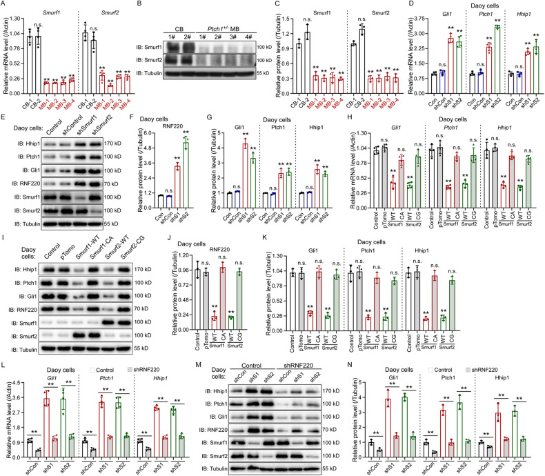
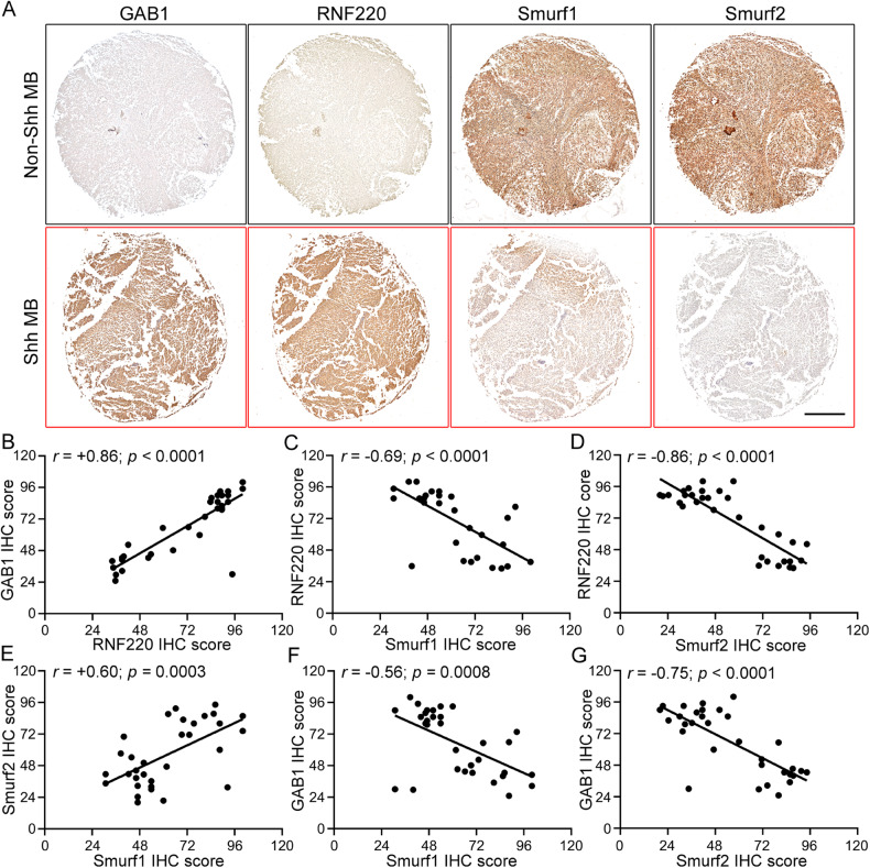
Sonic hedgehog (Shh)-group medulloblastoma (MB) (Shh-MB) encompasses a clinically and molecularly distinct group of cancers originating from the developing nervous system with aberrant high Shh signaling as a causative driver. We recently reported that RNF220 is required for sustained high Shh signaling during Shh-MB progression; however, how high RNF220 expression is achieved in Shh-MB is still unclear. In this study, we found that the ubiquitin E3 ligases Smurf1 and Smurf2 interact with RNF220, and target it for polyubiquitination and degradation. In MB cells, knockdown or overexpression of Smurf1 or Smurf2 promotes or inhibits cell proliferation, colony formation and xenograft growth, respectively, by controlling RNF220 protein levels, and thus modulating Shh signaling. Furthermore, in clinical human MB samples, the protein levels of Smurf1 or Smurf2 were negatively correlated with those of RNF220 or GAB1, a Shh-MB marker. Overall, this study highlights the importance of the Smurf1- and Smurf2-RNF220 axes during the pathogenesis of Shh-MB and provides new therapeutic targets for Shh-MB treatment.


Conflict of interest statement

The authors declare no competing interests.

Figures

Fig. 1
Fig. 1. RNF220 interacts with Smurf1 and Smurf2.
A, B Smurf1 (A) or Smurf2 (B) was co-immunoprecipitated with RNF220. C, D RNF220 was co-immunoprecipitated with Smurf1 (C) or Smurf2 (D). HEK293 cells were transiently transfected with different combinations of expression vectors of Smurf1, Smurf2, and RNF220, as indicated. Cell lysates were incubated with anti-Flag beads, washed, and subsequently analyzed by Western Blot. E, F Endogenous RNF220 could be immunoprecipitated by Smurf1 (E) or Smurf2 (F). Cell extracts of HEK293 cells were immunoprecipitated with antibody against Smurf1 or Smurf2, and endogenous RNF220 was detected by an anti-RNF220 antibody, with IgG as a negative control. G Endogenous Smurf1 and Smurf2 could be immunoprecipitated by RNF220. Cell extracts of HEK293 cells stably transfected with RNF220-Flag vectors were immunoprecipitated with anti-Flag beads, and endogenous Smurf1 or Smurf2 was detected by an anti-Smurf1 or anti-Smurf2 antibody, with IgG as a negative control. HL The ability of different RNF220 deletion constructs or mutants to pull down Smurf1 (I, K) or Smurf2 (J, L) in co-immuniprecipitation assays. Schematic representation of the structures of truncated or mutated mouse RNF220 with amino acid numbers indicated is shown in (H). IB immunoblot, IP immunoprecipitation, WCL whole cell lysate, WT wild-type, FL full-length, SA RN220S282/286A mutant, and KR RNF220K321/323/326R mutant.
Fig. 2
Fig. 2. Smurf1 regulates RNF220 protein stability.
A, B RNF220 protein was destabilized by overexpression of wild-type Smurf1 (A), but not their E3 ubiquitin ligase-defective mutants. Flag-tagged RNF220, myc-tagged wild-type or ligase-defective Smurf1 plasmids were transfected into HEK293 cells as indicated. After 48 h, cell lysates were analyzed by Western Blot. B Bar graphs, overlaid with the actual data points, show relative RNF220 protein expression (mean ± SD) normalized against the corresponding α-Tubulin. The control was set to 1. C, D Western Blot assays showing the protein level of RNF220 when co-expressed with Smurf1 in presence of MG132 or not. D Bar graphs, overlaid with the actual data points, show relative RNF220 protein expression (mean ± SD) normalized against the corresponding α-Tubulin. The respective controls were set to 1. E, F Effects of wild-type or ligase-defective Smurf1 or Smurf2 overexpression on the protein stability of RNF220. HEK293 cells were transiently transfected with the indicated plasmids. At 48 h posttransfection, cycloheximide was added to all samples, and cells were then harvested at the time points indicated. Level of endogenous RNF220 was determined by Western Blot with an anti-RNF220 antibody. In all cases, α-Tubulin was used as a loading control. The relative levels of RNF220 were quantified densitometrically and normalized against α-Tubulin. The data in F are the average of three independent experiments. G, H Western Blot results showing the effects of Smurf1 or Smurf2 knockdown on RNF220 protein level in HEK293 cells. HEK293 cells were transfected with the indicated siRNAs, and 72 h later, cells were harvested for Western Blot analysis. H Bar graphs, overlaid with the actual data points, show the relative expression (mean ± SD) of RNF220, normalized against the corresponding α-Tubulin. The control was set to 1. I, J Western Blot analysis showing the protein level of endogenous RNF220 when wild-type or E3 ubiquitin ligase activity defective Smurf1 was co-expressed with siRNAs against Smurf1 in HEK293 cells. The statistics of the result was shown in (J) with α-Tubulin as a loading control, and the control was set to 1. K, L Effect of Smurf1 knockdown on the protein stability of endogenous RNF220 in HEK293 cells. Cells were transiently transfected with the indicated siRNAs. At 72 h posttransfection, cycloheximide was added to all samples, and the cells were then harvested at the time points indicated. Protein level of RNF220 was determined by Western Blot with an anti-RNF220 antibody. The relative levels of RNF220 were quantified densitometrically and normalized against α-Tubulin. L The statistics showing the average of three independent experiments. IB immunoblot, WT wild-type, CA Smurf1 E3 ubiquitin ligase-defective mutant, CG Smurf2 E3 ubiquitin ligase-defective mutant, NC negative control, Chx cycloheximide, ns not significant. p > 0.05, *p < 0.05, **p < 0.01.
Fig. 3
Fig. 3. Smurf1 and Smurf2 target RNF220 for K48-linked polyubiquitination.
A, B In vivo ubiquitination assays showing the ubiquitination status of RNF220 when wild-type or E3 ubiquitin ligase inactive form of Smurf1 or Smurf2 was co-expressed in HEK293 cells. Overexpression of wild-type Smurf1 or Smurf2, but not their mutants promoted the ubiquitination of RNF220. The statistics of relative protein levels of polyubiquitinated RNF220 in each group was shown in (B), and the control group was set to 1. CF In vivo ubiquitination assays showing the ability of Smurf1 (C, D) or Smurf2 (E, F) to ubiquitinate RNF220 when the indicated ubiquitin mutants were used. Smurf1 and Smurf2 promote K48-linked ubiquitination of RNF220. For ubiquitin mutants: K48 the K48 ubiquitin mutant with all lysines except the K48 mutated to arginines, K48R the K48R ubiquitin mutant with only the K48 lysine mutated to arginine, WT the wild-type ubiquitin construct. The statistics of relative protein levels of polyubiquitinated RNF220 in each group was shown in (D, F), and the control group was set to 1. G, H In vivo ubiquitination analysis showing the level of endogenous ubiquitinated RNF220 protein when Smurf1 or Smurf2 was knocked down in HEK293 cells by siRNA transfection. The statistics of relative protein levels of ubiquitinated RNF220 in each group was shown in (H), and the control group was set to 1. I, J Western blot analysis showing the protein level of wild-type or KR mutated form of RNF220 protein when co-expressed with Smurf1 or Smurf2 in HEK293 cells. The statistics of relative protein levels of RNF220 in each group was shown in (J), and the respective controls were set to 1. KN In vivo ubiquitination analysis showing the ubiquitinated status of wild-type or KR mutated form of RNF220 protein when co-expressed with Smurf1 (K, L) or Smurf2 (M, N). OQ In vitro ubiquitination analysis showing that Smurf1 or Smurf2 promotes polyubiquitination of wild-type, but not KR mutated, RNF220 protein. The statistics of relative protein levels of RNF220 in each group was shown in (P, Q), and the respective controls were set to 1. IB immunoblot, IP immunoprecipitation, WCL whole cell lysate, WT wild type, KR RNF220K321/323/326R mutant; CA Smurf1 E3 ubiquitin ligase-defective mutant, CG Smurf2 E3 ubiquitin ligase-defective mutant; NC negative control, ns not significant. p > 0.05 and **p < 0.01.
Fig. 4
Fig. 4. Smurf1 and Smurf2 regulate Shh signaling through RNF220 in Daoy cells.
AC Real-time RT-PCR (A) Western Blot (B, C) assays showing Smurf1 and Smurf2 expression in control cerebellum and Ptch1± medulloblastoma tissues. The statistics showing relative Smurf1 and Smurf2 protein levels normalized against the corresponding α-Tubulin level was shown in (C), and the respective controls were set to 1. DG Real-time RT-PCR (D) and Western Blot (EG) assays showing the expression levels of RNF220 and Shh targets, including Gli1, Ptch1, and Hhip1, in Daoy cell stably transfected with shRNAs against Smurf1 or Smurf2. The statistics showing the relative protein levels of RNF220, Gli, Ptch1, and Hhip1, normalized against the corresponding α-Tubulin level was shown in (F, G), and the respective controls were set to 1. HK Real-time RT-PCR (H) and Western Blot (IK) assays showing the expression levels of RNF220 and Shh targets, including Gli1, Ptch1, and Hhip1, in Daoy cells transfected with expression plasmids for wild-type or E3 ubiquitin ligase-defective form of Smurf1 or Smurf2. The statistics showing the relative protein level of RNF220 (J), Gli, Ptch1, and Hhip1 (I) normalized against the corresponding α-Tubulin level. The respective controls were set to 1. LN Real-time PCR (L) and Western blot (M, N) assays showing the expression level of Shh targets, including Gli1, Ptch1, and Hhip1, in Daoy cells stably transfected with the indicated shRNAs against RNF220, Smurf1, or Smurf2. The statistics showing the relative protein levels of Gli, Ptch1, and Hhip1 normalized against the corresponding α-Tubulin level was shown in (N). The respective controls were set to 1. β-Actin was used as a loading control for real-time RT-PCR assays. IB immunoblot, CB cerebellum, MB medulloblastoma, ns not significant. p > 0.05, **p < 0.01.
Fig. 5
Fig. 5. Smurf1 or Smurf2 knockdown accelerates cell proliferation and tumor growth in Daoy cells.
A Growth curve for control, Smurf1 or Smurf2 knockdown Daoy cell line revealed by MTS assays. B, C EdU incorporation assays to evaluate DNA synthesis and proliferation rate of Daoy cells when Smurf1 or Smurf2 was knocked down. Scale bar, 50 µm. Quantification of EdU assay result was shown in (C). DF Soft agar colony formation assays for the indicated Daoy cell line. Scale bar, 120 µm. Quantification of colony number and size was shown in (E, F). GI Xenograft tumors from BALB/c nude mice subcutaneously injected with Daoy cells stably transfected with shRNAs against Smurf1 or Smurf2. Quantification of xenograft was shown in (H) for tumor size and (I) for tumor weight. JN The mRNA (J, K) and protein expression (LN) level of Shh targets (J, L, and M) and RNF220 (K, L, and N) in the indicated tumor xenografts. S1 Smurf1, S2 Smurf2, IB immunoblot, ns not significant p > 0.05, *p < 0.05 and **p < 0.01.
Fig. 6
Fig. 6. E3 ubiquitin ligase activity is required for cell proliferation and tumor growth inhibition by Smurf1 or Smurf2 overexpression in Daoy cells.
A Growth curve for control, wild-type or E3 ubiquitin ligase-defective Smurf1 or Smurf2 overexpressed Daoy cell line, revealed by MTS assays. B, C EdU incorporation assay to evaluate DNA synthesis and proliferation rates of UW228 cells when wild-type or E3 ubiquitin ligase-defective Smurf1 or Smurf2 was overexpressed. Scale bar, 50 µm. Quantification of the EdU assay results was shown in (C). DF Soft agar colony formation assays for the indicated Daoy cell line. Scale bar, 120 µm. Quantification of the colony number and size was shown in (E, F). GI Xenograft tumors from BALB/c nude mice subcutaneously injected with wild-type or E3 ubiquitin ligase inactive Smurf1 or Smurf2 overexpressed Daoy cells. Quantification of the xenografts was shown in (H) for tumor size and (I) for tumor weight. JN The mRNA (J, K) and protein expression (LN) level of Shh targets (J, L, and M) and RNF220 (K, L, and N) in the indicated tumor xenografts. IB immunoblot, WT wild-type, CA Smurf1 E3 ubiquitin ligase-defective mutant, CG Smurf2 E3 ubiquitin ligase-defective mutant, S1 Smurf1, S2 Smurf2, ns not significant. p > 0.05, *p < 0.05, and **p < 0.01.
Fig. 7
Fig. 7. Protein expression correlation analyzes among GAB1, RNF220, Smurf1, and Smurf2 in human clinical medulloblastoma samples.
A Representative immunohistochemical images of clinical Shh- and non-Shh medulloblastoma samples with the indicated antibodies. Scale bar, 5 mm. BG Statistical analysis of the correlation among GAB1-RNF220 (B), Smurf1-RNF220 (C), Smurf2-RNF220 (D), Smurf1-Smurf2 (E), Smurf1-GAB1 (F), and Smurf2-GAB1 (G) based on the immunohistochemical scores, Pearson product-moment correlation coefficient analysis was used for the statistics. MB medulloblastoma, IHC immunohistochemical staining.

Similar articles

Cited by

References

    1. Hatten ME, Roussel MF. Development and cancer of the cerebellum. Trends Neurosci. 2011;34:134–42. doi: 10.1016/j.tins.2011.01.002. - DOI - PMC - PubMed
    1. Northcott PA, Robinson GW, Kratz CP, Mabbott DJ, Pomeroy SL, Clifford SC, et al. Medulloblastoma. Nat Rev Dis Prim. 2019;5:11. doi: 10.1038/s41572-019-0063-6. - DOI - PubMed
    1. Northcott PA, Jones DT, Kool M, Robinson GW, Gilbertson RJ, Cho YJ, et al. Medulloblastomics: the end of the beginning. Nat Rev Cancer. 2012;12:818–34. doi: 10.1038/nrc3410. - DOI - PMC - PubMed
    1. Marshall GM, Carter DR, Cheung BB, Liu T, Mateos MK, Meyerowitz JG, et al. The prenatal origins of cancer. Nat Rev Cancer. 2014;14:277–89. doi: 10.1038/nrc3679. - DOI - PMC - PubMed
    1. Vaillant C, Monard D. SHH pathway and cerebellar development. Cerebellum. 2009;8:291–301. doi: 10.1007/s12311-009-0094-8. - DOI - PubMed

Publication types