. 2023 Aug 3;13(1):12631.
doi: 10.1038/s41598-023-38893-1.

Higher longitudinal brain white matter atrophy rate in aquaporin-4 IgG-positive NMOSD compared with healthy controls

Collaborators, Affiliations

Higher longitudinal brain white matter atrophy rate in aquaporin-4 IgG-positive NMOSD compared with healthy controls

Hiroki Masuda et al. Sci Rep. .

Abstract

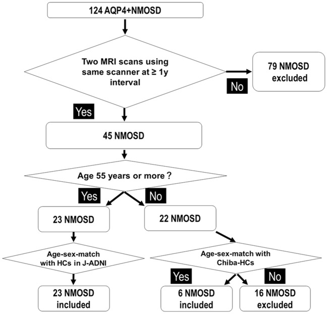
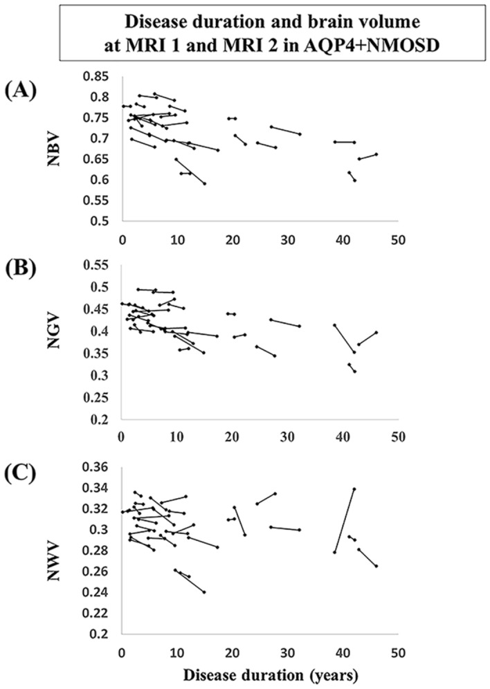
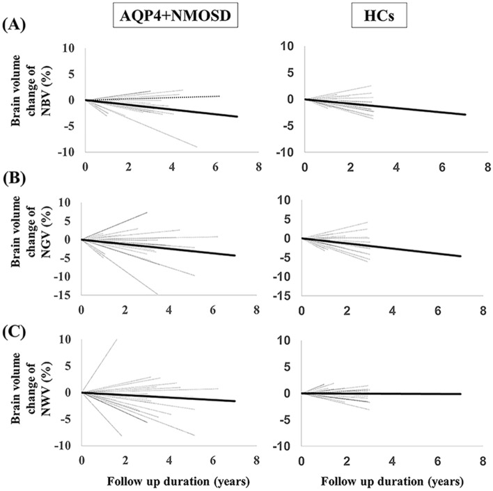
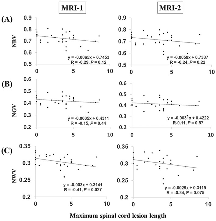
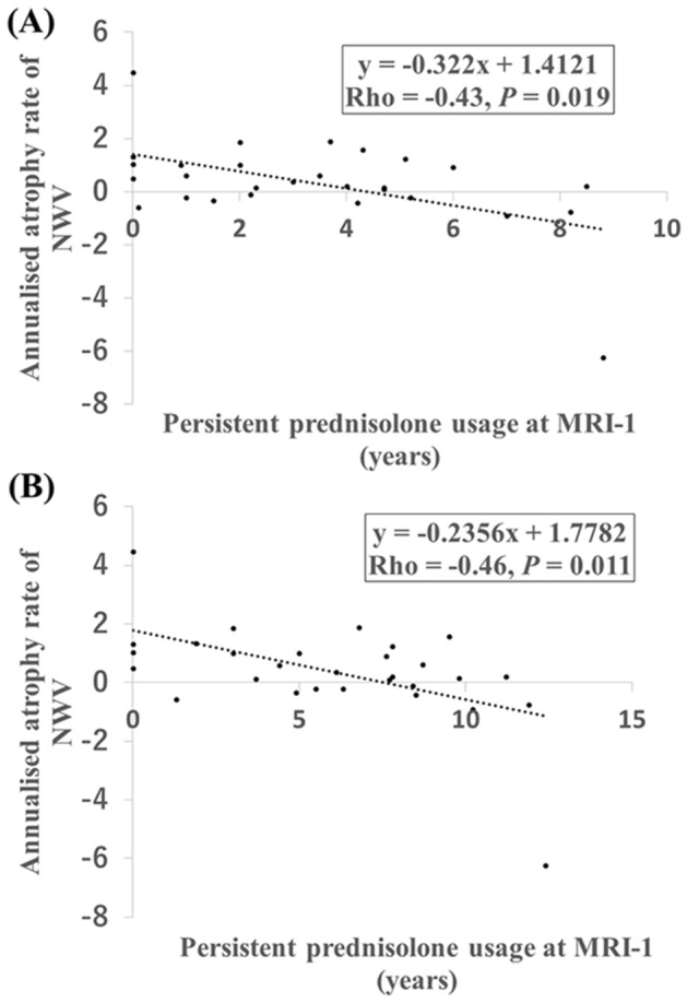
We aimed to compare longitudinal brain atrophy in patients with neuromyelitis optica spectrum disorder (NMOSD) with healthy controls (HCs). The atrophy rate in patients with anti-aquaporin-4 antibody-positive NMOSD (AQP4 + NMOSD) was compared with age-sex-matched HCs recruited from the Japanese Alzheimer's Disease Neuroimaging Initiative study and another study performed at Chiba University. Twenty-nine patients with AQP4 + NMOSD and 29 HCs were enrolled in the study. The time between magnetic resonance imaging (MRI) scans was longer in the AQP4 + NMOSD group compared with the HCs (median; 3.2 vs. 2.9 years, P = 0.009). The annualized normalized white matter volume (NWV) atrophy rate was higher in the AQP4 + NMOSD group compared with the HCs (median; 0.37 vs. - 0.14, P = 0.018). The maximum spinal cord lesion length negatively correlated with NWV at baseline MRI in patients with AQP4 + NMOSD (Spearman's rho = - 0.41, P = 0.027). The annualized NWV atrophy rate negatively correlated with the time between initiation of persistent prednisolone usage and baseline MRI in patients with AQP4 + NMOSD (Spearman's rho = - 0.43, P = 0.019). Patients with AQP4 + NMOSD had a greater annualized NWV atrophy rate than HCs. Suppressing disease activity may prevent brain atrophy in patients with AQP4 + NMOSD.


Conflict of interest statement

The authors declare no competing interests.

Figures

Figure 1
Figure 1
Flow chart and study design showing enrolment and age-sex-matching of patients with AQP4 + NMOSD and HCs. AQP4 + NMOSD anti-aquaporin-4 antibody-positive neuromyelitis optica spectrum disorders, HCs healthy controls. Chiba-HCs means another study performed in Chiba University by Shimizu et al.
Figure 2
Figure 2
Brain volume changes and disease duration in each patient with AQP4 + NMOSD. (A) NBV changes and disease durations. (B) NGV changes and disease durations. (C) NWV changes and disease durations. AQP4 + NMOSD anti-aquaporin-4 antibody-positive neuromyelitis optica spectrum disorders, NBV normalized brain volume, NGV normalized gray matter volume, NWV normalized white matter volume.
Figure 3
Figure 3
The percent brain volume changes between MRI-1 and MRI-2 in patients with AQP4 + NMOSD and HCs. (A) NBV changes and follow-up durations. (B) NGV changes and follow-up durations. (C) NWV changes and follow-up durations. The black dotted lines represent brain volume changes (in percentage) for each patient. The fitted average slopes in patients with AQP4 + NMOSD and HCs are shown by the black line. AQP4 + NMOSD anti-aquaporin-4 antibody-positive neuromyelitis optica spectrum disorders, HCs healthy controls, NBV normalized brain volume, NGV normalized gray matter volume, NWV normalized white matter volume.
Figure 4
Figure 4
Correlation between brain volumes and spinal lesion cord length in patients with AQP4 + NMOSD. (A) NBV changes and maximum spinal cord lesion length. (B) NGV changes and maximum spinal cord lesion length. (C) NWV changes and maximum spinal cord lesion length. AQP4 + NMOSD anti-aquaporin-4 antibody-positive neuromyelitis optica spectrum disorders, NBV normalized brain volume, NGV normalized gray matter volume, NWV normalized white matter volume.
Figure 5
Figure 5
Correlation between persistent prednisolone usage and the annualized NWV atrophy rate in patients with AQP4 + NMOSD. (A) The annualized NWV atrophy rates and persistent prednisolone usage durations at MRI-1. (B) The annualized NWV atrophy rates and persistent prednisolone usage durations at MRI-2. AQP4 + NMOSD anti-aquaporin-4 antibody-positive neuromyelitis optica spectrum disorders, NWV normalized white matter volume.

References

    1. Wingerchuk DM, et al. International consensus diagnostic criteria for neuromyelitis optica spectrum disorders. Neurology. 2015;85:177–189. doi: 10.1212/wnl.0000000000001729. - DOI - PMC - PubMed
    1. Jarius S, et al. Neuromyelitis optica. Nat. Rev. Dis. Primers. 2020;6:85. doi: 10.1038/s41572-020-0214-9. - DOI - PubMed
    1. Kawachi I, Lassmann H. Neurodegeneration in multiple sclerosis and neuromyelitis optica. J. Neurol. Neurosurg. Psychiatry. 2017;88:137–145. doi: 10.1136/jnnp-2016-313300. - DOI - PubMed
    1. Blanc F, et al. White matter atrophy and cognitive dysfunctions in neuromyelitis optica. PLoS ONE. 2012;7:e33878. doi: 10.1371/journal.pone.0033878. - DOI - PMC - PubMed
    1. Chanson JB, et al. White matter volume is decreased in the brain of patients with neuromyelitis optica. Eur. J. Neurol. 2013;20:361–367. doi: 10.1111/j.1468-1331.2012.03867.x. - DOI - PubMed

Publication types