. 2023 Aug 3;13(1):12610.
doi: 10.1038/s41598-023-39675-5.

Exploring the G-quadruplex binding and unwinding activity of the bacterial FeS helicase DinG

Affiliations

Exploring the G-quadruplex binding and unwinding activity of the bacterial FeS helicase DinG

Elisa De Piante et al. Sci Rep. .

Abstract

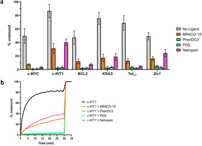
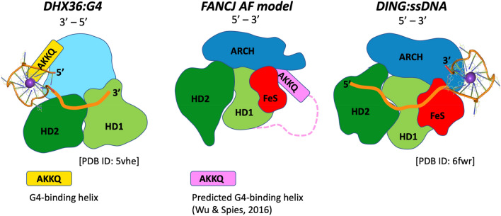
Despite numerous reports on the interactions of G-quadruplexes (G4s) with helicases, systematic analysis addressing the selectivity and specificity of each helicase towards a variety of G4 topologies are scarce. Among the helicases able to unwind G4s are those containing an iron-sulphur (FeS) cluster, including both the bacterial DinG (found in E. coli and several pathogenic bacteria) and the medically important eukaryotic homologues (XPD, FancJ, DDX11 and RTEL1). We carried out a detailed study of the interactions between the E. coli DinG and a variety of G4s, by employing physicochemical and biochemical methodologies. A series of G4-rich sequences from different genomic locations (promoter and telomeric regions), able to form unimolecular G4 structures with diverse topologies, were analyzed (c-KIT1, KRAS, c-MYC, BCL2, Tel23, T30695, Zic1). DinG binds to most of the investigated G4s with little discrimination, while it exhibits a clear degree of unwinding specificity towards different G4 topologies. Whereas previous reports suggested that DinG was active only on bimolecular G4s, here we show that it is also able to bind to and resolve the more physiologically relevant unimolecular G4s. In addition, when the G4 structures were stabilized by ligands (Pyridostatin, PhenDC3, BRACO-19 or Netropsin), the DinG unwinding activity decreased and in most cases was abolished, with a pattern that is not simply explained by a change in binding affinity. Overall, these results have important implications for the biochemistry of helicases, strongly suggesting that when analysing the G4 unwinding property of an enzyme, it is necessary to investigate a variety of G4 substrates.


Conflict of interest statement

The authors declare no competing interests.

Figures

Figure 1
Figure 1
Time evolution SPR sensorgrams obtained at 25 °C by injection of increasing concentrations of each investigated DNA (from 0.062 to 1 µM) on the chip-immobilized DinG helicase.
Figure 2
Figure 2
(a) Representative plots of fluorescence emission vs time for unwinding of selected DNA systems (S-c-MYC, S-c-KIT1, S-T30695, S-Tel23, S-KRAS, S-BCL2 and S-Zic1). ATP was added to begin the reaction (t = 0 min); the complementary strands (C-c-MYC, C-c-KIT1, C-T30695, C-Tel23, C-KRAS, C-BCL2 and C-Zic1 respectively) were added once the reactions reached a plateau (t = 30 min) as described. (b) Quantification of the helicase activity of E. coli DinG against selected DNA systems; error bars indicate standard deviation of three independent experiments.
Figure 3
Figure 3
(a) E. coli DinG helicase activity against DNA systems S-c-MYC, S-c-KIT1, S-BCL2, S-KRAS, S-Tel23 and S-Zic1 in the absence or presence of selected G4 ligands (BRACO-19, PhenDC3, PDS and Netropsin). Error bars indicate standard deviation of three independent experiments. (b) Example of typical real-time unfolding of S-c-KIT1 system in absence or presence of selected ligands (BRACO-19, PhenDC3, PDS and Netropsin). ATP was added at t = 0; the complementary strand (C-c-KIT1) was added at t = 30 min, as described in.
Figure 4
Figure 4
Time evolution SPR sensorgrams obtained at 25 °C by injection of increasing concentrations (from 0.062 to 1 µM) of c-KIT1 in the presence 1 mol equiv. of (a) PDS, (b) PhenDC3, (c) BRACO-19 and (d) Netropsin on the chip-immobilized DinG helicase.
Figure 5
Figure 5
Schematic representation of the architecture of potential helicase:G4 complexes for enzymes belonging to the SF2 family. The left panel shows the crystal structure of the helicase DHX36 bound to a G4, the central panel the AlphaFold predicted model for the human FancJ helicase, and the right panel the crystal structure of DinG bound to ssDNA. The HD1 and HD2 domains forming the helicase catalytic core are shown in light and dark green respectively; in other colors, the domains that are peculiar to each family, and in orange the path of ssDNA. The DHX36 helix which plays a major role in G4 recognition (including the AKKQ sequence motif) is shown in yellow. It has been suggested that an equivalent helix is present in a FancJ-specific and partially unstructured insertion (shown in pink), that also contains a AKKQ motif, and was predicted to be involved in G4 recognition; the AlphaFold model positions this helix between the FeS and the ARCH domains. This location is consistent with the crystal structure of the complex between DinG and ssDNA: a G4 has been placed on the [DinG:ssDNA] schematic diagram to illustrate the putative G4 binding region.

Similar articles

Cited by

References

    1. Bedrat A, Lacroix L, Mergny J-L. Re-evaluation of G-quadruplex propensity with G4Hunter. Nucleic Acids Res. 2016;44:1746–1759. doi: 10.1093/nar/gkw006. - DOI - PMC - PubMed
    1. Adrian M, Heddi B, Phan AT. NMR spectroscopy of G-quadruplexes. Methods. 2012;57:11–24. doi: 10.1016/j.ymeth.2012.05.003. - DOI - PubMed
    1. Criscuolo A, et al. Insights into the small molecule targeting of biologically relevant G-quadruplexes: An overview of NMR and crystal structures. Pharmaceutics. 2022;14:2361. doi: 10.3390/pharmaceutics14112361. - DOI - PMC - PubMed
    1. Campbell N, Collie GW, Neidle S. Crystallography of DNA and RNA G-quadruplex nucleic acids and their ligand complexes. Curr. Protoc. Nucleic Acid Chem. 2012 doi: 10.1002/0471142700.nc1706s50. - DOI - PubMed
    1. Neidle S, Parkinson GN. Quadruplex DNA crystal structures and drug design. Biochimie. 2008;90:1184–1196. doi: 10.1016/j.biochi.2008.03.003. - DOI - PubMed

Publication types