

Hypothalamus volumes in adolescent Myalgic Encephalomyelitis/Chronic Fatigue Syndrome (ME/CFS): impact of self-reported fatigue and illness duration
- PMID: 37537279
- PMCID: PMC10471696
- DOI: 10.1007/s00429-023-02682-3
Hypothalamus volumes in adolescent Myalgic Encephalomyelitis/Chronic Fatigue Syndrome (ME/CFS): impact of self-reported fatigue and illness duration
Abstract
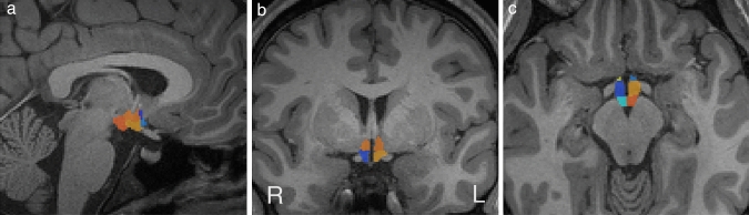
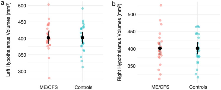
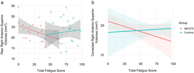
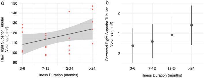
Adolescent Myalgic Encephalomyelitis/Chronic Fatigue Syndrome (ME/CFS) is a complex illness of unknown aetiology. Emerging theories suggest ME/CFS may reflect a progressive, aberrant state of homeostasis caused by disturbances within the hypothalamus, yet few studies have investigated this using magnetic resonance imaging in adolescents with ME/CFS. We conducted a volumetric analysis to investigate whether whole and regional hypothalamus volumes in adolescents with ME/CFS differed compared to healthy controls, and whether these volumes were associated with fatigue severity and illness duration. 48 adolescents (25 ME/CFS, 23 controls) were recruited. Lateralised whole and regional hypothalamus volumes, including the anterior-superior, superior tubular, posterior, anterior-inferior and inferior tubular subregions, were calculated from T1-weighted images. When controlling for age, sex and intracranial volume, Bayesian linear regression models revealed no evidence for differences in hypothalamus volumes between groups. However, in the ME/CFS group, a weak linear relationship between increased right anterior-superior volumes and fatigue severity was identified, which was absent in controls. In addition, Bayesian quantile regression revealed a likely-positive association between illness duration and right superior tubular volumes in the ME/CFS group. While these findings suggest overall comparability in regional and whole hypothalamus volumes between adolescents with ME/CFS and controls, preliminary evidence was identified to suggest greater fatigue severity and longer illness duration were associated with greater right anterior-superior and superior-tubular volumes, respectively. These regions contain the anterior and superior divisions of the paraventricular nucleus, involved in the neuroendocrine response to stress, suggesting involvement in ME/CFS pathophysiology. However, replication in a larger, longitudinal cohort is required.
Keywords: Adolescence; Chronic Fatigue Syndrome; Hypothalamus; Myalgic Encephalomyelitis; Structural magnetic resonance imaging; Volumetric analysis.
© 2023. The Author(s).
Conflict of interest statement
The authors have no relevant financial or non-financial interests to disclose. The funders had no role in the design of the study, in collection, analyses, or interpretation of data, in the writing of the manuscript, or in the decision to publish the results.
Figures
References
-
- Bakken IJ, Tveito K, Gunnes N, Ghaderi S, Stoltenberg C, Trogstad L, Håberg SE, Magnus P. Two age peaks in the incidence of chronic fatigue syndrome/myalgic encephalomyelitis: a population-based registry study from Norway 2008–2012. BMC Med. 2014;12:167. doi: 10.1186/s12916-014-0167-5. - DOI - PMC - PubMed
MeSH terms
Grants and funding
LinkOut - more resources
Full Text Sources
Medical
