Review
. 2023 Aug;4(8):1063-1082.
doi: 10.1038/s43018-023-00595-y. Epub 2023 Aug 3.

Cancer cell plasticity during tumor progression, metastasis and response to therapy

Affiliations
Review

Cancer cell plasticity during tumor progression, metastasis and response to therapy

Andrea Pérez-González et al. Nat Cancer. 2023 Aug.

Abstract

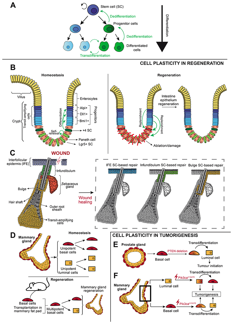
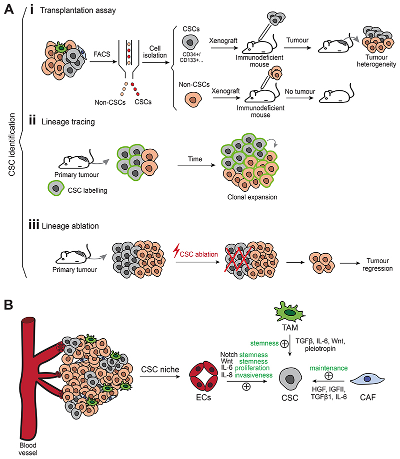
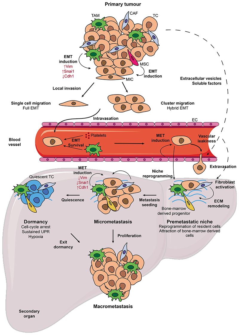
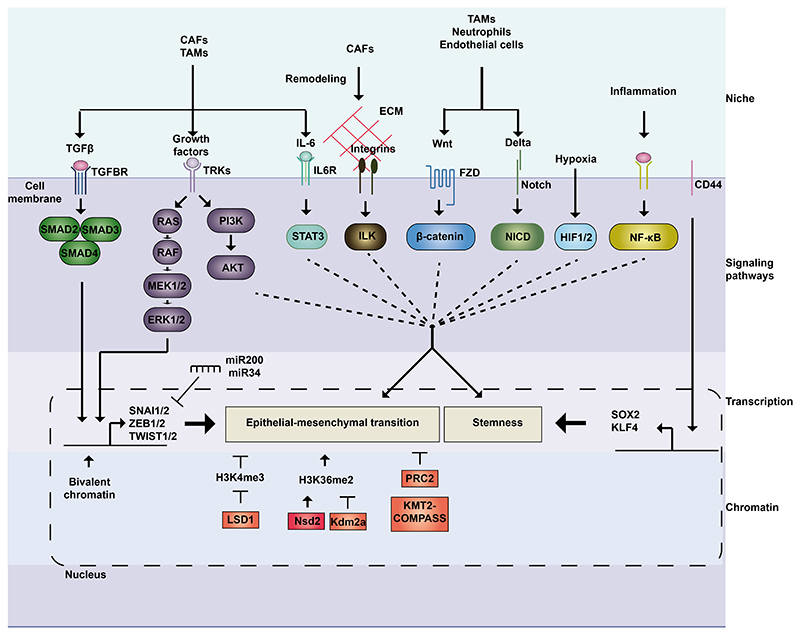
Cell plasticity represents the ability of cells to be reprogrammed and to change their fate and identity, enabling homeostasis restoration and tissue regeneration following damage. Cell plasticity also contributes to pathological conditions, such as cancer, enabling cells to acquire new phenotypic and functional features by transiting across distinct cell states that contribute to tumor initiation, progression, metastasis and resistance to therapy. Here, we review the intrinsic and extrinsic mechanisms driving cell plasticity that promote tumor growth and proliferation as well as metastasis and drug tolerance. Finally, we discuss how cell plasticity could be exploited for anti-cancer therapy.


Conflict of interest statement

Conflict of Interest

The authors declare that they have no conflict of interest.

Figures

Figure 1
Figure 1. Cell plasticity during homeostasis, regeneration and tumorigenesis.
(A) Stem cell differentiation, dedifferentiation and transdifferentiation occurring during cell plasticity. (B) Lgr5+ intestinal stem cells self-renew and give rise to the distinct intestinal lineages during homeostasis. Following stem cell lineage ablation, more committed progenitors can replenish the pool of stem cells, enabling epithelium regeneration. (C) During homeostasis, the different epidermal compartments are sustained by distinct pools of unipotent SCs whereas during wound healing, interfollicular epidermis stem cells contribute to skin repair but also stem cells from the infundibulum and bulge can migrate upwards, proliferate, and be reprogrammed into interfollicular epidermis stem cells to contribute to regeneration. (D) Under homeostatic conditions, basal and luminal cells in the mammary gland are unipotent. Following transplantation into the mammary fat pad, basal cells become multipotent and can give rise to luminal cells, enabling the generation of a functional mammary gland. (E) PTEN deletion in basal cells of the prostate gland promotes basal-to-luminal transdifferentiation and leads to tumor initiation. (F) Pik3caH1047R expression in basal cells in the mammary gland leads to a transdifferentiation into luminal cells, while its expression in luminal cells enables a transdifferentiation into basal cells. Both basal and luminal cells expressing Pik3caH1047R can initiate tumorigenesis. IFE, interfollicular epidermis; SC, stem cell.
Figure 2
Figure 2. Defining cancer stem cells and their niche.
(A) Functional strategies to identify CSCs include: (i) transplantation assays (tumor subpopulations isolated by fluorescence-activated cell sorting are transplanted into immunodeficient mice. If CSCs are grafted, a tumor will appear and will recapitulate tumor heterogeneity, while non-CSCs will be less efficient to propagate the tumor following transplantation), (ii) lineage tracing of CSCs (which allows to follow their fate during tumor progression and to assess clonal expansion) and (iii) lineage ablation (which allows the elimination of a specific subpopulation. If CSCs are eliminated, the remaining subpopulations will not be able to sustain tumor growth, and tumor regression will occur). (B) A crosstalk between CSCs and their microenvironment is essential to sustain tumor growth. CSCs are supported by a niche composed by cancer-associated fibroblasts, endothelial cells and immune cells, which extrinsically promote tumor stemness. CAF, cancer-associated fibroblast; CSC, cancer stem cell; EC, endothelial cell; FACS, fluorescence-activated cell sorting; TAM, tumor-associated macrophage.
Figure 3
Figure 3. Cell plasticity along the metastatic cascade.
T umor cells can acquire metastasis-initiating properties through the induction of EMT by intrinsic and extrinsic stimuli. EMT allows MICs to detach from the primary tumor and the vascular niche facilitates MIC intravasation into the bloodstream, where single or clustered CTCs exhibit high plasticity and hybrid EMT. Interaction of CTCs with platelets and macrophages can promote plasticity, while platelet coating protects CTCs from the shredding force. The secondary organ is prepared by the primary tumor through the secretion of extracellular vesicles and soluble factors which create a permissive microenvironment. Colonizing the metastatic site involves the reversion of tumor cells to the epithelial state in response to signals coming from the metastatic niche. Following seeding, tumor cells can enter dormancy, which confers them with immune evasion traits and resistance to therapy, or proliferate and give rise to macroscopic metastases. CAF, cancer-associated fibroblast; CTC, circulating tumor cell; EC, endothelial cell; ECM: extracellular matrix; EMT, epithelial-to-mesenchymal transition; MET, mesenchymal-to-epithelial transition; MIC, metastasis-initiating cell; MSC: mesenchymal stem cell; SC, stem cell; TAM, tumor-associated macrophage; TC, tumor cell.
Figure 4
Figure 4. Molecular mechanisms regulating cancer cell plasticity.
Cancer cell plasticity is regulated extracellularly, by signals coming from the microenvironment, and intrinsically, through signaling pathways, transcriptional programs, and chromatin remodeling. TFGβ and RAS-MAPK pathways can act jointly to induce EMT. CD44 and Wnt regulate stemness, while Notch, JAK-STAT and integrins act on stemness and EMT in a context-dependent manner. Hypoxia induces stemness, while NF-κB is involved in plasticity by its role in inflammation. These pathways activate transcriptional programs regulated by key transcription factors involved in EMT (e.g., SNAI1/2, ZEB1/2, TWIST1/2) and stemness (e.g., SOX2, KLF4). Their action can be modulated by negative feedback loops involving miRNAs (e.g., ZEB/miR-200 and SNAI1/- miR-34) and depends on the chromatin landscape. LSD1 can remove the transcriptionally active H3K4me3 histone mark and collaborate with Snai1 to silence epithelial genes. Nsd2 and Kdm2a exhibit antagonist actions, as writer and eraser of H3K36me2, histone mark increased during EMT. PRC2 and KMT2-COMPASS are critical to regulate the epithelial state. CAF, cancer-associated fibroblast; ECM: extracellular matrix; FZD, frizzled; HIF, Hypoxia-inducible factor; IL6R, interleukin-6 receptor; TAM, tumor-associated macrophage; TGFBR, Transforming Growth Factor Receptor; TRK, Tyrosine receptor kinase.
Figure 5
Figure 5. Genetic induced drug resistance and non-genetic drug tolerance in anti-cancer therapy.
Pre-existing (A) or acquired (B) mutations can confer intrinsic genetic drug resistance, by which mutated tumor cells can display a clonal selection, survive, and proliferate under a particular therapeutic regimen. (C) Non-genetic drug tolerance can occur through transcriptional selection of primed cells that acquire a DTP dormant state during therapy and can lead to tumor relapse after therapy. (D) Non-genetic drug tolerance can occur through an adaptation to the therapeutic pressure, by which plastic tumor cells acquire a DTP quiescent state following therapy and can lead to tumor relapse after therapy. (E) Targeting the signaling pathways activated in the DTP state enables its eradication. The DTP state induced upon BRAFi/MEKi treatment in melanoma relies on FAK signaling and the transcriptional program of this state is largely driven by the nuclear receptor RXR. Consistently, the DTP state can be targeted by FAK inhibition and RXR antagonism. However, de novo mutations could still lead to genetic resistance and tumor relapse,. DTP, drug tolerant persister; RAR, retinoic acid receptor; RXR, retinoid X receptor; SC, stem cell.

References

    1. Mills JC, Stanger BZ, Sander M. Nomenclature for cellular plasticity: are the terms as plastic as the cells themselves? EMBO J. 2019;38:e103148. - PMC - PubMed
    1. Yuan S, Norgard RJ, Stanger BZ. Cellular Plasticity in Cancer. Cancer Discov. 2019;9:837–851. - PMC - PubMed
    1. Gurdon JB. The Developmental Capacity of Nuclei taken from Intestinal Epithelium Cells of Feeding Tadpoles. Development. 1962;10:622–640. - PubMed
    1. Takahashi K, Yamanaka S. Induction of Pluripotent Stem Cells from Mouse Embryonic and Adult Fibroblast Cultures by Defined Factors. Cell. 2006;126:663–676. - PubMed
    1. Le Magnen C, Shen MM, Abate-Shen C. Lineage Plasticity in Cancer Progression and Treatment. Annu Rev Cancer Biol. 2018;2:271–289. - PMC - PubMed

Publication types