. 2023 Sep;10(27):e2207672.
doi: 10.1002/advs.202207672. Epub 2023 Aug 3.

Structural Basis for the Enzymatic Activity of the HACE1 HECT-Type E3 Ligase Through N-Terminal Helix Dimerization

Affiliations

Structural Basis for the Enzymatic Activity of the HACE1 HECT-Type E3 Ligase Through N-Terminal Helix Dimerization

Sunil Singh et al. Adv Sci (Weinh). 2023 Sep.

Abstract

HACE1 is an ankyrin repeat (AKR) containing HECT-type E3 ubiquitin ligase that interacts with and ubiquitinates multiple substrates. While HACE1 is a well-known tumor suppressor, its structure and mode of ubiquitination are not understood. The authors present the cryo-EM structures of human HACE1 along with in vitro functional studies that provide insights into how the enzymatic activity of HACE1 is regulated. HACE1 comprises of an N-terminal AKR domain, a middle (MID) domain, and a C-terminal HECT domain. Its unique G-shaped architecture interacts as a homodimer, with monomers arranged in an antiparallel manner. In this dimeric arrangement, HACE1 ubiquitination activity is hampered, as the N-terminal helix of one monomer restricts access to the C-terminal domain of the other. The in vitro ubiquitination assays, hydrogen-deuterium exchange mass spectrometry (HDX-MS) analysis, mutagenesis, and in silico modeling suggest that the HACE1 MID domain plays a crucial role along with the AKRs in RAC1 substrate recognition.

Keywords: HACE1; HECT E3 ligases; RAC1; cancer; ubiquitination.


Conflict of interest statement

The authors declare no conflict of interest.

Figures
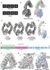
Figure 1
Figure 1
The cryo‐EM structure of HACE1. A) A representative 2D class averages of HACE1 monomer and dimer. B) Coulomb potential map of HACE1 monomer. Red dotted circle indicates the additional density that could not be modeled. C) Coulomb potential map of the HACE1 dimer. A 4.6 Å map was used for modeling. D) Overall structure of HACE1 monomer, including the N‐terminal helix (magenta), the ankyrin repeat (AKR) repeat domain (light green), the middle (MID) domain (pink), and the HECT domain (sky blue). E) Domain organization of HACE1 monomer. The square and the number inside represent helices counted from the N‐terminus. F) The N‐terminal helix and AKR domain model (aa 1–252) fit in monomer map. G) The MID domain model (aa 253–522) fit in monomer map. The insertion loop (aa 386–441) could not be modeled. H) The HECT domain (aa 523–909) fit in monomer map. The last C‐terminal three residues (aa 907–909) could not be modeled.
Figure 2
Figure 2
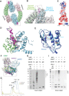
Dimerization of HACE1. A) HACE1 dimer made of two head‐to‐tail contacts between identical subunits. The boxed area is zoomed in on (B). B) The N‐terminal helix plus linker docked with the HECT domain small N‐lobe. The boxed area is zoomed in on (D). C) Electrostatic surface potential map of HACE1 monomer. D) The arginine‐rich N‐terminal helix plus loop (magenta, aa 1–21) is inserted onto the HECT domain small N‐lobe (sky blue) at dimerization interface. The first 19 residues contain six arginine residues as shown. E) Expansion of the HECT domain N‐lobe upon dimerization. Monomer is shown in dark blue. Dimer is shown in sky blue. F) Structural alignment of the HECT domain small N‐lobe from HACE1 (blue), NEDD4 (purple) and E6AP (green). Small N‐lobe is boxed. The small N‐lobes of HACE1 and NEDD4 matched with RMSD 1.035 Å. The small N‐lobes of HACE1 and E6AP matched with RMSD 1.043 Å. G) Analytical gel‐filtration chromatogram of WT HACE1 (aa 1–909) and N‐terminal 21 aa deletion mutant (∆21). H) Western blot analysis of in vitro RAC1 ubiquitination assay using anti‐ubiquitin antibody. WT HACE1 (aa 1–909) and the N‐terminal 21 aa deletion mutant (∆21, aa 22–909) were added at equal concentration. I) Western blot analysis of in vitro autoubiquitination assay with WT HACE1 and ∆21 HACE mutant using anti‐ubiquitin antibody.
Figure 3
Figure 3
Mapping of HACE1–RAC1 interaction sites. A) Woods’ differential plot showing hydrogen‐deuterium exchange (HDX) differences between WT HACE1 + RAC1 and WT HACE1 alone at 1 min. Peptides (represented as horizontal bars) showing deprotection are in red, while those showing decreased deuterium exchange in WT HACE1 + RAC1 complex are in blue, based on a 99% confidence interval. Differences in deuterium exchange (ΔHDX) were then mapped onto WT HACE1 structure solved in this study and colored as indicated in key (bottom panel). B) Woods’ differential plot showing HDX differences between ∆21 HACE1 + RAC1 and ∆21 HACE1 alone at 1 min. Peptides showing predominantly decreased deuterium exchange (protected, blue lines) were mapped onto structure of ∆21 HACE1 (bottom panel). C) SDS–PAGE and western blot analysis of in vitro RAC1 ubiquitination by WT HACE1 and its middle (MID) domain mutants detected using an anti‐ubiquitin antibody. D) SDS–PAGE and western blot analysis of RAC1 in vitro ubiquitination by WT HACE1 and its ankyrin repeat (AKR) domain mutants detected using an anti‐ubiquitin antibody. E) SDS–PAGE and western blot analysis of in vitro RAC1 ubiquitination by ∆21 HACE1 and its MID domain mutants detected using an anti‐ubiquitin antibody. F) SDS–PAGE and Western blot analysis of RAC1 in vitro ubiquitination by ∆21 HACE1 and its AKR domain mutants detected using an anti‐ubiquitin antibody. G) HDX difference heatmap on in silico model of the WT HACE1 + RAC1 complex. H) HDX difference heatmap on in silico model of the ∆21 HACE1 + RAC1 complex.
Figure 4
Figure 4
The role of the C‐terminal tail on ubiquitination activity. A) Pair‐wise structural alignment of the HECT domains of HACE1 monomer and dimer. The catalytic cysteine (C876) is highlighted with red. The experimental models are matched on the helix (aa 685–693) in the small N‐lobe. The tilt angles were determined between the planes along the strand (aa 872–875) and the helix (aa 825–836) in the C‐lobe. B) Western blot analysis of RAC1 ubiquitination by full length (FL) WT HACE1 and its E782A mutant using anti‐RAC1 antibody. C) Pair‐wise structural alignment of HUWE1 HECT domain (pink, aa 3991–4374, PDB:5LP8‐B) in closed conformation and HUWE1 HECT domain (green, aa 3991–4374, PDB:6XZ1‐A) in open conformation. D) Western blot analysis of RAC1 ubiquitination by FL WT HACE1 and its Δ3 CT mutant (aa 1–906) using anti‐RAC1 antibody. E) Multiple sequence alignment of the C‐terminal regions in human HECT E3 ligases. The arrow indicates the aromatic residue at −4 position. F) Western blot analysis of RAC1 ubiquitination by FL WT HACE1 and its Y906A mutant using anti‐RAC1 antibody. G) Western blot analysis of in vitro autoubiquitination by WT HACE1 extended HECT domain (aa 483–909) and its Δ3 CT mutant (aa 483–906) using anti‐ubiquitin antibody. H) Western blot analysis of in vitro autoubiquitination by WT HACE1 extended HECT domain (aa 483–909) and its Y906A mutant using anti‐ubiquitin antibody. I) E3‐Ub linkage of WT HACE1 extended HECT domain and its Δ3 CT and Y906A mutants detected by nonreducing SDS–PAGE and J) reducing SDS–PAGE visualized by Coomassie staining. Asterisk indicates the band corresponding to a mono‐Ub linked HACE1 extended HECT domain (aa 483–909).
Figure 5
Figure 5
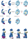
Schematic representation of autoinhibition, E2 binding, RAC1 binding and ubiquitination. HACE1 monomer (blue), HACE1 dimer (sky blue), ubiquitin conjugating enzyme E2 (purple), ubiquitin (pink), and RAC1 (yellow).

References

    1. Komander D., Rape M., Annu. Rev. Biochem. 2012, 81, 203. - PubMed
    1. Ulrich H. D., Walden H., Nat. Rev. Mol. Cell Biol. 2010, 11, 479. - PubMed
    1. Kerscher O., Felberbaum R., Hochstrasser M., Annu. Rev. Cell Dev. Biol. 2006, 22, 159. - PubMed
    1. Singh S., Ng J., Sivaraman J., Pharmacol. Ther. 2021, 224, 107809. - PubMed
    1. Scheffner M., Nuber U., Huibregtse J. M., Nature 1995, 373, 81. - PubMed

Publication types

LinkOut - more resources