. 2023 Oct;531(14):1425-1442.
doi: 10.1002/cne.25520. Epub 2023 Jul 8.

Members of the CUGBP Elav-like family of RNA-binding proteins are expressed in distinct populations of primary sensory neurons

Affiliations

Members of the CUGBP Elav-like family of RNA-binding proteins are expressed in distinct populations of primary sensory neurons

Eliza Grlickova-Duzevik et al. J Comp Neurol. 2023 Oct.

Abstract

Primary sensory dorsal root ganglia (DRG) neurons are diverse, with distinct populations that respond to specific stimuli. Previously, we observed that functionally distinct populations of DRG neurons express mRNA transcript variants with different 3' untranslated regions (3'UTRs). 3'UTRs harbor binding sites for interaction with RNA-binding proteins (RBPs) for transporting mRNAs to subcellular domains, modulating transcript stability, and regulating the rate of translation. In the current study, analysis of publicly available single-cell RNA-sequencing data generated from adult mice revealed that 17 3'UTR-binding RBPs were enriched in specific populations of DRG neurons. This included four members of the CUG triplet repeat (CUGBP) Elav-like family (CELF): CELF2 and CELF4 were enriched in peptidergic, CELF6 in both peptidergic and nonpeptidergic, and CELF3 in tyrosine hydroxylase-expressing neurons. Immunofluorescence studies confirmed that 60% of CELF4+ neurons are small-diameter C fibers and 33% medium-diameter myelinated (likely Aδ) fibers and showed that CELF4 is distributed to peripheral termini. Coexpression analyses using transcriptomic data and immunofluorescence revealed that CELF4 is enriched in nociceptive neurons that express GFRA3, CGRP, and the capsaicin receptor TRPV1. Reanalysis of published transcriptomic data from macaque DRG revealed a highly similar distribution of CELF members, and reanalysis of single-nucleus RNA-sequencing data derived from mouse and rat DRG after sciatic injury revealed differential expression of CELFs in specific populations of sensory neurons. We propose that CELF RBPs may regulate the fate of mRNAs in populations of nociceptors, and may play a role in pain and/or neuronal regeneration following nerve injury.

Keywords: 3′UTR; CELF; RNA-binding protein; dorsal root ganglion; nociceptor; sensory.


Conflict of interest statement

CONFLICT OF INTEREST STATEMENT

The authors declare no competing financial interests.

Figures
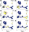
Figure 1:
Figure 1:. Utilization of publicly available single cell RNA sequencing (scRNA-Seq) data, derived from adult mouse sensory neurons, to generate population-specific gene expression profiles.
To generate gene expression profiles for specific populations of mouse adult sensory neurons, publicly available scRNA-seq data (GSE59739) were downloaded and processed with the latest release of SEURAT software using up to date gene annotations. Graphs are Uniform Manifold Approximation and Projections (UMAPs) of gene expression profiles. Each data point is a cell in UMAP space. Yellow shading = high expression, Blue shading = low expression. Expression profiles distributed into clusters as expected: A-D Peptidergic neurons were identified by CALCA (CGRP) expression (PEP, purple cluster), non-peptidergic by MRGPRD expression (NP, green cluster), tyrosine hydroxylase–expressing neurons (TH, red cluster) and myelinated neurons by neurofilament heavy chain expression (NF, blue cluster). E Sub-cluster of TRPV1-expressing nociceptors. F Sub-cluster of parvalbumin (PVALB) -expressing proprioceptors.
Figure 2:
Figure 2:. The CELF family of RNA-binding proteins are expressed in distinct populations of sensory neurons.
A Mean vs variance plot of mRNA expression values for 3’UTR-binding RNA-binding protein genes in adult mouse sensory neurons. X’s are individual genes. FXR1 and CELF4 are highlighted for reference. B Expression of a representative RBP, FXR1 is broadly expressed in all clusters of DRG neurons. Yellow circles are individual neurons that express FXR1 to high levels, blue circles = cells with low FXR1 expression C Heatmap of expression values of 3’UTR-binding RBPs significantly differentially expressed (FDR<0.01, top-10) in distinct populations of sensory neurons. Each line shows expression values in a single cell. Yellow = high expression, Purple = low expression. D Expression of significantly differentially expressed CELF-family RBPs in DRG neuron clusters. Yellow = high expression, blue = low expression. E Violin plots showing the distribution of expression values for significantly differentially expressed CELF family proteins. Yellow triangles denote significant enrichment, purple triangles denote significant exclusion. TH = tyrosine hydroxylase–expressing, NP = non-peptidergic, NF = neurofilament heavy chain-expressing and PEP = peptidergic neurons.
Figure 3:
Figure 3:. Re-analysis of publicly available single cell RNA-sequencing datasets confirm that CELF4 members are expressed in distinct populations of cells in sensory ganglia of adult mouse and macaque.
A Expression of CELF member mRNAs in populations of neuronal and non-neuronal adult mouse sensory ganglia cells. Data from GSE154659 (Renthal et al., 2020). CELF1 and 2 mRNAs are detected in non-neuronal populations, whereas CELF4 is enriched in peptidergic neurons. B In the adult macaque, neuronal expression of CELF members is highly similar to mouse. Data from (Kupari et al., 2021).
Figure 4:
Figure 4:. CELF4 protein is expressed in small and medium diameter sensory neurons.
A CELF4 gene deletion abolishes CELF4 immunoreactivity, confirming the specificity of the CELF4 antibody. B Representative immunofluorescent micrograph of a 14μm DRG section stained with CELF4 antibody. C Soma size distribution of CELF4+ neurons. Data are collated from 3 sections, from 3 DRG from 3 replicate animals (approx. 100 soma per section). Somata were categorized according to cross-sectional area - small (<300μm), medium (300–700μm) or large (>700μm). C i Proportion of small, medium or large cross-sectional area DRG neurons that express CELF4. ii Proportion of CELF4-expressing neurons with small, medium or large cross-sectional areas.
Figure 5:
Figure 5:. CELF4 protein expression in peptidergic (CGRP+), non-peptidergic (IB4-binding) and myelinated (NF-H+) neurons.
A-C 14μm DRG sections stained with CELF4 antibody, co-stained with established histological markers. Peptidergic neurons were identified with CGRP antibody, non-peptidergic with isolectin IB4 and myelinated with NFH. Scatter plots show distribution of fluorescence intensities of somata collated from DRG from 3 replicate animals, 2 non-overlapping cross sections per animal (approx. 100 soma per section). Dashed lines indicate background signals levels. D Pie charts showing the proportion of double positive somata from each of the 3 stains. E Mean CELF4 fluorescence values. Stats = ANOVA with post hoc t-tests, n=3 animals, *=p<.05, **=p<.01.
Figure 6:
Figure 6:. CELF4 is co-expressed with the TRPV1 capsaicin receptor in DRG neurons.
(A) Pearson’s r correlation coefficients were calculated for CELF4 mRNA concentration vs all other transcripts. The 3 most positively and negatively correlated genes are shown. Also shown are reference values for a known co-expressed pair of mRNAs (CGRP vs NTRK1) and a pair of mRNA’s know to be expressed in different neurons (CGRP vs MRGPRD). (B) 14μm DRG sections were stained with CELF4 and TRPV1 antibody, and fluorescence intensity measured in soma of 2 sections from 3 animals. Representative micrographs are shown. (C) To support the mRNA correlations, we identified muscle afferents that co-express TRPV1 and CELF4 protein by using retrograde neuronal tracing from the gastrocnemius and tibialis anterior muscles combined with immunohistochemistry. Data collected from 3 DRG from 8 replicate animals (lumbar level 4). Representative micrographs are shown along with the no primary controls.
Figure 7:
Figure 7:. CELF4 is distributed to the peripheral termini of sensory neurons.
The location of lanceolate endings in mouse hind paw hairy skin sections was identified by NFH immunoreactivity. CELF4 was detected in 49.2% of lanceolate sensory nerve endings in hair follicles within both the longitudinal and circumferential portions of the sensory terminals (arrow heads). Representative micrographs are shown along with no primary control. n=5 animals.
Figure 8:
Figure 8:. CELF transcripts are downregulated in sensory ganglia in rodent models of neuropathic pain.
Single nucleus sequencing data GSE154659 (Renthal et al., 2020) from mouse DRG (A) and bulk homogenate RNA-Seq from rat DRG (Harrison et al., 2019) (B) showed differential expression of CELF members post sciatic injury. * = q<0.05, ** = q<0.01 where q values are determined by Student’s t-tests with Bonferroni multiple testing correction.

References

    1. Abdelhamid RE, Kovács KJ, Honda CN, Nunez MG, & Larson AA (2013). Resiniferatoxin (RTX) Causes a Uniquely Protracted Musculoskeletal Hyperalgesia in Mice by Activation of TRPV1 Receptors. The Journal of Pain, 14(12), 1629–1641. 10.1016/j.jpain.2013.07.021 - DOI - PMC - PubMed
    1. An JJ, Gharami K, Liao G-Y, Woo NH, Lau AG, Vanevski F, Torre ER, Jones KR, Feng Y, Lu B, & Xu B (2008). Distinct role of long 3’ UTR BDNF mRNA in spine morphology and synaptic plasticity in hippocampal neurons. Cell, 134(1), 175–187. 10.1016/j.cell.2008.05.045 - DOI - PMC - PubMed
    1. Arora A, Castro-Gutierrez R, Moffatt C, Eletto D, Becker R, Brown M, Moor AE, Russ HA, & Taliaferro JM (2022). High-throughput identification of RNA localization elements in neuronal cells. Nucleic Acids Research, 50(18), 10626–10642. 10.1093/nar/gkac763 - DOI - PMC - PubMed
    1. Ashburner M, Ball CA, Blake JA, Botstein D, Butler H, Cherry JM, Davis AP, Dolinski K, Dwight SS, Eppig JT, Harris MA, Hill DP, Issel-Tarver L, Kasarskis A, Lewis S, Matese JC, Richardson JE, Ringwald M, Rubin GM, & Sherlock G (2000). Gene Ontology: Tool for the unification of biology. Nature Genetics, 25(1), Article 1. 10.1038/75556 - DOI - PMC - PubMed
    1. Bolger AM, Lohse M, & Usadel B (2014). Trimmomatic: A flexible trimmer for Illumina sequence data. Bioinformatics, 30(15), 2114–2120. 10.1093/bioinformatics/btu170 - DOI - PMC - PubMed

Publication types

Substances