. 2023 Aug 2;13(33):23285-23307.
doi: 10.1039/d3ra04007k. eCollection 2023 Jul 26.

Identification of new theobromine-based derivatives as potent VEGFR-2 inhibitors: design, semi-synthesis, biological evaluation, and in silico studies

Affiliations

Identification of new theobromine-based derivatives as potent VEGFR-2 inhibitors: design, semi-synthesis, biological evaluation, and in silico studies

Ibrahim H Eissa et al. RSC Adv. .

Abstract

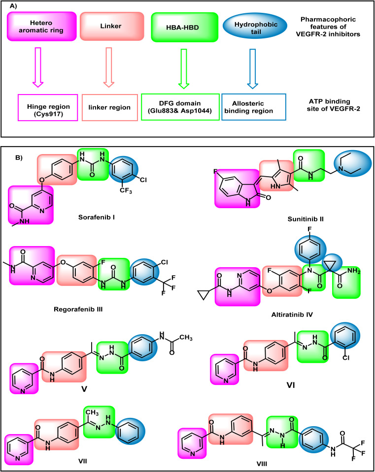
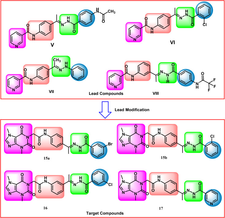
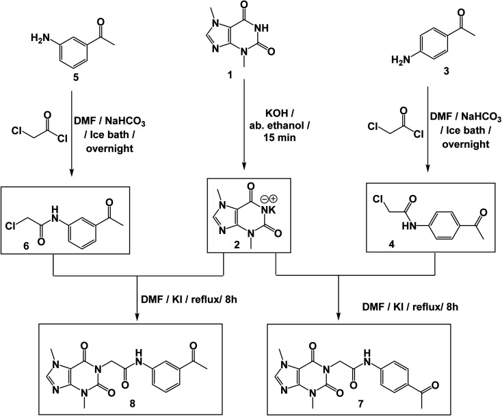
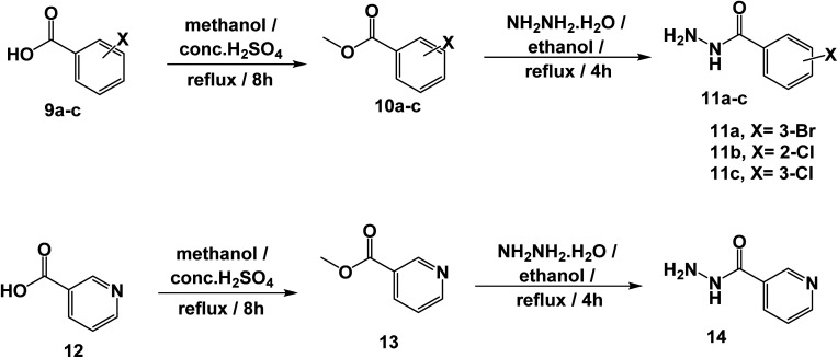
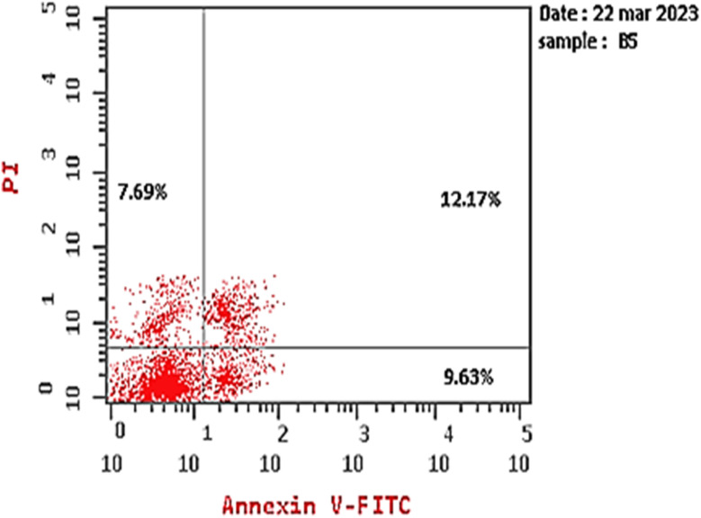
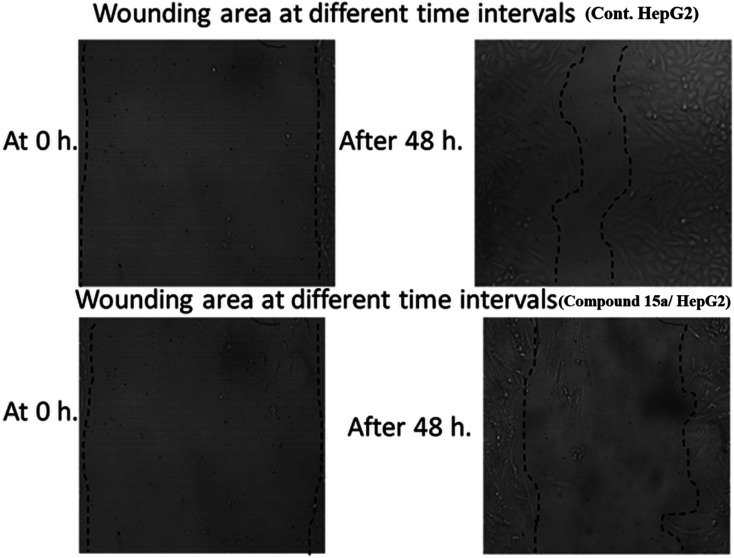
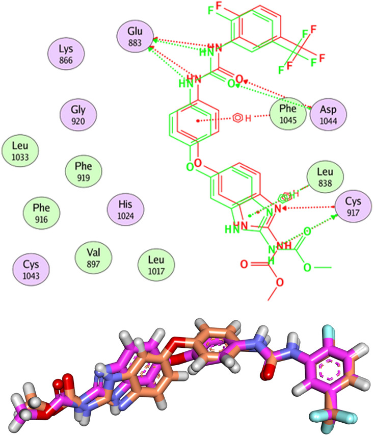
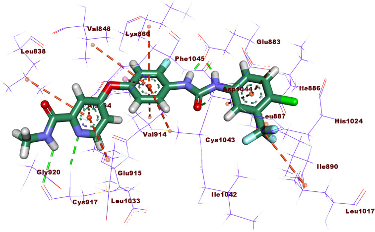
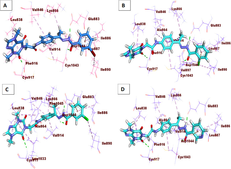
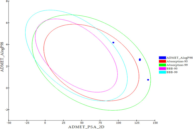
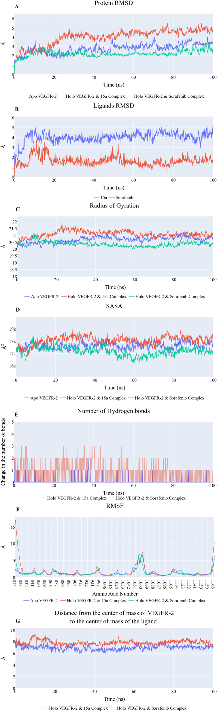
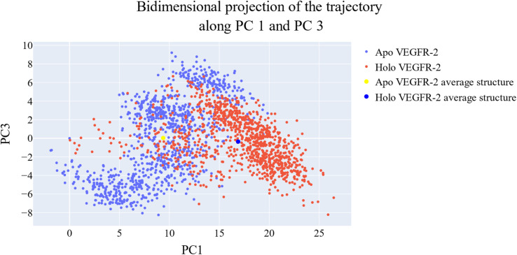
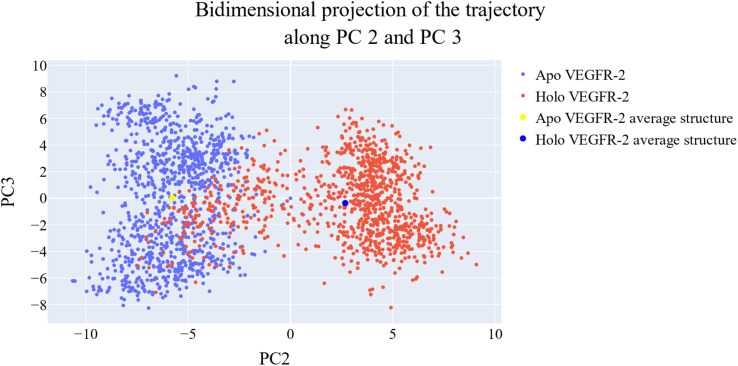
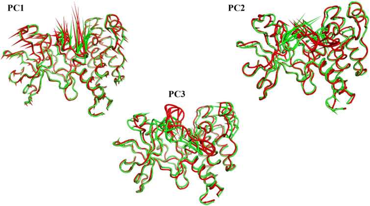
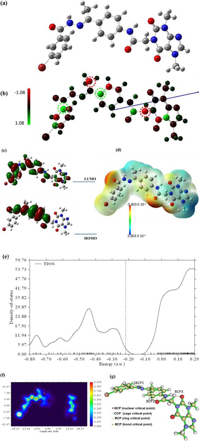
This study aimed to design anticancer theobromine derivatives inhibiting VEGFR-2. The new compounds were tested in vitro to evaluate their effectiveness against MCF-7 and HepG2 cancer cell lines. Among these compounds, 15a showed the highest cytotoxicity against HepG2, with an IC50 value of 0.76 μM, and significant anti-proliferative effects on MCF-7, with an IC50 value of 1.08 μM. Notably, the selectivity index of 15a against the two cancer cells was 98.97 and 69.64, respectively. Moreover, 15a demonstrated potent VEGFR-2 inhibitory activity (IC50 = 0.239 μM). Further investigations revealed that 15a induced apoptosis in HepG2 cells, significantly increasing early-stage and late-stage apoptosis percentages from 3.06% and 0.71% to 29.49% and 9.63%, respectively. It also upregulated caspase-3 and caspase-9 levels by 3.45-fold and 2.37-fold, respectively compared to control HepG2 cells. Additionally, 15a inhibited the migration and wound healing ability of HepG2 cells. Molecular docking confirmed the binding affinities of the semi-synthesized compounds to VEGFR-2, consistent with in vitro results. Several computational analyses (DFT, MD simulations, MM-GBSA, PLIP, and essential dynamics) supported the stability of the 15a-VEGFR-2 complex. Overall, the biological and computational findings suggest that compound 15a could be a promising lead compound for the development of a novel apoptotic anticancer agent.


Conflict of interest statement

There are no conflicts of interest to declare.

Figures

Fig. 1
Fig. 1. (A) Pharmacophoric requirements for VEGFR-2 inhibitors. (B) Reported VEGFR-2 inhibitors.
Fig. 2
Fig. 2. Rationale design of the work.
Scheme 1
Scheme 1. Synthesis of the key intermediates 7 and 8.
Scheme 2
Scheme 2. Synthesis of the key intermediates 11a–d and 14.
Scheme 3
Scheme 3. Synthesis of the target compounds 15a,b, 16, and 17.
Fig. 3
Fig. 3. Flow cytometry chart of apoptosis in HepG2 cells exposed to compound 15a.
Fig. 4
Fig. 4. The effect of compound 15a on HepG2's migration and healing after 48 hours.
Fig. 5
Fig. 5. Validation of the docking process. The RMSD value between the original ligand (pink) and the docked one (orange) is 1.19 Å.
Fig. 6
Fig. 6. 2D interaction of sorafenib with the active site of VEGFR-2.
Fig. 7
Fig. 7. 3D interactions of the synthesized compounds in the VEGFR-2 binding site. (A) Compound 15a, (B) compound 15b, (C) compound 16, (D) compound 17.
Fig. 8
Fig. 8. The executed ADMET study.
Fig. 9
Fig. 9. (A) RMSD values from the trajectory for the VEGFR-2 protein in apo form (blue line) and holo forms (red line for 15a system and green for sorafenib system). (B) Ligand RMSD values (15a: blue line, sorafenib: green line). (C) Radius of gyration for the VEGFR-2 protein in apo form (blue line) and holo forms (red line). (D) SASA for the VEGFR-2 protein in apo form (blue line) and holo forms (red line for 15a system and green for sorafenib system). (E) Change in the number of hydrogen bonds between 15a and VEGFR-2 protein (blue line) and sorafenib and VEGFR-2 protein (red line). (F) RMSF for the VEGFR-2 protein in apo form (blue line) and holo forms (red line for 15a system and green for sorafenib system). (G) Distance from the center of mass of compound 15a or sorafenib and VEGFR-2 protein.
Fig. 10
Fig. 10. Different energetic components of MM-GBSA. Bars represent the standard deviations.
Fig. 11
Fig. 11. Comparison between the binding free energy decomposition of the VEGFR-2_15a complex and VEGFR-2_sorafenib complex.
Fig. 12
Fig. 12. The amino acids, the types of interactions with 15a, and their occurrence during the whole simulation time using the ProLIF python library. (A) Amino acids panel from HSD814 to VAL897, (B) amino acids panel from VAL912 to ILE1023 and (C) amino acids panel from HES1024 to TYR1052.
Fig. 13
Fig. 13. The three clusters representative obtained from TTClust and their 3D interactions with 15a. Grey dashed lines: hydrophobic interactions, blue solid lines: H-bonds, green dashed line: Pi-stacking interaction, orange sticks: 15a, blue sticks: amino acids of VEGFR-2 protein.
Fig. 14
Fig. 14. The amino acids, the types of interactions with compound sorafenib, and their occurrence during the whole simulation time using the ProLIF python library. (A) Amino acids panel from HSD814 to LEU887, (B) amino acids panel from ILE890 to CYS1022 and (C) amino acids panel from ILE1023 to LEU1047.
Fig. 15
Fig. 15. The five clusters representative obtained from TTClust and their 3D interactions with sorafenib. Grey dashed lines: hydrophobic interactions, blue solid lines: H-bonds, green dashed line: Pi-stacking interaction, cyan solid line: halogen bond, orange sticks: sorafenib, blue sticks: amino acids of VEGFR-2 protein.
Fig. 16
Fig. 16. The change in the eigenvalues with increasing the eigenvectors (blue line) and the cumulative variance retained in the eigenvectors is shown (red line).
Fig. 17
Fig. 17. The projection of each trajectory on the first two eigenvectors.
Fig. 18
Fig. 18. The projection of each trajectory on the first and third eigenvectors.
Fig. 19
Fig. 19. The projection of each trajectory on the second and third eigenvectors.
Fig. 20
Fig. 20. The porcupine figures of each of the first three eigenvectors for both the apo and VEGFR-2_15a systems. Green cartoon: apo protein trajectory, red cartoon: VEGFR-2_15a trajectory.
Fig. 21
Fig. 21. The optimized geometry (a), the Mullikan atomic charge distribution (b), the frontier molecular orbitals (c), the electrostatic potential (d), the total density of states (e), and the QTAIM maps (f and g) at B3LYB/6-311++G(d,p) for 15a.

References

    1. Bray F. Ferlay J. Soerjomataram I. Siegel R. L. Torre L. A. Jemal A. Global cancer statistics 2018: GLOBOCAN estimates of incidence and mortality worldwide for 36 cancers in 185 countries. CA Cancer J Clin. 2018;68(6):394–424. - PubMed
    1. Belal A. Abdel Gawad N. M. Mehany A. B. Abourehab M. A. Elkady H. Al-Karmalawy A. A. Ismael A. S. Design, synthesis and molecular docking of new fused 1 H-pyrroles, pyrrolo [3,2-d] pyrimidines and pyrrolo [3,2-e][1,4] diazepine derivatives as potent EGFR/CDK2 inhibitors. J. Enzyme Inhib. Med. Chem. 2022;37(1):1884–1902. - PMC - PubMed
    1. Khalifa M. M. Al-Karmalawy A. A. Elkaeed E. B. Nafie M. S. Tantawy M. A. Eissa I. H. Mahdy H. A. Topo II inhibition and DNA intercalation by new phthalazine-based derivatives as potent anticancer agents: design, synthesis, anti-proliferative, docking, and in vivo studies. J. Enzyme Inhib. Med. Chem. 2022;37(1):299–314. - PMC - PubMed
    1. DeVita Jr V. T. Chu E. A history of cancer chemotherapy. Cancer Res. 2008;68(21):8643–8653. - PubMed
    1. Fidler I. J. Ellis L. M. Chemotherapeutic drugs—more really is not better. Nat. Med. 2000;6(5):500–502. - PubMed