. 2023 Jul 19:14:1196731.
doi: 10.3389/fimmu.2023.1196731. eCollection 2023.

ChemR23 activation reprograms macrophages toward a less inflammatory phenotype and dampens carcinoma progression

Affiliations

ChemR23 activation reprograms macrophages toward a less inflammatory phenotype and dampens carcinoma progression

Margot Lavy et al. Front Immunol. .

Abstract

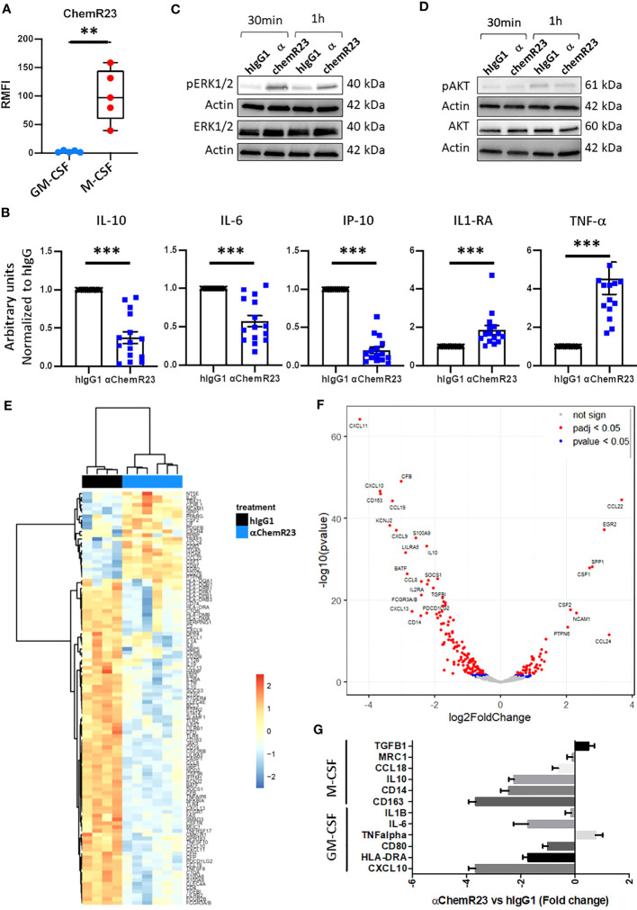
Introduction: Tumor Associated Macrophages (TAM) are a major component of the tumor environment and their accumulation often correlates with poor prognosis by contributing to local inflammation, inhibition of anti-tumor immune response and resistance to anticancer treatments. In this study, we thus investigated the anti-cancer therapeutic interest to target ChemR23, a receptor of the resolution of inflammation expressed by macrophages, using an agonist monoclonal antibody, αChemR23.

Methods: Human GM-CSF, M-CSF and Tumor Associated Macrophage (TAM)-like macrophages were obtained by incubation of monocytes from healthy donors with GM-CSF, M-CSF or tumor cell supernatants (Breast cancer (BC) or malignant pleural mesothelioma (MPM) cells). The effects of αChemR23 on macrophages were studied at the transcriptomic, protein and functional level. Datasets from The Cancer Genome Atlas (TCGA) were used to study CMKLR1 expression, coding for ChemR23, in BC and MPM tumors. In vivo, αChemR23 was evaluated on overall survival, metastasis development and transcriptomic modification of the metastatic niche using a model of resected triple negative breast cancer.

Results: We show that ChemR23 is expressed at higher levels in M-CSF and tumor cell supernatant differentiated macrophages (TAM-like) than in GM-CSF-differentiated macrophages. ChemR23 activation triggered by αChemR23 deeply modulates M-CSF and TAM-like macrophages including profile of cell surface markers, cytokine secretion, gene mRNA expression and immune functions. The expression of ChemR23 coding gene (CMKLR1) strongly correlates to TAM markers in human BC tumors and MPM and its histological detection in these tumors mainly corresponds to TAM expression. In vivo, treatment with αChemR23 agonist increased mouse survival and decreased metastasis occurrence in a model of triple-negative BC in correlation with modulation of TAM phenotype in the metastatic niche.

Conclusion: These results open an attractive opportunity to target TAM and the resolution of inflammation pathways through ChemR23 to circumvent TAM pro-tumoral effects.

Keywords: ChemR23 receptor; agonist; antibody; cancer; macrophage.


Conflict of interest statement

CT, VG, CM, and NP are inventors on a patent application (no. WO 2019/193029, filed 3 April 2019, published 8 October 2019) and product (WO2021/069709, filed 09 october 2019, published 15 April 2021) related to this work filed by OSE Immunotherapeutics and employees of OSE Immunotherapeutics with IG and SN. The remaining authors declare that the research was conducted in the absence of any commercial or financial relationships that could be construed as a potential conflict of interest. This study was partially supported by OSE Immunotherapeutics funding.

Figures

Figure 1
Figure 1
ChemR23 expression and activation in M-CSF macrophages. (A) Human monocytes from 5 healthy donors were differentiated in vitro using GM-CSF (20ng/ml) or M-CSF (50ng/ml) for 3 days, then ChemR23 expression was analyzed by flow cytometry. RFMI=Ratio of Median fluorescent intensity **, p < 0.01. (B) M-CSF macrophages were treated with αChemR23 or control hIgG1 for 24h. Then, cells were stimulated with LPS (200 ng/ml) for 24h and supernatants were collected. The indicated cytokines were quantified using multiplex ELISA in the corresponding supernatants. n= 15. *, p < 0.05; **, p < 0.01; ***, p < 0.001. pERK1/2(Thr202/Tyr204) (C) and pAKT(Ser473)/AKT (D) ChemR23 signaling assessed by immunoblot in M-CSF macrophages treated with either 10µg/ml of αChemR23 agonist antibody or with control hIgG1 for 30min or 1h. (E-G) M-CSF differentiated macrophages were treated with αChemR23 agonist or control hIgG1 for 24h followed by LPS stimulation (200ng/ml) for 6h. mRNA were extracted and analyzed using NanoString Technology (Human immunology panel). (E) Heatmap of genes differentially expressed between αChemR23 agonist versus control hIgG1 treated macrophages from 4 and 6 donors respectively with a p value adjusted ≤ 0,05 and absolute log2 fold change ≥ 1. (F) Volcano plot showing differentially expressed genes following αChemR23 agonist treatment, and genes with absolute value of the log2 fold change ≥ 2 and p value adjusted ≤ 0.05 are highlighted with their gene code name. (G) Graphic showing variation of expression of a panel of genes specific of GM-CSF or M-CSF macrophages. n=4-6.
Figure 2
Figure 2
αChemR23 agonist modifies M-CSF macrophages cell surface markers expression. M(M-CSF) macrophages were treated for 24h with αChemR23 or control hIgG1 and cells were collected and labelled with a panel of 10 antibodies including: CD209, CD163, CD14, CD16, CD206, HLA-DR and CD45 and analyzed using flow cytometry. (A) Macrophage subpopulations represented using t-SNE. (B) Phenotype of the different macrophage subpopulations impacted by treatment with αChemR23. Experiments were performed using monocytes from 3 different healthy donors and results include data from the 3 donors.
Figure 3
Figure 3
αChemR23 modulates functional properties of M-CSF macrophages. For phagocytosis assay, (A) breast cancer or (B) mesothelioma (MPM) apoptotic cells were incubated with untreated GM-CSF macrophages or M-CSF macrophages (left panels) and M-CSF macrophages (M(GM-CSF)) pretreated or not with human hIgG1 control or αChemR23 mAb for 24h (right panels). Efferocytic activity was quantified by live-cell imaging (Incucyte®). Results represent the maximum fluorescence intensity normalized to M(GM-CSF) used as reference. n=4. *, p < 0.05. (C, D) For T cell proliferation assay, CPD stained CD3+ T-cells were incubated for 5 days with CD3/CD28 beads and either IL-2 (75 U/ml), or IL-10 (50 ng/ml), or supernatants of M(M-CSF) macrophages (MΦ) treated with αChemR23 agonist (10µg/ml) or hIgG1 control (10µg/ml) and T-cells were assessed by flow cytometry. Frequency of non-fluorescent proliferating cells was evaluated after gating on CD3+ cells (C) and after gating on CD8+ cells (D) and normalized to the unstimulated condition. n=4. *, p < 0.05.
Figure 4
Figure 4
CMKLR1 expression in breast cancer and mesothelioma tumors correlates to TAM markers and ChemR23 is expressed in in vitro TAM-like models. Correlation of CMKLR1 expression with macrophage markers CD14, CD163, MRC1 and HLA-DRA in breast cancers (n=1034) (A) and mesothelioma (n=87) (B) tumors using transcriptomic data from the TCGA database. (C) IHC staining of ChemR23 and Iba1 in breast tumors and MPM from 9 patients in tumor and peritumoral areas. (D) Quantification of ChemR23 IHC staining by HPF (high power field, 1590μm2) for each patient.
Figure 5
Figure 5
αChemR23 agonist modulates TAM-like macrophages cytokine secretion and mRNA expression profiles. Monocytes from healthy donors were incubated with conditioned media of BC cells (A, C) or MPM cells (B, D) for 3 days were treated with 10µg/ml of αChemR23 agonist or control hIgG1 for 24h. For cytokine quantification (A, B), supernatants were collected after 24h stimulation with LPS (200ng/ml) then the indicated cytokines were quantified using multiplex ELISA in the supernatants. n= 6. *, p < 0.05. For mRNA analyses (C, D), cells were lysed after 6h stimulation with LPS (200ng/ml) then mRNA were extracted and gene expressions were measured using RT-PCR. n= 5. *, p < 0.05; **, p < 0.01.
Figure 6
Figure 6
αChemR23 decreased phagocytic properties of TAM-like macrophages. Phagocytosis of apoptotic (A) breast cancer (BC) or (B) mesothelioma (MPM) cell lines by untreated GM-CSF and M-CSF macrophages or TAM-like macrophages pretreated with αChemR23 or hIgG1 control (10µg/ml) for 24h and quantified by live-cell imaging (Incucyte®). Results represent the maximum fluorescence intensity normalized to GM-CSF macrophages condition. n=4. *, p < 0.05.
Figure 7
Figure 7
αChemR23 monotherapy increases survival and reduces metastasis in a murine orthotopic breast tumor model. Survival (A) and lung metastasis count by bioluminescence (BLI) (B) of 4T1-luc2 bearing mice treated i.p. with hIgG1 control or αChemR23 mAb (1mg/kg, 3 times a week from d7 to d28). Representative heatmap of clustered differential gene expression (C) and scores of transcriptomic analysis (D) of lungs in the αChemR23 (blue; n=7) or isotype control (black; n=4) treated 4T1-luc2-bearing mice that have been resected at d13 using the Nanostring mouse PCIP panel. (E) Osteopontin mRNA expression, measured using RT-PCR, in lung tumors of mice treated with hIgG1 control (black, n=8) or αChemR23 non responders (red, n=4) or αChemR23 responders (blue, n=4) mAb. (F) M1 score signature of transcriptomic analysis of lungs from αChemR23 and isotype hIgG1 (black; n=4 or 8) mAb-treated mice. (G) Neutrophil chemotaxis scores and (H) IFNγ scores of transcriptomic analysis of lungs in the αChemR23 (red for non-responders n =3-4; blue for responders n=3) mAb-treated mice. ns, non-significant; *, p < 0.05; **, p < 0.01. NR, non-responders n=3-4; R, responders n= 3-4.

References

    1. Hanahan D, Weinberg RA. Hallmarks of cancer: the next generation. Cell (2011) 144(5):646−74. doi: 10.1016/j.cell.2011.02.013 - DOI - PubMed
    1. Mantovani A, Allavena P, Marchesi F, Garlanda C. Macrophages as tools and targets in cancer therapy. Nat Rev Drug Discov (2022) 21(11):799–820. doi: 10.1038/s41573-022-00520-5 - DOI - PMC - PubMed
    1. Greten FR, Grivennikov SI. Inflammation and cancer: triggers, mechanisms, and consequences. Immunity (2019) 51(1):27−41. doi: 10.1016/j.immuni.2019.06.025 - DOI - PMC - PubMed
    1. Sulciner ML, Gartung A, Gilligan MM, Serhan CN, Panigrahy D. Targeting lipid mediators in cancer biology. Cancer Metastasis Rev (2018) 37(2−3):557−72. doi: 10.1007/s10555-018-9754-9 - DOI - PubMed
    1. Fattori V, Zaninelli TH, Rasquel-Oliveira FS, Casagrande R, Verri WA. Specialized pro-resolving lipid mediators: a new class of non-immunosuppressive and non-opioid analgesic drugs. Pharmacol Res (2020) 151:104549. doi: 10.1016/j.phrs.2019.104549 - DOI - PubMed

Publication types

Substances