

A Case Report of Rare and Lethal Methicillin-Resistant Staphylococcus aureus (MRSA) Peritonitis in Infancy
- PMID: 37539401
- PMCID: PMC10394718
- DOI: 10.7759/cureus.41303
A Case Report of Rare and Lethal Methicillin-Resistant Staphylococcus aureus (MRSA) Peritonitis in Infancy
Abstract
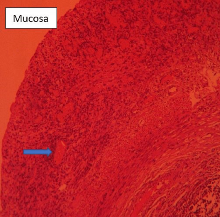
Peritoneal inflammation without a discernible intraperitoneal source is referred to as primary peritonitis. Only 2% of pediatric acute abdominal crises are diagnosed preoperatively. Association with other infections is uncommon and is often limited to hepatic and urinary pathogens. Here, we describe a case of primary peritonitis in a one-month-old child who had laparotomy and appendicectomy as per the recommended treatment plan. There were no accompanying hepatic and urinary diseases. In this instance, methicillin-resistant Staphylococcus aureus (MRSA) was the responsible bacteria. The use of linezolid, as per the culture sensitivity report of intraperitoneal pus, ensured a smooth recovery in this case.
Keywords: acute abdominal emergencies; appendicectomy; methicillin resistant staphylococcus aureus (mrsa); perforation peritonitis; primary peritonitis.
Copyright © 2023, Kumar et al.
Conflict of interest statement
The authors have declared that no competing interests exist.
Figures


Similar articles
-
[A multicentric study on clinical characteristics and antibiotic sensitivity in children with methicillin-resistant Staphylococcus aureus infection].Zhonghua Er Ke Za Zhi. 2020 Aug 2;58(8):628-634. doi: 10.3760/cma.j.cn112140-20200505-00469. Zhonghua Er Ke Za Zhi. 2020. PMID: 32842382 Chinese.
-
Methicillin-Resistant Staphylococcus aureus Peritonitis due to Hematogenous Dissemination from Central Venous Catheter in a Maintenance Dialysis Patient.Case Rep Nephrol Dial. 2021 Sep 13;11(3):281-285. doi: 10.1159/000517143. eCollection 2021 Sep-Dec. Case Rep Nephrol Dial. 2021. PMID: 34703828 Free PMC article.
-
Successful salvage of peritoneal catheter in unresolved methicillin-resistant staphylococcus aureus peritonitis by combination treatment with daptomycin and rifampin.Blood Purif. 2011;32(4):249-52. doi: 10.1159/000328028. Epub 2011 Aug 12. Blood Purif. 2011. PMID: 21846982
-
Antibiotic management of methicillin-resistant Staphylococcus aureus--associated acute pulmonary exacerbations in cystic fibrosis.Ann Pharmacother. 2015 Apr;49(4):458-68. doi: 10.1177/1060028014567526. Epub 2015 Jan 12. Ann Pharmacother. 2015. PMID: 25583881 Review.
-
Methicillin-resistant Staphylococcus aureus intracranial abscess: An analytical series and review on molecular, surgical and medical aspects.Indian J Med Microbiol. 2018 Jan-Mar;36(1):97-103. doi: 10.4103/ijmm.IJMM_17_41. Indian J Med Microbiol. 2018. PMID: 29735835 Review.
References
-
- Peritonitis in children with nephrotic syndrome. Gorensek MJ, Lebel MH, Nelson JD. https://pubmed.ncbi.nlm.nih.gov/3368284/ Pediatrics. 1988;81:849–856. - PubMed
-
- Primary peritonitis: changing aspects 1956-1970. Fowler R. https://pubmed.ncbi.nlm.nih.gov/5093237/ Aust Paediatr J. 1971;7:73–83. - PubMed
-
- Primary peritonitis in the newborn: a recently recognised disease. Lainakis N, Koulopoulos K, Kourakos A, Kostopoulos E, Trigas B, Skanavis K, Trapalis B. https://pubmed.ncbi.nlm.nih.gov/19537122/ Ann Ital Chir. 2009;80:39–41. - PubMed
Publication types
LinkOut - more resources
Full Text Sources