Review
. 2023 Nov 21;74(21):6448-6467.
doi: 10.1093/jxb/erad310.

Cell wall dynamics: novel tools and research questions

Affiliations
Review

Cell wall dynamics: novel tools and research questions

Luis Alonso Baez et al. J Exp Bot. .

Abstract

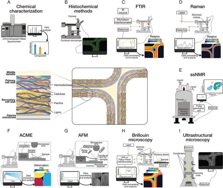
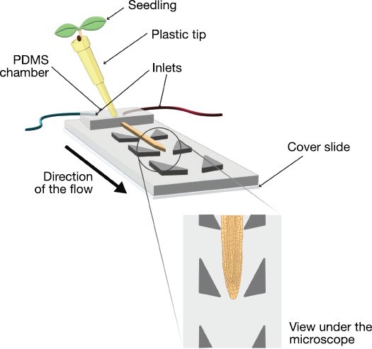
Years ago, a classic textbook would define plant cell walls based on passive features. For instance, a sort of plant exoskeleton of invariable polysaccharide composition, and probably painted in green. However, currently, this view has been expanded to consider plant cell walls as active, heterogeneous, and dynamic structures with a high degree of complexity. However, what do we mean when we refer to a cell wall as a dynamic structure? How can we investigate the different implications of this dynamism? While the first question has been the subject of several recent publications, defining the ideal strategies and tools needed to address the second question has proven to be challenging due to the myriad of techniques available. In this review, we will describe the capacities of several methodologies to study cell wall composition, structure, and other aspects developed or optimized in recent years. Keeping in mind cell wall dynamism and plasticity, the advantages of performing long-term non-invasive live-imaging methods will be emphasized. We specifically focus on techniques developed for Arabidopsis thaliana primary cell walls, but the techniques could be applied to both secondary cell walls and other plant species. We believe this toolset will help researchers in expanding knowledge of these dynamic/evolving structures.

Keywords: Biophysics; cell wall composition; cell wall structure; live-imaging; mechanics; plant cell wall.


Conflict of interest statement

The authors have reviewed the potential for conflicts of interest and declare that no financial, personal, or academic affiliations exist that could influence the objective discussion of techniques or the scientific discussions presented in this paper.

Figures

Fig. 1.
Fig. 1.
The plant cell wall is a chemically diverse structure composed mainly of polysaccharides (cellulose, hemicelluloses, and pectin), with secondary cell walls also including lignin. These molecules are arranged in a complex 3D structure that gives the cell wall its strength and rigidity. New techniques for studying plant cell walls provide insights into cell wall composition (A–E) and mechanical characteristics (F–I). (A) Chemical characterization provides accurate absolute quantification of the different cell wall components, although with no possibilities of in situ studies. (B) Histochemical methods such as using monoclonal antibodies against cell wall epitopes or click chemistry carbohydrate probes allow the location of specific components on the cell walls (e.g. hemicelluloses are marked in green on the figure). (C) Fourier-transform infrared (FTIR) spectroscopy enables relative quantifications of specific structural components (e.g. hemicelluloses) but the resolution is limited. (D) Raman spectroscopy also enables relative quantifications, in this case with higher resolution. (E) Solid-state NMR (ssNMR) allows a high level of detail in the study of chemical composition, but the data interpretation is complicated, and it is not compatible with in situ analysis. (F) An automated confocal-microextensometer (ACME) enables the measurement of extensibility and other mechanical properties in vivo in individual cells. (G) Atomic force microscopy (AFM) uses a probe (cantilever) to determine cell wall stiffness and topography, and generates an image with nanometric resolution, although it is restricted to the first cell layer. (H) Brillouin microscopy can be used to study mechanical properties in different cell layers with a micrometric resolution. The frequency shift caused by the Brillouin effect is a measurement of stiffness, whereas the amplitude of the signal can be related to viscosity. (I) Ultrastructural microscopy techniques can be used to image macromolecules at submolecular resolution, allowing the observation of cell wall microstructure.
Fig. 2.
Fig. 2.
Microfluidics devices allow the study of dynamic responses. The schema represents a basic implementation for a microfluidics device with two inlets, which permits changing of the growing conditions and/or the delivery of specific treatments. The growth of the root tip can be observed under a microscope.

References

    1. Adichtchev SV, Karpegina YA, Okotrub KA, Surovtseva MA, Zykova VA, Surovtsev NV.. 2019. Brillouin spectroscopy of biorelevant fluids in relation to viscosity and solute concentration. Physical Review E 99, 062410. - PubMed
    1. Allison DP, Mortensen NP, Sullivan CJ, Doktycz MJ.. 2010. Atomic force microscopy of biological samples. WIREs Nanomedicine and Nanobiotechnology 2, 618–634. - PubMed
    1. Alonso-Simón A, Garcia-Angulo P, Mélida H, Encina A, Alvarez JM, Acebes JL.. 2011. The use of FTIR spectroscopy to monitor modifications in plant cell wall architecture caused by cellulose biosynthesis inhibitors. Plant Signaling and Behaviour 6, 1104–1110. - PMC - PubMed
    1. Altartouri B, Bidhendi AJ, Tani T, Suzuki J, Conrad C, Chebli Y, Liu N, Karunakaran C, Scarcelli G, Geitmann A.. 2019. Pectin chemistry and cellulose crystallinity govern pavement cell morphogenesis in a multi-step mechanism. Plant Physiology 181, 127–141. - PMC - PubMed
    1. Amsbury S, Hunt L, Elhaddad N, Baillie A, Lundgren M, Verhertbruggen Y, Scheller HV, Knox JP, Fleming AJ, Gray JE.. 2016. Stomatal function requires pectin de-methyl-esterification of the guard cell wall. Current Biology 26, 2899–2906. - PMC - PubMed

Publication types