Review
. 2023 Oct;65(10):1425-1438.
doi: 10.1007/s00234-023-03204-y. Epub 2023 Aug 4.

The ptotic tongue-imaging appearance and pathology localization along the course of the hypoglossal nerve

Affiliations
Review

The ptotic tongue-imaging appearance and pathology localization along the course of the hypoglossal nerve

Vineet Vijay Gorolay et al. Neuroradiology. 2023 Oct.

Abstract

CT and MRI findings of tongue ptosis and atrophy should alert radiologists to potential pathology along the course of the hypoglossal nerve (cranial nerve XII), a purely motor cranial nerve which supplies the intrinsic and extrinsic muscles of the tongue. While relatively specific for hypoglossal nerve pathology, these findings do not accurately localize the site or cause of denervation. A detailed understanding of the anatomic extent of the nerve, which crosses multiple anatomic spaces, is essential to identify possible underlying pathology, which ranges from benign postoperative changes to life-threatening medical emergencies. This review will describe key imaging findings of tongue denervation, segmental anatomy of the hypoglossal nerve, imaging optimization, and comprehensive imaging examples of diverse pathology which may affect the hypoglossal nerve. Armed with this knowledge, radiologists will increase their sensitivity for detection of pathology and provide clinically relevant differential diagnoses when faced with findings of tongue ptosis and denervation.

Keywords: Denervation; Hypoglossal; Hypoglossal palsy; MRI; Ptosis.


Conflict of interest statement

The authors have no relevant financial or non-financial interests to disclose.
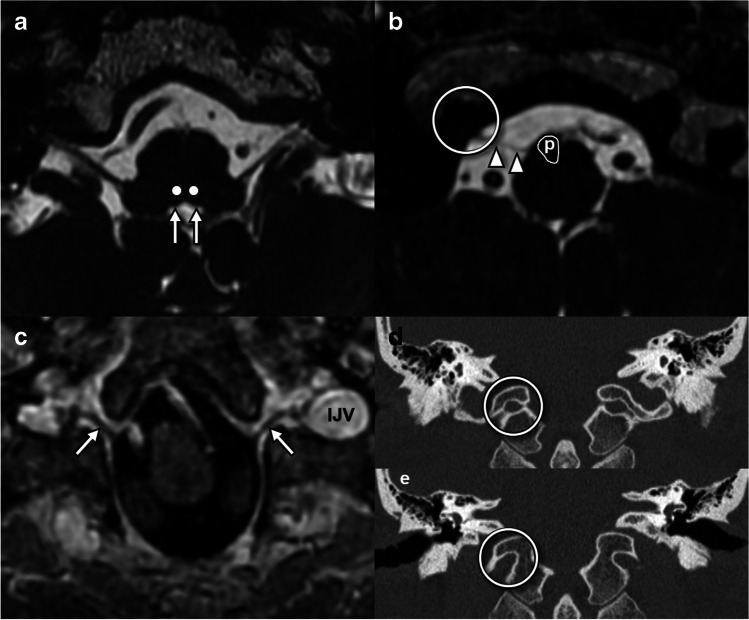
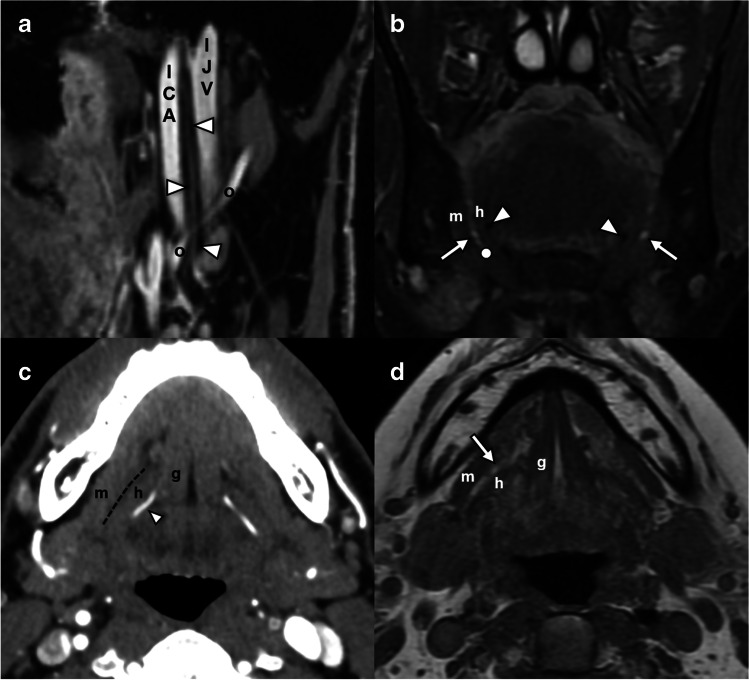
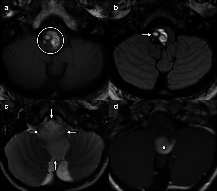
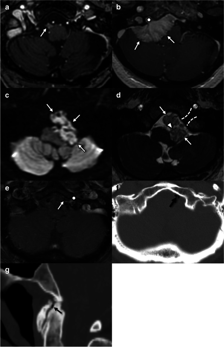
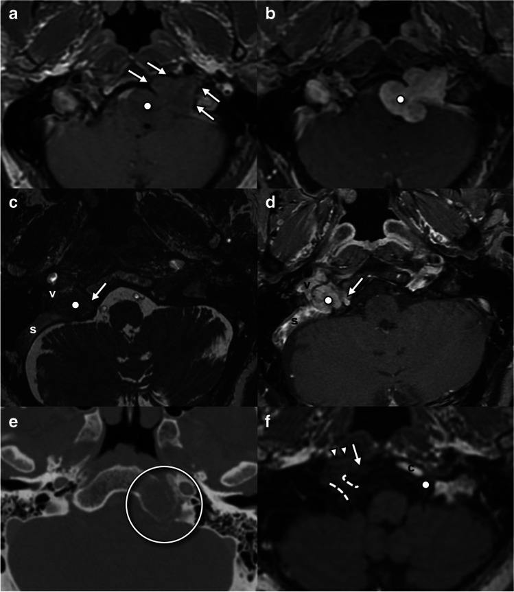
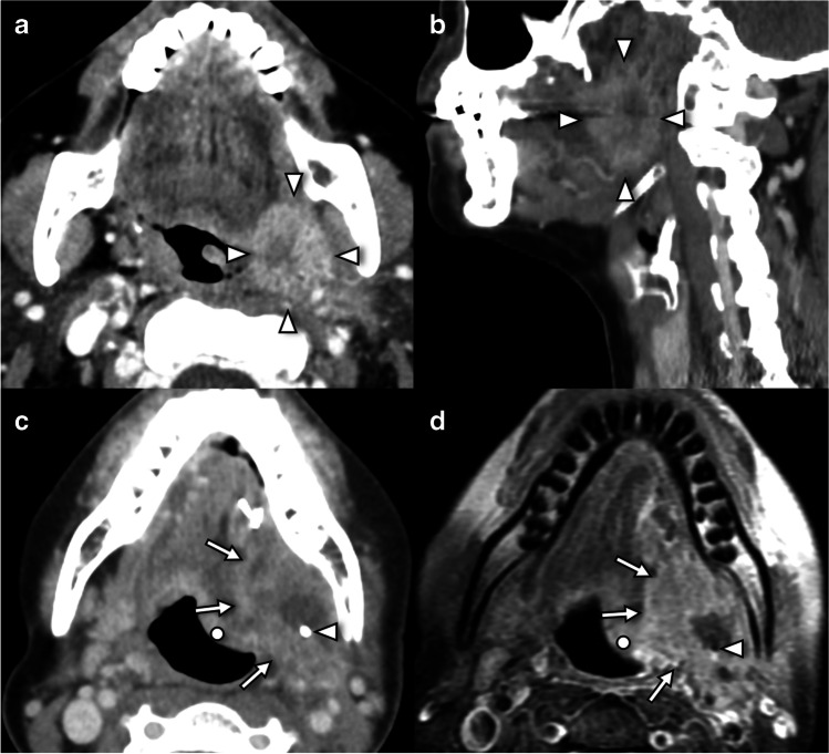
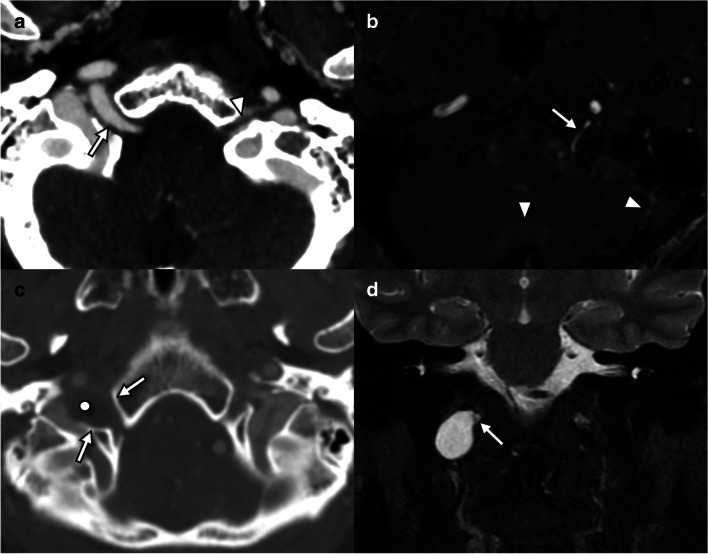
Figures

Fig. 1
Fig. 1
Imaging features of hypoglossal denervation. a Axial T1W sequence in a patient with acute tongue ptosis due to cranio-cervical junction degenerative disease (CCJD) (see Fig. 5) demonstrates posterior displacement of the affected right hemitongue (arrowheads), which remains isointense in signal and bulk. b Axial T2FS sequence in the same patient as a demonstrates posterior displacement (arrowheads) and T2 hyperintensity of the affected right hemitongue. Note the sharply marginated signal change along the median raphe (arrows) which distinguishes this from a mass lesion. c Axial contrast-enhanced CT in a 44-year-old patient presenting with neck pain and tongue deviation demonstrates posterior displacement of the left hemitongue (arrowheads) without change in attenuation. A high cervical carotid dissection was identified (not shown). d Axial T1W sequence in a 29-year-old patient with chronic tongue ptosis due to a jugular paraganglioma (circle). The affected right hemitongue demonstrates volume loss and T1 shortening (dashed triangle) consistent with fatty atrophy. e Axial T2FS sequence in the same patient as d demonstrates signal dropout (dashed triangle) within areas of fatty atrophy, but also some areas of high signal attributed to edema. Note how signal changes respect the median raphe (arrows), distinguishing denervation from a mass lesion. In this case, denervation is caused by extrinsic compression by a vagal paraganglioma (circle) in the carotid space, with characteristic T2 hyperintensity and internal flow voids. f Axial post-gadolinium T1FS sequence in the same patient as d and e demonstrates enhancement of the right hemitongue. Note how signal changes respect the median raphe (arrows), distinguishing denervation from a mass lesion. The vagal paraganglioma (circle) enhances markedly. g Axial T1W sequence in a 54-year-old patient with tongue weakness and episodic dizziness demonstrates T1 shortening and volume loss in the right hemitongue respecting the median raphe (arrows) compatible with fatty atrophy. In this case, denervation is caused by extrinsic compression by a T1 hypointense schwannoma (circle). h Axial T2W sequence in the same patient as g demonstrates T2 prolongation and volume loss in the right hemitongue respecting the median raphe (arrows) compatible with fatty atrophy. The causative schwannoma (circle) is of intermediate T2 signal with a T2 hyperintense cystic component. i Axial CT in a patient with chronic tongue ptosis due to osseous metastasis (not shown) demonstrates posterior displacement (arrowheads), hypoattenuation, and volume loss in the right hemitongue respecting the median raphe (arrows) compatible with fatty atrophy
Fig. 2
Fig. 2
Imaging anatomy of the hypoglossal nuclei and intramedullary fascicles and cisternal and skull base segments. a Axial FIESTA sequence through the upper medulla at the level of the inferior olivary nucleus demonstrates the hypoglossal eminence (arrows) at the floor of the fourth ventricle. The expected location of the hypoglossal nuclei (dots) can be inferred from this landmark. b Axial FIESTA sequence through the lower medulla below the obex demonstrates HN rootlets (arrowheads) exiting lateral to the pyramidal tracts (p) and into the hypoglossal canal (open circle). c Axial post-gadolinium volumetric T1 sequence through the hypoglossal canal demonstrates the canalicular segment of the HN (arrows) as a filling defect surrounded by venous enhancing venous plexus, passing toward the internal jugular vein (IJV) at the upper margin of the carotid sheath. d Coronal CT at the level of the internal (posterior) os of the hypoglossal canal where the canal has a circular shape (circle). e Coronal CT at the level of the external (anterior) os of the hypoglossal canal with a “bird’s beak” appearance (circle)
Fig. 3
Fig. 3
Imaging anatomy of the carotid and sublingual segments of the hypoglossal nerve. a Sagittal oblique volumetrically acquired T1FS sequence through the suprahyoid neck demonstrates the path of the HN (arrowheads) in the carotid sheath, between the internal carotid artery (ICA) and the internal jugular vein (IJV) before crossing the occipital artery (o) and turning anteriorly to enter the sublingual space. b Coronal T2FS sequence through the floor of mouth at the level of the angle of the mandible demonstrates the anticipated location of the hypoglossal nerve (dot) in the sublingual plane between mylohyoid (m) and hyoglossus (h) muscles and inferior to the submandibular duct (arrow). Note the lingual artery flow voids (arrowheads) deep to hyoglossus. c Axial contrast-enhanced CT angiogram demonstrates the plane of the hypoglossal nerve (dashed line) between the genioglossus (g), hyoglossus (h), and mylohyoid (m). Note how the lingual artery (arrowhead) lies deep to hyoglossus. d Axial T1W sequence through the floor of mouth demonstrates key landmarks including the genioglossus (g), hyoglossus (h), and mylohyoid (m) and the sublingual fat plane (arrowhead) through which the hypoglossal nerve traverses beneath the submandibular duct
Fig. 4
Fig. 4
Lesions involving the hypoglossal nuclei and intramedullary fascicles. a Axial T1W through the lower medulla in a 56-year-old presenting with headaches, nausea, and tongue weakness demonstrates a multilobulated lesion with intrinsic T1 shortening compatible with subacute blood products (circle). b Axial T2W in the same patient as a shows the multilobulated lesion contents are mostly T2 hyperintense with “shading” (arrow) suggesting blood products of varying ages and a peripheral T2 hypointense rim, typical of a cavernous venous malformation. c Axial T2W in a 4-year-old with headaches and lower cranial neuropathies demonstrates an expansile T2 hyperintense mass (arrows) occupying the medulla. d Axial T1W+C in the same patient as c demonstrates solid nodular enhancement at the periphery of the mass (dot). No reduced diffusion (not shown). Operative pathology confirmed pilocytic astrocytoma (WHO CNS grade 1)
Fig. 5
Fig. 5
Lesions involving the cisternal segment of the hypoglossal nerve. a Axial post-gadolinium T1W SPGR sequence in a 44-year-old with history of NF2 and prior left vestibular schwannoma resection demonstrates an enhancing nodule arising from the right lateral medulla from a hypoglossal nerve root. b Axial post-gadolinium T1W SPGR sequence in a 66-year-old with multiple right-sided cranial nerve palsies demonstrates a homogenously enhancing lobulated dural-based mass (arrow) arising from the right petrous face and extending into the hypoglossal canal (dot). Surgical pathology confirmed WHO grade 1 meningioma. c Axial DWI (b = 1000) in a 14-year-old female with headache and slowly progressive lower cranial neuropathies demonstrates a lobulated extra-axial cystic lesion in the left cerebellomedullary cistern with markedly reduced diffusion (arrows) consistent with an epidermoid cyst. d Axial FIESTA in the same patient as c clearly shows left hypoglossal nerve rootlets (arrowhead) traversing the epidermoid cyst (arrows) en route to the hypoglossal canal (dashed lines). e Axial post-gadolinium T1W in a 54-year-old with chronic left-sided tongue weakness is notable for a non-enhancing T1 hypointense extradural lesion (arrow) at the orifice of the left hypoglossal canal (dot). This was of low signal intensity on T2W (not shown). f Axial unenhanced CT in the same patient as e reveals the lesion is of gas density (black arrow). g Sagittal reformatted unenhanced CT image of the midline skull base in the same patient as e demonstrates marked degenerative changes at the cranio-cervical junction with intra-articular gas (black arrow). This was thought to have migrated to the extradural space in f and resulted in cisternal impingement of the hypoglossal nerve
Fig. 6
Fig. 6
Lesions involving the intracanalicular segment of the hypoglossal nerve. a Axial T1W in a 44-year-old patient with chronic left-sided tongue weakness demonstrates a dumbbell-shaped mass (dot) extending from the left cerebellomedullary cistern causing smooth remodeling of the hypoglossal canal (arrows). b Axial post-gadolinium T1W in the same patient as a confirms marked, homogenous enhancement of the mass (dot), subsequently proven to be a schwannoma by fine needle aspiration. c Axial SSFP in a 55-year-old male with multiple right lower cranial neuropathies demonstrates a mass (dot) occupying the right jugular bulb, with involvement of the right hypoglossal nerve (arrow) in within the hypoglossal canal and with partial occlusion of the sigmoid sinus (s) and the internal jugular vein (v). d Axial post-gadolinium T1W in the same patient as c confirms enhancement of the mass, which was later biopsy proven to be a jugular paraganglioma. e Axial CT bone reconstructions following intravenous iodinated contrast in a patient with history of metastatic NSCLC demonstrates permeative erosion of the left basi-occiput involving the left hypoglossal canal (circle) due to a skull base metastasis. f Axial T1W in a 66-year-old patient with metastatic NSCLC demonstrates hypointense T1 signal in the right clivus and cortical destruction (arrow) due to a metastatic deposit with extraosseous extension (arrowheads). T1 hypointense mass involves the right hypoglossal canal (dashed lines). The normal left hypoglossal canal (dot) is hyperintense on T1W and surrounded by T1 hyperintense clival fatty marrow (c)
Fig. 7
Fig. 7
Lesions involving the carotid segment of the hypoglossal nerve. Further tumors of the carotid sheath have been presented in Fig. 1. a Axial unenhanced CT in a 76-year-old female with prior cervical fusion surgery presents with right tongue weakness after trigger point injections reveals an osteophyte (arrow) overlying the external os of the right hypoglossal canal (dashed line). b Coronal unenhanced CT in the same patient as a reveals ankylosis of the right C1-C2 articulation contiguous with the osteophyte (dot) causing stenosis of the right hypoglossal canal (arrow). The left hypoglossal canal (circle) is normal in caliber. c Axial contrast-enhanced CT angiogram in a 44-year-old presenting with neck pain and tongue weakness demonstrates irregular narrowing of the left cervical internal carotid artery lumen consistent with a dissection flap (arrow). Left tongue ptosis was shown in Fig. 1c. d Coronal T1W in the same patient as Fig. 1f, a 54-year-old patient with tongue weakness and episodic dizziness, demonstrates a fusiform T1 hypointense schwannoma (s) arising from pars nervosa of the right jugular foramen (arrow) and causing extrinsic mass effect on the right hypoglossal canal (dot). Further tumors of the carotid sheath have been presented in Fig. 1. e Axial contrast-enhanced CT neck in a 62-year-old 3 months after salvage right neck dissection for nodal recurrence of oropharyngeal SCC demonstrates surgical clips in the right carotid space (arrows) and effaced suprahyoid fat planes due to post-treatment changes as well as acute right hypoglossal denervation (arrowhead). There was no evidence of perineural disease on MRI (not shown). f Axial contrast-enhanced CT angiogram of the neck in a 61-year-old 1 month after right carotid endarterectomy demonstrates linear scarring (arrows) and loss of fat planes (circle) in the right carotid sheath due to post-surgical changes and acute right hypoglossal denervation (arrowhead). Follow-up CT angiogram for planning of contralateral endarterectomy demonstrated resolution of the right hypoglossal palsy (not shown)
Fig. 8
Fig. 8
Lesions involving the sublingual segment of the hypoglossal nerve. a Axial contrast-enhanced CT in a 61-year-old female smoker with left neck pain and swallowing difficulty reveals a centrally necrotic, enhancing and infiltrative mass in the left glossotonsillar sulcus (arrowheads) consistent with squamous cell carcinoma. b Sagittal contrast-enhanced CT in the same patient as a demonstrates the mass (arrowheads) occupies the expected location of the hypoglossal nerve at the junction of carotid and sublingual space segments. c Axial contrast-enhanced CT in a 65-year-old male smoker status post partial glossectomy, neck dissection, and radiotherapy for left oral tongue SCC demonstrates a bulky, necrotic recurrent mass (arrows) around surgical clips (arrowhead) and accompanying left tongue ptosis (dot). d Axial contrast-enhanced T1W in the same patient as c better demonstrates extent of soft tissue involvement by recurrent tumor (arrows) around the surgical resection clips (arrowhead) with associated denervation ptosis (dot)
Fig. 9
Fig. 9
Lesions of the hypoglossal canal which are not commonly associated with denervation. a Axial CT angiography in an adult patient presenting with transient ischemic attack demonstrates an artery coursing through the right hypoglossal canal (arrow) forming an anastomosis between the right internal carotid artery and the basilar trunk (not shown), a persistent primitive hypoglossal artery, and a congenital anomaly. Note the normal left hypoglossal nerve (arrowhead), seen as a filling defect surrounded by venous plexus. This patient did not have hypoglossal palsy. b Axial time-of-flight MR angiogram in a 73-year-old (male) with pulsatile tinnitus demonstrates a high flow vessel traversing the left hypoglossal canal (arrow) and with arterialized flow in the dural venous sinuses (arrowheads). DSA (not shown) revealed multiple dural arteriovenous fistulas. This patient did not have hypoglossal palsy. c Axial CT angiography in a 59-year-old (female) presenting with an episode of dizziness reveals smooth remodeling of the right hypoglossal canal (arrows) due to a hypoattenuating cystic lesion (dot). d Coronal T2W MRI in the same patient as c demonstrates a T2 hyperintense circumscribed lesion protruding through the right hypoglossal canal (arrow), consistent with perineural sleeve cyst. It was isointense to CSF on all sequences and did not demonstrate any enhancement or reduced diffusion (not shown). This patient did not have hypoglossal palsy

References

    1. Thompson EO, Smoker WR. Hypoglossal nerve palsy: a segmental approach. Radiographics. 1994;14:939–958. doi: 10.1148/radiographics.14.5.7991825. - DOI - PubMed
    1. Bademci G, Yasargil MG. Microsurgical anatomy of the hypoglossal nerve. J Clin Neurosci. 2006;13:841–847. doi: 10.1016/j.jocn.2005.12.028. - DOI - PubMed
    1. Mu L, Chen J, Li J, et al. Innervation of human soft palate muscles. Anat Rec (Hoboken). 2021;304:1054–1070. doi: 10.1002/ar.24531. - DOI - PubMed
    1. Loh C, Maya MM, Go JL. Cranial nerve XII: the hypoglossal nerve. Semin Ultrasound CT MR. 2002;23:256–265. doi: 10.1016/S0887-2171(02)90050-8. - DOI - PubMed
    1. Stino AM, Smith BE, Temkit M, Reddy SN. Hypoglossal nerve palsy: 245 cases. Muscle Nerve. 2016;54:1050–1054. doi: 10.1002/mus.25197. - DOI - PubMed