. 2023 Nov;46(6):1170-1185.
doi: 10.1002/jimd.12667. Epub 2023 Sep 11.

Beyond genetics: Deciphering the impact of missense variants in CAD deficiency

Affiliations

Beyond genetics: Deciphering the impact of missense variants in CAD deficiency

Francisco Del Caño-Ochoa et al. J Inherit Metab Dis. 2023 Nov.

Abstract

CAD is a large, 2225 amino acid multienzymatic protein required for de novo pyrimidine biosynthesis. Pathological CAD variants cause a developmental and epileptic encephalopathy which is highly responsive to uridine supplements. CAD deficiency is difficult to diagnose because symptoms are nonspecific, there is no biomarker, and the protein has over 1000 known variants. To improve diagnosis, we assessed the pathogenicity of 20 unreported missense CAD variants using a growth complementation assay that identified 11 pathogenic variants in seven affected individuals; they would benefit from uridine treatment. We also tested nine variants previously reported as pathogenic and confirmed the damaging effect of seven. However, we reclassified two variants as likely benign based on our assay, which is consistent with their long-term follow-up with uridine. We found that several computational methods are unreliable predictors of pathogenic CAD variants, so we extended the functional assay results by studying the impact of pathogenic variants at the protein level. We focused on CAD's dihydroorotase (DHO) domain because it accumulates the largest density of damaging missense changes. The atomic-resolution structures of eight DHO pathogenic variants, combined with functional and molecular dynamics analyses, provided a comprehensive structural and functional understanding of the activity, stability, and oligomerization of CAD's DHO domain. Combining our functional and protein structural analysis can help refine clinical diagnostic workflow for CAD variants in the genomics era.

Keywords: developmental and epileptic encephalopathy; dihydroorotase; epilepsy; functional validation assay; inborn metabolic disease; molecular dynamics; protein structure-function; pyrimidine metabolism; treatment; uridine; variant of uncertain significance; x-ray crystallography.


Conflict of interest statement

Competing interest statement

Francisco del Caño-Ochoa, Bobby G. Ng, Antonio Rubio-del-Campo, Sonal Mahajan, Matthew P. Wilson, Marçal Vilar, Daisy Rymen, Paula Sánchez-Pintos, Janna Kenny, Myriam Ley Martos, Teresa Campos, Saskia B. Wortmann, Hudson H. Freeze and Santiago Ramón-Maiques declare that they have no conflict of interest related to this work. Saskia B. Wortmann receives funding from Paracelsus Medical University (PMU-FFF A-20/01/040-WOS) and ERAPERMED2019-310 – Personalized Mitochondrial Medicine (PerMiM): Optimizing diagnostics and treatment for patients with mitochondrial diseases (Austrian Science Funds (4704-B)). S.B. Wortmann declares that over the last 3 years in the area of inherited metabolic diseases she has received accommodation support from Nutricia Metabolics.

Figures

Figure 1.
Figure 1.. Functional validation of CAD variants.
A) Growth complementation assay of CAD-knockout (KO) cells grown without uridine and transfected with GFP-CAD WT (grey) or bearing point variants in the GLN, CPS-2, DHO, or ATC domains. Cell proliferation is shown as % confluence to KO-cells transfected with GFP-CAD WT. Each point represents the mean and standard deviation of two independent experiments, counted in duplicates, except for L56V, Q195K, and P2137L, which were assayed once and counted in triplicates. B) Scheme of CAD mapping all missense changes functionally validated. Pathogenic and likely benign changes are shown in red and black, respectively. Those reported for the first time in this study are underscored, whereas others reported in different studies are indicated with superscripts: A, B, C, D, E, F,, and G. Variants p.V762I and p.Y967C, shown with a red frame, were reported as pathogenic but reconsidered as likely benign based on the cell assay. Variant p.Q677R rescues cell growth and is shown in black, although the nucleotide change is pathogenic for inducing aberrant splicing.
Figure 2.
Figure 2.. Performance of computational predictors contrasted with results of the KO-rescue assay.
Scatter plots of values calculated with computational predictors CADD (A) and VARITY (B,C) for variants validated as pathogenic (red dots) or likely benign (white dots) using the KO-rescue assay. The horizontal bar represents the median of the values.
Figure 3.
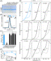
Figure 3.. Characterization of pathogenic variants in CAD's DHO domain.
A) SDS-PAGE of purified recombinant DHO domains bearing point mutations. St, molecular weight standards (kDa). B) Size exclusion chromatography of purified DHO proteins eluting as a single peak at the position expected for a homodimer. The tailed peak of R1810Q is indicated. On top, column calibration with proteins of known molecular weight (ferritin, 440 kDa; aldolase, 158 kDa; conalbumin, 75 kDa; carbonic anhydrase 29 kDa), indicating in blue the positions for the DHO dimer (closed circle) and the expected position for the monomer (open circle). C) Enzymatic activity of WT and mutants measured at 25 °C. The scattered plot shows three independent measurements. Activity units (U) are nmol of dihydroorotate per min. D) Denaturing curves in the absence (APO) and presence of fluoroorotate (FOA) measured by differential scanning fluorimetry. The midpoint temperatures (Tm) for the curves are indicated. E) Activity assay at different temperatures for the WT and R1810Q.
Figure 4.
Figure 4.. Characterization of DHO variants by X-ray crystallography.
A) Cartoon representation of the crystal structure of CAD's DHO subunit and dimer (PDB ID 4C6C). B) Detail of the DHO's active site with Zn2+ shown as cyan spheres and a molecule of FOA depicted with carbon atoms in pink. The carboxylated lysine (KCX) and the other residues bearing pathogenic substitutions are depicted with carbon atoms in yellow. Dashed lines represent relevant electrostatic interactions. C–G) Details of the crystal structures for mutants K1556T (C), R1475Q (D), K1482M (E), R1617Q (F), and R1722W (G). The 2Fobs-Fcalc electron density map for regions of interest is shown as a blue mesh contoured at 1.0 σ. Flexible disordered loops are represented as thick dashed lines. Panel G) shows the structure of the WT and R1722W side by side for comparison.
Figure 5.
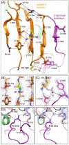
Figure 5.. Structure of DHO variants in the adjacent domain and C-terminal extension.
A) Cartoon representation of the WT DHO adjacent domain and beginning of the C-terminal extension. B–D) Detailed views of the crystal structures of mutants R1785C (B), R1789Q (C), and R1810Q (D). 2Fobs-Fcalc electron density maps are shown as a blue mesh contoured at 1.0 σ. Panel D) shows the structure of the WT and R1810Q side by side for comparison.

References

    1. Biesecker LG, Green RC. Diagnostic Clinical Genome and Exome Sequencing. N Engl J Med. 2014;370:2418–2425. - PubMed
    1. Richards S, Aziz N, Bale S, et al. Standards and guidelines for the interpretation of sequence variants: a joint consensus recommendation of the American College of Medical Genetics and Genomics and the Association for Molecular Pathology. Genet Med. 2015;17:405–424. - PMC - PubMed
    1. Ernst C, Hahnen E, Engel C, et al. Performance of in silico prediction tools for the classification of rare BRCA1/2 missense variants in clinical diagnostics. BMC Med Genomics. 2018;11:35. - PMC - PubMed
    1. Niroula A, Vihinen M. How good are pathogenicity predictors in detecting benign variants? PLoS Comput Biol. 2019;15:e1006481. - PMC - PubMed
    1. Rodenburg RJ. The functional genomics laboratory: functional validation of genetic variants. J Inherit Metab Dis. 2018;41:297–307. - PMC - PubMed

Publication types