. 2023 Aug 29;42(8):112935.
doi: 10.1016/j.celrep.2023.112935. Epub 2023 Aug 2.

Angiotensin AT1A receptor signal switching in Agouti-related peptide neurons mediates metabolic rate adaptation during obesity

Affiliations

Angiotensin AT1A receptor signal switching in Agouti-related peptide neurons mediates metabolic rate adaptation during obesity

Kirthikaa Balapattabi et al. Cell Rep. .

Abstract

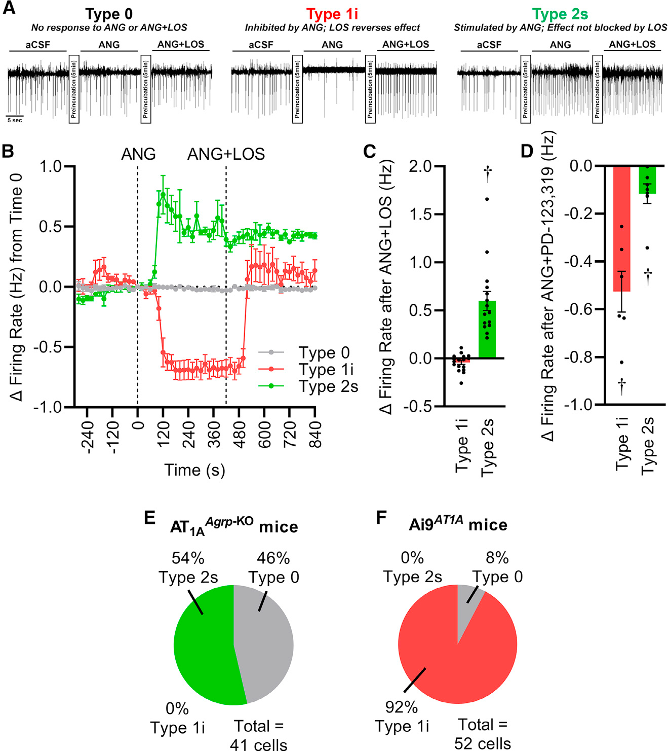
Resting metabolic rate (RMR) adaptation occurs during obesity and is hypothesized to contribute to failed weight management. Angiotensin II (Ang-II) type 1 (AT1A) receptors in Agouti-related peptide (AgRP) neurons contribute to the integrative control of RMR, and deletion of AT1A from AgRP neurons causes RMR adaptation. Extracellular patch-clamp recordings identify distinct cellular responses of individual AgRP neurons from lean mice to Ang-II: no response, inhibition via AT1A and Gαi, or stimulation via Ang-II type 2 (AT2) receptors and Gαq. Following diet-induced obesity, a subset of Ang-II/AT1A-inhibited AgRP neurons undergo a spontaneous G-protein "signal switch," whereby AT1A stop inhibiting the cell via Gαi and instead begin stimulating the cell via Gαq. DREADD-mediated activation of Gαi, but not Gαq, in AT1A-expressing AgRP cells stimulates RMR in lean and obese mice. Thus, loss of AT1A-Gαi coupling within the AT1A-expressing AgRP neuron subtype represents a molecular mechanism contributing to RMR adaptation.

Keywords: CP: Metabolism; CP: Neuroscience; G-protein signaling; arcuate nucleus; obesity; resting metabolic rate.


Conflict of interest statement

Declaration of interests The authors declare no competing interests.

Figures

Figure 1.
Figure 1.. Ang-II causes distinct electrical responses in individual AgRP neurons of chow-fed mice
(A) Initial analyses of firing-rate responses of individual AgRP neurons to application of Ang-II (n = 18 cells). (B) Cells were grouped into three subtypes according to electrical responses to Ang-II: no response, inhibited, or stimulated by Ang-II (n = 15–17 per response). (C) Pie chart illustrating the relative distribution of AgRP neuron subtypes in chow-fed mice of each sex. (D) Example tracings of three subtypes under baseline and Ang-II-stimulated conditions. (E) Current clamping demonstrates a similar distribution of subtypes of AgRP neuron within the ARC (n = 7–8 per response). *p < 0.05 by Šidák multiple comparisons procedure (A, B, E); summary data are presented as mean ± SEM. Replicates are indicated by individual dots or summaries within each panel. See also Figure S1.
Figure 2.
Figure 2.. Ang-II inhibits Type 1i AgRP neurons via AT1R, and stimulates Type 2s AgRP neurons via AT2R
(A) Example tracings from Type 0, Type 1i, and Type 2s neurons after application of Ang-II, and subsequently Ang-II in the presence of the AT1R antagonist losartan (LOS). (B) Example quantification of changes in firing rate of Type 0, Type 1i, and Type 2s neurons (n = 4–5 each) in response to Ang-II versus Ang-II + LOS. Note the normalization of firing rate of Type 1i neurons by LOS, but the lack of effect of LOS in Type 2s neurons. (C) Quantification of the firing rate of Type 1i and Type 2s neurons after Ang-II + LOS application, relative to baseline firing rates (n = 17, 15). (D) Quantification of the firing rate of Type 1i and Type 2s neurons (n = 6, 7) after application of Ang-II in the presence of the AT2R antagonist PD-123319, relative to baseline firing rates. (E) Pie chart illustrating the relative abundance of Type 0 and Type 2s neurons, and the lack of Type 1i neurons, in mice with conditional genetic deletion of Agtr1a (AT1A receptor) from all AgRP neurons (AT1AAgrp-KO mice). (F) Pie chart illustrating firing-rate responses of neurons within the ARC that express AT1A (Ai9AT1A mice) to acute Ang-II application. †p < 0.05 versus zero by one-sample t test (C and D); summary data are presented as mean ± SEM. Replicates are indicated by individual dots or summaries within each panel. See also Figure S2.
Figure 3.
Figure 3.. Ang-II inhibits Type 1i neurons through a cascade involving Gαi, inward-rectifier, and voltage-gated potassium channels, and L-type calcium channels
(A–C) Changes in firing rates of Type 1i neurons from the ARC of Ai9Agrp mice in response to Ang-II after pharmacological blockade of Gαi via pertussis toxin (PTX), or Gαq via BIM-46187. (A) Time course of responses to Ang-II before and after incubation with vehicle (n = 6) or PTX (n = 15). (B) Time course of responses to Ang-II before and after application of vehicle (n = 6) or BIM-46187 (n = 9). (C) Quantification of responses after preincubation with vehicle (n = 9), losartan (LOS, n = 17), PTX (n = 15), or BIM (n = 9). (D) Changes in firing rate of Type 1i neurons (identified by inhibitory response to Ang-II, n = 13) from the ARC of mice expressing the Gαi-coupled hM4Di DREADD in AgRP neurons (hM4DiAgrp mice), in response to clozapine N-oxide (CNO) with or without pretreatment with PTX. (E) Summary of responses of Type 1i neurons from the ARC of Ai9Agrp mice in response to Ang-II after pretreatment with vehicle or selected channel inhibitors (n = 9–11 per inhibitor). *p < 0.05 as indicated by Šidák multiple comparisons procedure, p < 0.05 versus zero by one-sample t test; summary data are presented as mean ± SEM. Replicates are indicated by individual dots. See also Figure S3.
Figure 4.
Figure 4.. Type 1i neurons project to a subset of brain regions that are known to receive ARC AgRP inputs
(A) Schematic drawing of the strategy for unilateral anterograde tracing of AT1A-expressing AgRP (Type 1i) neurons. (B) Representative image showing the precise unilateral targeting of Type 1i AgRP neurons by ChR2-eYFP. (C) Representative images showing the distribution of Type 1i AgRP neurons across the rostrocaudal ARC. (D–I) Representative images showing the projections of ARC Type 1i neurons to (D) bed nucleus of the stria terminalis (BNST), (E) ventromedial preoptic nucleus (VMPO), (F) medial preoptic nucleus (MPO), (G) paraventricular nucleus of hypothalamus (PVN), (H) supraoptic nucleus (SON), and (I) paraventricular nucleus of the thalamus (PVT). (J) Schematic diagram depicting the projections of ARC AgRP neurons (red) versus the Type 1i subtype (green). Additional structures: DMH, dorsomedial hypothalamic nucleus; LHA, lateral hypothalamic area; ARC, arcuate hypothalamic nucleus; AMY, amygdala; PBN, parabrachial nucleus; PAG, periaqueductal gray; ac, anterior commissure; f, fornix; opt, optic tract. Scale bars represent 200 μm (B–G), 50 μm (H), and 100 μm (I). See also Figure S4.
Figure 5.
Figure 5.. Ten weeks of high-fat diet (HFD; 45% kcal from fat) induces a G-protein signal switch within a subset of Type 1i AgRP neurons
(A) Ten weeks of HFD altered the relative proportion of AgRP neurons that were unaffected, inhibited, or stimulated by Ang-II in Ai9AgRP mice (distribution p < 0.05 versus Figure 1C), with an increased representation of AgRP neurons that were stimulated by Ang-II. (B) Time course of firing-rate responses of ARC AgRP neurons from HFD-fed Ai9Agrp mice to acute application of Ang-II before and after AT1R blockade by losartan (LOS). Notably, a subset of cells (Type 1s) are stimulated by Ang-II through a mechanism that is sensitive to blockade by LOS. Type 1i, n = 11; Type 1s, n = 12; Type 2s, n = 20. (C and D) Summary of firing-rate responses of Type 1i, Type 1s, and Type 2s neurons to Ang-II after pretreatment with LOS (C; n = 11, 12, 20) or the AT2R antagonist PD-123319 (D; n = 6, 6, 9). (E) Pie chart summarizing the distribution of AgRP neuron subtypes in chow-fed male Ai9AgRP mice. (F) Pie chart summarizing the distribution of AgRP neuron subtypes in HFD-fed male Ai9AgRP mice. (G and H) Summary of firing-rate responses of Type 1i (G; n = 10–11 each) or Type 1s (H; n = 8–12 each) neurons (identified by LOS-dependent inhibition or stimulation, respectively) to acute Ang-II application after pretreatment with the Gαi inhibitor, pertussis toxin (PTX) or the Gαq inhibitor, BIM-46187. *p < 0.05 as indicated by Šidák multiple comparisons procedure, p < 0.05 versus zero by one-sample t test (B, C, D, G, H); summary data are presented as mean ± SEM. Replicates are indicated by individual dots or summaries within each panel. See also Figure S5.
Figure 6.
Figure 6.. Activation of Gαi within Type 1 AgRP neurons is sufficient to stimulate RMR
(A) Example tracings from Ang-II-inhibited (Type 1i) AgRP neurons from hM4DiARC-AT1A and hM3DqARC-AT1A mice, demonstrating pertussis toxin (PTX)-sensitive Gαi-mediated inhibition or BIM-46187 (BIM)-sensitive Gαq-mediated stimulation, respectively, in response to clozapine N-oxide (CNO). (B and C) Quantification of effects of activating Gαi or Gαq within multiple Type 1i neurons from hM4DiARC-AT1A (B; n = 15) and hM3DqARC-AT1A (C; n = 17) mice. (D and E) Fat-free mass (FFM) (D) and fat mass (E) of mice after 10 weeks of chow or HFD feeding. (F) RMR immediately preceding injection of CNO, corrected for body composition by GLM. (G) Change in RMR with injection of CNO. (H) Change in RMR with injection of CNO, with sexes combined, corrected for body composition by GLM. For (D) to (G), control: n = 4 males + 2 females each diet; hM4DiARC-AT1A: n = 4 males + 4 females fed chow versus 5 males and 5 females fed HFD; hM3DqARC-AT1A: n = 4 males + 4 females each diet. For all panels except (A), *p < 0.05 as indicated by Šidák multiple comparisons procedure, p < 0.05 versus zero by one-sample t test; summary data are presented as mean ± SEM. Replicates are indicated by individual dots within each panel. See also Figure S6.
Figure 7.
Figure 7.. Implication of β-arrestin in AT1R signaling within Type 1i neurons, and overall working model
(A) Firing rates of Type 1i neurons from the ARC of Ai9Agrp mice in response to Ang-II, TRV027, or TRV027 in the presence of Ang-II (n = 12). Summary data are presented as mean ± SEM. (B) Quantification of change in firing rates of Type 1i neurons in response to TRV027 after preincubation with vehicle (n = 27), losartan (LOS, n = 15), or pertussis toxin (PTX, n = 12). *p < 0.05 as indicated by Šidák multiple comparisons procedure, p < 0.05 versus zero by one-sample t test; summary data are presented as mean ± SEM (C) Pie charts illustrating the relative distribution of ARC AgRP neuron responses to Ang-II in chow-fed Arrb1Agrp-KO mice. (D) Pie charts illustrating the relative distribution of ARC AgRP neuron responses to Ang-II in chow-fed Arrb2Agrp-KO mice. (E) Under normal physiological conditions, Ang-II acts on the Type 1i subtype of AgRP neuron within the ARC via its AT1R and a second-messenger cascade involving both β-arrestin-1 and Gαi to cause inhibition of the cell. This results in reduced inhibitory neurotransmission to postsynaptic neurons, ultimately resulting in increased resting metabolism. Following prolonged obesity, a fraction of Type 1i AgRP neurons undergoes a spontaneous G-protein “signal switch,” whereby AT1R stops coupling via this cascade to inhibit the cell and instead begins coupling via the Gαq cascade to stimulate the cell. Resulting net increases in inhibitory neurotransmission to postsynaptic targets are expected to contribute to the pathological adaptation of resting metabolic rate control that is often observed with obesity.

References

    1. Blundell JE, Caudwell P, Gibbons C, Hopkins M, Naslund E, King N, and Finlayson G (2012). Role of resting metabolic rate and energy expenditure in hunger and appetite control: a new formulation. Dis. Model. Mech. 5, 608–613. 10.1242/dmm.009837. - DOI - PMC - PubMed
    1. Oliveira V, Kwitek AE, Sigmund CD, Morselli LL, and Grobe JL (2021). Recent advances in hypertension: intersection of metabolic and blood pressure regulatory circuits in the central nervous system. Hypertension 77, 1061–1068. 10.1161/HYPERTENSIONAHA.120.14513. - DOI - PMC - PubMed
    1. Fothergill E, Guo J, Howard L, Kerns JC, Knuth ND, Brychta R, Chen KY, Skarulis MC, Walter M, Walter PJ, and Hall KD (2016). Persistent metabolic adaptation 6 years after “The Biggest Loser” competition. Obesity 24, 1612–1619. 10.1002/oby.21538. - DOI - PMC - PubMed
    1. Mark AL (2013). Selective leptin resistance revisited. Am. J. Physiol. Regul. Integr. Comp. Physiol. 305, R566–R581. 10.1152/ajpregu.00180.2013. - DOI - PMC - PubMed
    1. Morselli LL, Claflin KE, Cui H, and Grobe JL (2018). Control of energy expenditure by AgRP neurons of the arcuate nucleus: neurocircuitry, signaling pathways, and angiotensin. Curr. Hypertens. Rep. 20, 25. 10.1007/s11906-018-0824-8. - DOI - PMC - PubMed

Publication types