. 2023 Aug 4;17(8):e0011535.
doi: 10.1371/journal.pntd.0011535. eCollection 2023 Aug.

Immunogenic mapping of rDyn-1 and rKDDR-plus proteins and selection of oligopeptides by immunoblotting for the diagnosis of Leishmania infantum-infected dogs

Affiliations

Immunogenic mapping of rDyn-1 and rKDDR-plus proteins and selection of oligopeptides by immunoblotting for the diagnosis of Leishmania infantum-infected dogs

Williane Fernanda Siqueira et al. PLoS Negl Trop Dis. .

Abstract

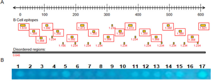
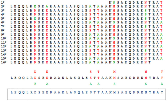
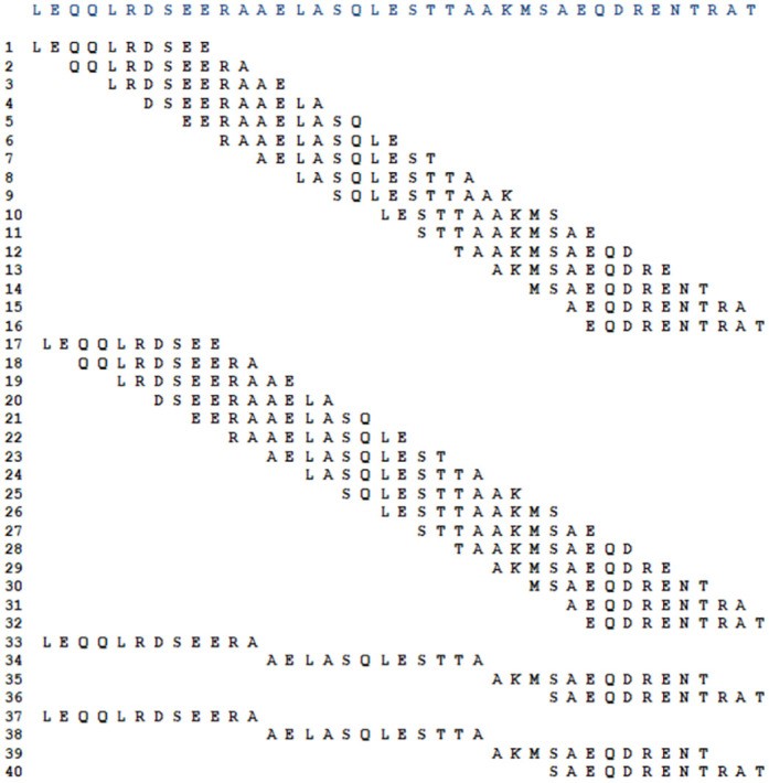
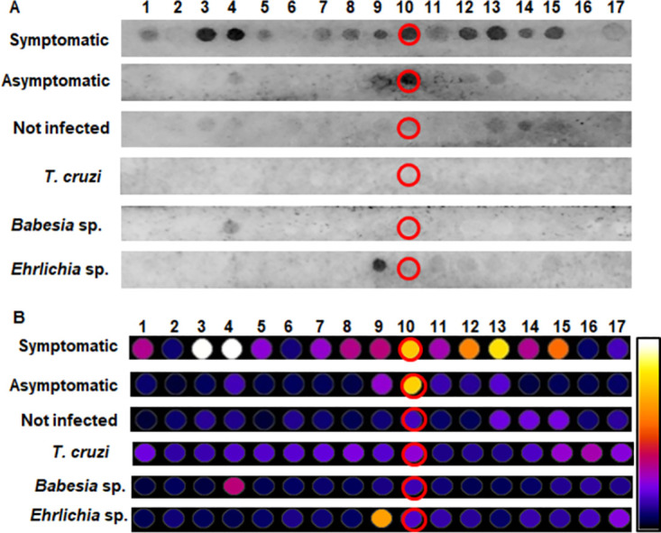
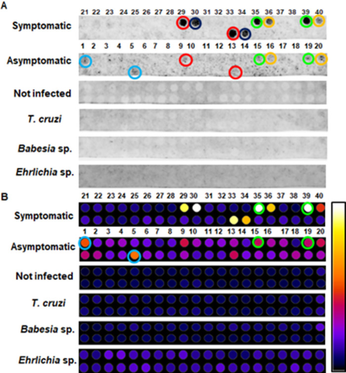
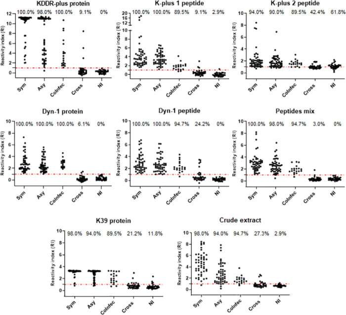
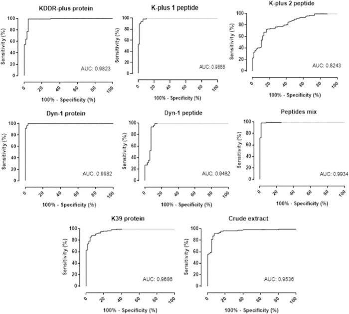
Endemic in Brazil, visceral leishmaniasis (VL) is a zoonotic infection that is among the most important parasitic diseases transmitted by vectors. Dogs are the main reservoirs of canine leishmaniasis (CanL) and their identification is used in some countries as part of disease prevention and control measures in the canine and human population. In this context, serological tests are necessary, composed of antigens capable of correctly identifying infected dogs, minimizing the number of false-negative cases. This study aimed to identify more immunoreactive peptides derived from two previously described whole proteins (rDyn-1 and rKDDR-plus) and compare their performance to the control antigens rK39 and the crude extract for the detection of dogs infected with L. infantum, especially the asymptomatic ones. The three selected peptides and a mixture of them, along with the rDyn-1, rKDDR-plus, rK39, and crude extract antigens were evaluated using indirect ELISA with sera samples from 186 dogs with CanL, being asymptomatic (n = 50), symptomatic (n = 50), co-infected (n = 19), infected with Babesia sp. (n = 7), Ehrlichia sp. (n = 6), T. cruzi (n = 20) and uninfected (n = 34). The results showed that the rDyn-1 protein and the peptide mixture had the highest sensitivity (100% and 98.32%, respectively) and specificity (97.01 and 98.51, respectively). A high degree of kappa agreement was found for rDyn-1 protein (0.977), mixed peptides (0.965), rKDDR-plus protein (0.953), K-plus peptide 1 (0.930) and Dyn-1 peptide (0.893). The mixture of peptides showed the highest likelihood (65.87). The ELISA using the mixture of peptides and the rDyn-1 protein showed high performance for CanL serodiagnosis. More mix combinations of the peptides and additional extended field tests with a larger sample size are recommended.


Conflict of interest statement

The authors have declared that no competing interests exist.

Figures

Fig 1
Fig 1. Predictions of B-cell linear e and intrinsically unstructured/disordered regions (entropy) from L. infantum rDyn-1 gene.
(A) Each point inside the red boxes represents a B-cell epitopes predicted by BepiPred defined as sequence regions with values above the threshold score of 0.6 and the numbers below indicate the prediction score obtained by each epitope. The black box indicates disordered regions predicted by IUPred with a score of 0.5. Predicted regions as B-cell linear epitopes that are associated with a high degree of structural disorder also exhibit a high antigenicity score, as observed in the graph at the center of the figure. (B) Cellulose membrane containing the 17 spots with the peptide sequences derived from the rDun-1 protein selected after bioinformatics analyses visualized by ultraviolet light.
Fig 2
Fig 2. Alignment of the repetitive array of amino acids that make up the rKDDR-plus protein.
The 15.3 repetitive blocks containing the 39 amino acids were aligned for better visualization of the least repeated amino acids (green amino acid) and the most repeated (red amino acid) in the seven degenerate sites in black this sequence of the 39 amino acids that make up the 15.3 motifs of the rKDDR-plus protein. In blue is the final sequence used as a template used in the sliding window.
Fig 3
Fig 3. Sliding window technique.
The first ten amino acids of the template sequence formed the first set of oligopeptides. Then after a jump of 2 amino acids plus 10 amino acids were selected forming another set of oligopeptides. Following this logic, 16 oligopeptide sequences were selected. To complete the oligopeptide catalog 4 sequences composed of 12 amino acids with random sliding windows were included, completing the 20 sequences of oligopeptides that were synthesized on the membrane in duplicate.
Fig 4
Fig 4. Membrane immunoblotting analysis containing peptides derived from the rDyn-1 protein.
(A) Chemiluminescence image from spot-synthesis technique to evaluate the reactivity and specificity of rDyn-1-derived peptides incubated with pools of canine sera from symptomatic and asymptomatic infected with L. infantum and sera from control dogs; healthy dogs without leishmaniasis other pathogens of interest (not infected), dogs infected with Babesia sp. and Ehrlichia sp. (B) Densitometry analysis of previously synthesized peptide spots on the cellulose membrane for selection of the most reactive spot in incubations with pools of dogs infected with L. infantum, not recognized by sera from dogs without CanL. The closer to the white hue of the scale, the more reactive the spot.
Fig 5
Fig 5. Analysis of the membrane containing peptides derived from the rKDDR-plus protein.
(A) Chemiluminescence image from membrane immunoblotting assays incubated with pools of sera from symptomatic and asymptomatic dogs infected with L. infantum and sera from control dogs; healthy dogs without leishmaniasis other pathogens of interest (not infected), dogs infected with Babesia sp. and Ehrlichia sp. Red, green, and yellow spots were lit for both groups of infected dogs. Dark blue spot lit only for the pool of symptomatic dogs and light blue spot lit only for the pool of asymptomatic sera. (B) Densitometric analysis for selection of the most reactive spots in incubations with pools of dogs infected with L. infantum, not recognized by sera from dogs without CanL. The closer to the white hue of the scale, the more reactive the spot. Green circle; reactive spots with symptomatic and asymptomatic sera. Blue circle; more reactive spots only with asymptomatic sera.
Fig 6
Fig 6. Comparing the reactivity of canine sera using rKDDR-plus, rDyn-1 and K39 proteins, their derived peptides K-plus 1, K-plus 2, Dyn-1 and mixtures of these three peptides and the crude extract of L. infantum in an ELISA protocol.
Reactivity index results obtained under Leishmania complete recombinant proteins Dyn-1 and KDDR-plus, the synthetic peptides derived from each recombinant protein (Dyn-1, K-plus 1 and K-plus 2 peptides), a mixture (Mix peptides) of 0.3 μg/well of the three peptides and control antigens rK39 and crude extract against a canine serological panel composed by serum from infected dogs presenting symptoms (Sym), infected asymptomatic dogs (Asy), dogs co-infected with Leishmania and Babesia sp. (CoInfec), dogs with other infections (Babesia sp., Ehrlichia sp. and T. cruzi) (Cross) and not infected (NI). The index above each column in the plot indicates the percentage of samples that are above the cut-off.
Fig 7
Fig 7. ROC curve analysis of the area under the curve (AUC), considering the results from ELISA.
Comparation of the ROC curve obtained employing Leishmania complete recombinant proteins Dyn-1 and KDDR-plus, the synthetic peptides derived from each recombinant protein (Dyn-1, K-plus 1 and K-plus 2 peptides), a mixture (Mix peptides) of 0.3 μg/well of the three peptides and control antigens rK39 and crude extract was established by GraphPad Prism 8.0 using serum samples of negative and positive samples in each plate. Abbreviations: (AUC) area under curve.

References

    1. Organization WH. Neglected tropical diseases 2021 [https://www.who.int/news-room/questions-and-answers/item/neglected-tropi....
    1. Organization WH. Leishmaniasis 2022 [cited 2022. https://www.who.int/news-room/fact-sheets/detail/leishmaniasis.
    1. Valero NNH, Uriarte M. Environmental and socioeconomic risk factors associated with visceral and cutaneous leishmaniasis: a systematic review. Parasitol Res. 2020;119(2):365–84. doi: 10.1007/s00436-019-06575-5 - DOI - PubMed
    1. Dantas-Torres F. The role of dogs as reservoirs of Leishmania parasites, with emphasis on Leishmania (Leishmania) infantum and Leishmania (Viannia) braziliensis. Vet Parasitol. 2007;149(3–4):139–46. doi: 10.1016/j.vetpar.2007.07.007 - DOI - PubMed
    1. Paltrinieri S, Solano-Gallego L, Fondati A, Lubas G, Gradoni L, Castagnaro M, et al.. Guidelines for diagnosis and clinical classification of leishmaniasis in dogs. J Am Vet Med Assoc. 2010;236(11):1184–91. doi: 10.2460/javma.236.11.1184 - DOI - PubMed

Publication types