. 2023 Aug 4;9(31):eadh4057.
doi: 10.1126/sciadv.adh4057. Epub 2023 Aug 4.

Single-dose mucosal replicon-particle vaccine protects against lethal Nipah virus infection up to 3 days after vaccination

Affiliations

Single-dose mucosal replicon-particle vaccine protects against lethal Nipah virus infection up to 3 days after vaccination

Stephen R Welch et al. Sci Adv. .

Abstract

Nipah virus (NiV) causes a highly lethal disease in humans who present with acute respiratory or neurological signs. No vaccines against NiV have been approved to date. Here, we report on the clinical impact of a novel NiV-derived nonspreading replicon particle lacking the fusion (F) protein gene (NiVΔF) as a vaccine in three small animal models of disease. A broad antibody response was detected that included immunoglobulin G (IgG) and IgA subtypes with demonstrable Fc-mediated effector function targeting multiple viral antigens. Single-dose intranasal vaccination up to 3 days before challenge prevented clinical signs and reduced virus levels in hamsters and immunocompromised mice; decreases were seen in tissues and mucosal secretions, critically decreasing potential for virus transmission. This virus replicon particle system provides a vital tool to the field and demonstrates utility as a highly efficacious and safe vaccine candidate that can be administered parenterally or mucosally to protect against lethal Nipah disease.


Figures
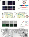
Fig. 1.
Fig. 1.. A NiV virus replicon particle can be efficiently grown in a cell line supplying F protein in trans.
(A) Constitutive expression of NiV nucleoprotein (N), phosphoprotein (P), matrix protein (M), fusion protein (F), and glycoprotein (G) in Vero and CHO cells, confirmed by immunofluorescent staining showing viral proteins (red). Cells were also stained with 4′,6-diamidino-2-phenylindole (blue) to confirm viability. (B) Schematic showing design of NiV∆F genome. GE, gene end; GS, gene start; RdRp, RNA-dependant-RNA-polymerase; CDS, coding sequence; Le, leader sequence; Tr, trailer sequence. (C) Schematic showing the generation of NiV∆F. All required plasmids were transfected into BSR-T7/5 cells, and 2 days after transfection, cells were overlayed with the Vero-Fco cell line constitutively expressing codon-optimized NiV F protein. Five days after transfection, cell culture supernatants were harvested and clarified by centrifugation to collect NiV∆F VRP. (D) Both Vero and Vero-Fco cells were infected at multiplicity of infection (MOI) 1 with NiV∆F; 3 days postinfection (dpi), viral growth was confirmed by immunofluorescent staining (green). Clarified supernatants were passed onto fresh cells (P2), and 3 dpi, viral growth was again confirmed by immunofluorescent staining (green). IFA, immunofluorescence assay; BF, bright field. (E) Vero cells were infected with NiV (red squares), while Vero-Fco cells were infected with either NiV (blue triangles) or NiV∆F (green circles) (all MOI, 0.1). Viral titers were determined by TCID50 calculation 24 to 96 hours after infection. (F) Vero and Vero-Fco cells were infected with NiV and NiV∆F, respectively, at MOI of 0.01. After 4 days, the cells were processed for transmission electron microscopy to determine virion morphology. Particles of equivalent size and morphology were seen budding from both samples (arrows). No such particles were seen in mock-inoculated control samples. Inset shows a negatively stained electron microscopy image of a NiV∆F particle. ns, not significant.
Fig. 2.
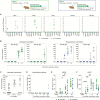
Fig. 2.. NiV∆F is nonspreading and noninfectious and confers a high safety margin in both suckling mice and hamsters.
Kaplan-Meier survival curves of animals inoculated with NiV∆F: (A) Two- to 3-day-old suckling mice were intracerebrally (IC) inoculated with either 105 or 103 TCID50 of NiV∆F or one of seven dilutions of wild-type NiV ranging from 106 to 100 TCID50. (B) Five- to 7-week-old Syrian hamsters were IN inoculated with either 106 TCID50 of NiV∆F or one of three dilutions of wild-type NiV ranging from 106 to 103 TCID50. Survival significance calculated by log-rank (Mantel-Cox test): ****P ≤ 0.0001; ***P ≤ 0.001; **P ≤ 0.01; *P ≤ 0.05. (C) Differential mRNA expression of select innate immune response genes in lungs of animals (D to G) was determined by RT-qPCR. (D) and (E) Lung and brain tissues and (F) and (G) mucosal swab samples (oral and rectal) were taken at euthanasia from hamsters serially euthanized 1, 3, or 7 days after inoculation with 106 TCID50 of NiV∆F (green circles) or 1, 3, or 5 to 7 days after inoculation with wild-type NiV (red squares) to determine both vRNA levels (RT-qPCR) and quantify infectious virus titers (TCID50) in sample types in which NiV∆F RNA was detected. For samples analyzed in (C) to (G), a subset of wild-type NiV-infected animals did not survive to predetermined 7 dpi time point and were sampled earlier when end point criteria were reached (5 to 6 dpi). Significance was calculated by multiple t test: *P ≤ 0.05; **P ≤ 0.01.
Fig. 3.
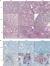
Fig. 3.. Absence of histopathology and nonspreading phenotype of NiV∆F in lung tissue from Syrian hamsters.
Lung tissues from hamsters serially euthanized following inoculation with 106 TCID50 of NiV∆F (1, 3, or 7 days), wild-type NiV [1, 3, or 5 to 7 days; subset of wild-type NiV-infected animals did not survive to predetermined 7 dpi time point and were sampled earlier when end point criteria were reached (5 to 6 dpi)], or DMEM only (mock-infected at 1, 3, or 7 days) were formalin fixed and evaluated via (A) hematoxylin and eosin staining to characterize tissue pathology and via (B) ISH to detect vRNA. Lungs of NiV∆F- and mock-inoculated animals showed no notable histopathologic changes at any time point, while lungs from NiV-inoculated animals displayed progressive inflammation, with epithelial syncytia (insets) compatible with paramyxoviral pneumonia. NiV∆F vRNA was detected by ISH 1 dpi and decreased by 7 days. In contrast, NiV vRNA was detected at relatively higher levels starting 1 day after inoculation and increased over time with severity of pneumonia.
Fig. 4.
Fig. 4.. Humoral immunity after single-dose mucosal or subcutaneous delivery of NiV∆F.
Five- to 7-week-old Syrian hamsters were inoculated either IN (green circles) or SC (blue triangles) with 106 TCID50 of NiV∆F and euthanized 1, 3, 7, 14, or 28 days postvaccination (dpv; n = 10 per group). (A) NiV∆F vRNA tissue levels at each time point were determined by RT-qPCR. (B) IgG antibody titers against NiV N and G proteins were determined by enzyme-linked immunosorbent assay (ELISA) at all time points after vaccination. (C) For IN-vaccinated hamsters, IgA titers against NiV F, N, and G were determined by ELISA at 28 days postvaccination. (D) Neutralizing antibody titers against NiV strain Malaysia were evaluated 1, 3, 7, 14, and 28 days postvaccination. (E) Antibody-dependent complement deposition (ADCD) assay showing complement fixing activity of antibodies and (F) antibody-dependent cellular phagocytosis (ADCP) assay depicting phagocytic activity of antibodies were performed on plasma collected 7, 14, and 28 days postvaccination. In all panels, individual values are shown, with bars representing mean and SD. Significance calculated by multiple t test: *P ≤ 0.05; **P ≤ 0.01; ***P ≤ 0.001; ****P ≤ 0.0001. ND, not detected; BLD, below limit of detection; MFI, mean fluorescence intensity.
Fig. 5.
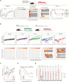
Fig. 5.. Mucosal NiV∆F vaccination protects Syrian hamsters from NiV clinical signs and lethality up to 3 days before IN challenge.
Groups of 10 hamsters were vaccinated IN with 100 μl containing 106 TCID50 of NiV∆F: once 28 (black), 14 (orange), 7 (purple), or 3 (green) days before challenge; or twice (blue) both 3 and 1 days before challenge. Mock-vaccinated animals (red) were given an equivalent IN volume of DMEM 28 days before challenge. All hamsters were challenged IN with 106 TCID50 NiV strain Malaysia. (A) Percent weight change from baseline (taken −1 dpi); mean daily body temperatures; and individual daily clinical scores (from 0 to 10), with severity depicted by increased intensity of red. Animals scoring ≥ 10 were humanely euthanized; any animal that succumbed to disease before euthanasia was allocated a score of 10. Gray boxes indicate the end of monitoring/scoring due to euthanasia/death. Individual animals are represented as circles, with the solid line representing the daily mean. (B) Mean percent weight changes from baseline (taken −1 dpi; significance calculated by two-way analysis of variance (ANOVA): *P ≤ 0.05). (C) Kaplan-Meier curves showing survival of vaccinated and mock-vaccinated animals challenged with NiV strain Malaysia. Significance calculated by log-rank (Mantel-Cox test): *P ≤ 0.05; ns, not significant. (D) RT-qPCR detection of NiV vRNA in select tissues from hamsters vaccinated at indicated times and subsequently challenged. Individual animals from each group are represented. Error bars represent the means and SD. Significance calculated by t test: ****P ≤ 0.0001; ***P ≤ 0.001; **P ≤ 0.01; *P ≤ 0.05.
Fig. 6.
Fig. 6.. Mucosal NiV∆F vaccination confers high-level protection against disease and lethality in robust Syrian hamster intraperitoneal challenge model.
Groups of 9 and 10 hamsters were vaccinated IN with 100 μl containing 106 TCID50 of NiV∆F: once 28 (black), 14 (orange), 7 (purple), or 3 (green) days before challenge; or twice (blue) both 3 and 1 days before challenge. Mock-vaccinated animals (red) were given an equivalent IN volume of DMEM 28 days before challenge. All hamsters were challenged IP with 104 TCID50 NiV strain Malaysia. (A) Percent weight change from baseline (taken −1 dpi); mean daily body temperature; and individual daily clinical scores (scored from 0 to 10), with severity depicted by increased intensity of red. Animals scoring ≥ 10 were humanely euthanized; any animal that succumbed to disease before euthanasia was allocated a score of 10. Gray boxes indicate the end of monitoring/scoring due to euthanasia or death. Individual animals are represented as circles, with the solid line representing the daily mean. (B) Mean percent weight changes from baseline (taken at −1 dpi; significance calculated by two-way ANOVA: *P ≤ 0.05). (C) Kaplan-Meier curves showing survival of vaccinated and mock-vaccinated animals challenged with NiV strain Malaysia. Significance calculated by log-rank (Mantel-Cox test): ****P ≤ 0.0001; **P ≤ 0.01. (D) RT-qPCR detection of NiV vRNA in select tissues from hamsters vaccinated at indicated times and subsequently challenged. Individual animals from each group are represented. Error bars represent the means and SD. Significance calculated by t test: ****P ≤ 0.0001; ***P ≤ 0.001; **P ≤ 0.01.
Fig. 7.
Fig. 7.. NiV∆F vaccination reduces NiV replication in tissues and mucosal membranes early in infection.
Groups of hamsters were vaccinated IN once 28 (blue), 14 (orange), 7 (green), or 3 (purple) days before challenge or mock-vaccinated with DMEM (red) 28 days before challenge (total n = 120; n = 24 in each vaccination group; 12 males and 12 females; all age matched). Animals were challenged either IN with 106 TCID50 (n = 60) or IP with 104 TCID50 (n = 60) with NiV strain Malaysia. At 1, 4, or 6 dpi, select animals were euthanized for analysis (n = 40 per dpi; n = 4 per vaccination period per vaccination route per day; two males and two females per group). Represented are the NiV vRNA titers in select tissues (A and B) and in oral and rectal swabs (C and D) at each time point. In all panels, individual animals are represented as circles, with a solid line indicating the mean at each time point. In all panels, significance is indicated in relation to mock-vaccinated animals (red dots) from the same time point after challenge, calculated by log two-way ANOVA with Dunnett’s test to correct for multiple comparisons: ****P ≤ 0.0001; ***P ≤ 0.001; **P ≤ 0.01; *P ≤ 0.05.
Fig. 8.
Fig. 8.. NiV∆F vaccination enhances Fc-mediated antibody effector functions.
Groups of hamsters were vaccinated IN once 28 (blue), 14 (orange), 7 (green), or 3 (purple) days before challenge, or mock-vaccinated with DMEM (red) 28 days before challenge (total, n = 120; for each vaccination group, n = 24; 12 males and 12 females; all age matched). Animals were challenged either IN with 106 TCID50 (n = 60) or IP with 104 TCID50 (n = 60) with NiV strain Malaysia. At 1, 4, and 6 dpi, select animals were euthanized for analysis (n = 40 per dpi; n = 4 per vaccination period per vaccination route per day; two males and two females per group). (A and B) ADCD and (C and D) ADCP against N and G antigens. In all panels, individual animals are represented as circles, with a solid line indicating the mean at each time point. In all panels, significance is indicated in relation to mock-vaccinated animals (red dots) from the same time point after challenge, calculated by log two-way ANOVA with Dunnett’s test to correct for multiple comparisons: ****P ≤ 0.0001; ****P ≤ 0.001; **P ≤ 0.01; *P ≤ 0.05; ns, non significant. MFI, mean fluorescence intensity. (E) Single-cell suspensions of splenocytes collected 6 days after NiV challenge (IP or IN) from hamsters either mock-vaccinated (DMEM only) or VRP-vaccinated IN for indicated vaccine intervals before challenge were stained for fluorescence-activated cell sorting analysis. T cells were identified and stained for expression of CD3, CD4, and CD8, with levels depicted as % of viable cells. NK cells were identified and stained for expression of CD94 (gating strategy shown in fig. S4). Samples from NiV-infected hamsters were compared to those obtained from naïve (unvaccinated and uninfected) hamsters.
Fig. 9.
Fig. 9.. A single dose of NiV∆F provides complete protection from clinical signs and lethality in mouse model of disease up to 3 days before challenge.
(A) Safety study: Groups of eight Ifnar−/− mice were challenged IP with either NiV∆F (106 TCID50, green), or NiV strain Malaysia (106 TCID50, red; or 104 TCID50, blue), and (B) efficacy study: Groups of 8 Ifnar−/− mice were vaccinated IP with 106 TCID50 NiV∆F either 28 (black), 7 (purple), 3 (green), or 1 (blue) day before challenge; or mock-vaccinated with DMEM 28 (red) days before challenge. Mice were challenged IP with 107 TCID50 of NiV strain Malaysia. Graphs show the percent weight change from baseline (taken −1 dpi; significance calculated by two-way ANOVA: *P ≤ 0.05); individual daily clinical scores (from 0 to 10), with severity depicted by increased intensity of red. Animals scoring ≥ 10 were humanely euthanized; any animals that succumbed to disease before euthanasia were allocated a score of 10. Gray boxes indicate the end of monitoring/scoring due to euthanasia or death. (C) Mean percentage weight changes from baseline (taken −1 dpi; significance calculated by two-way ANOVA: *P ≤ 0.05). (D) Kaplan-Meier curves show survival in challenged animals; significance calculated by log-rank (Mantel-Cox test): **P ≤ 0.01. (E) RT-qPCR detection of NiV vRNA in select tissues. Individual animals from each group are represented. Error bars represent the means and SD. Significance calculated by t test: ***P ≤ 0.001; **P ≤ 0.01; *P ≤ 0.05.

References

    1. C. F. Spiropoulou, Nipah virus outbreaks: Still small but extremely lethal. J Infect Dis 219, 1855–1857 (2019). - PubMed
    1. N. I. Paton, Y. S. Leo, S. R. Zaki, A. P. Auchus, K. E. Lee, A. E. Ling, S. K. Chew, B. Ang, P. E. Rollin, T. Umapathi, I. Sng, C. C. Lee, E. Lim, T. G. Ksiazek, Outbreak of Nipah-virus infection among abattoir workers in Singapore. Lancet 354, 1253–1256 (1999). - PubMed
    1. V. P. Hsu, M. J. Hossain, U. D. Parashar, M. M. Ali, T. G. Ksiazek, I. Kuzmin, M. Niezgoda, C. Rupprecht, J. Bresee, R. F. Breiman, Nipah virus encephalitis reemergence, Bangladesh. Emerg. Infect. Dis. 10, 2082–2087 (2004). - PMC - PubMed
    1. M. S. Chadha, J. A. Comer, L. Lowe, P. A. Rota, P. E. Rollin, W. J. Bellini, T. G. Ksiazek, A. Mishra, Nipah virus-associated encephalitis outbreak, Siliguri, India. Emerg. Infect. Dis. 12, 235–240 (2006). - PMC - PubMed
    1. K. Halpin, A. D. Hyatt, R. Fogarty, D. Middleton, J. Bingham, J. H. Epstein, S. A. Rahman, T. Hughes, C. Smith, H. E. Field, P. Daszak; Henipavirus Ecology Research Group , Pteropid bats are confirmed as the reservoir hosts of henipaviruses: A comprehensive experimental study of virus transmission. Am. J. Trop. Med. Hyg. 85, 946–951 (2011). - PMC - PubMed