. 2023 Aug 3;83(15):2810-2828.e6.
doi: 10.1016/j.molcel.2023.07.004.

FACS-based genome-wide CRISPR screens define key regulators of DNA damage signaling pathways

Affiliations

FACS-based genome-wide CRISPR screens define key regulators of DNA damage signaling pathways

Min Huang et al. Mol Cell. .

Abstract

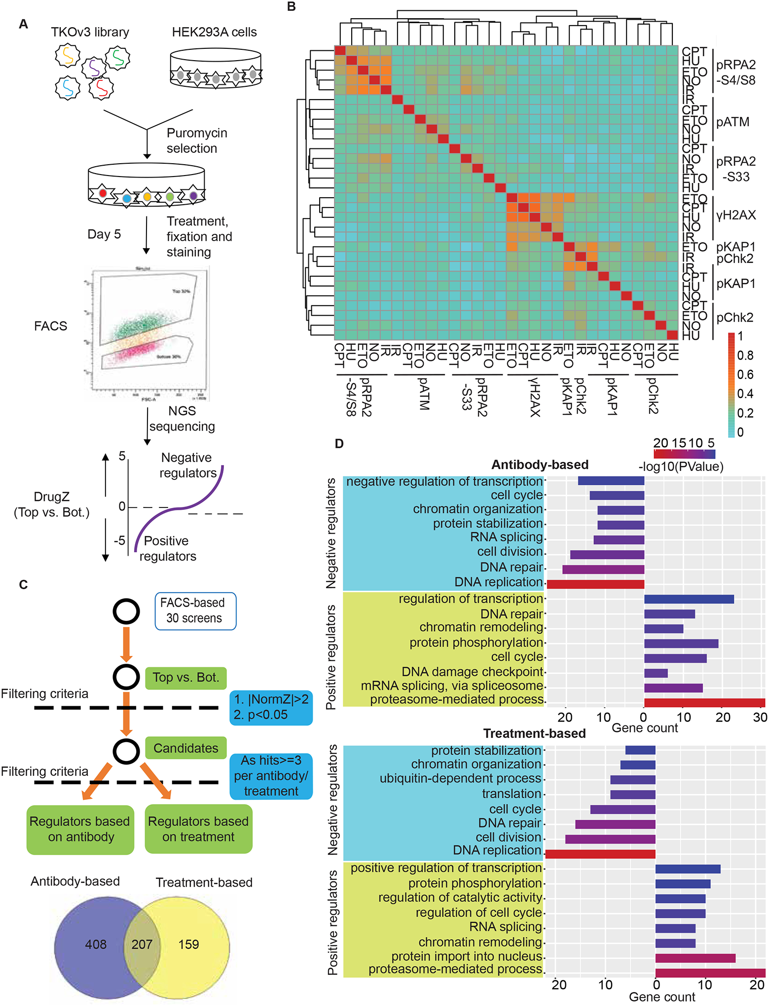
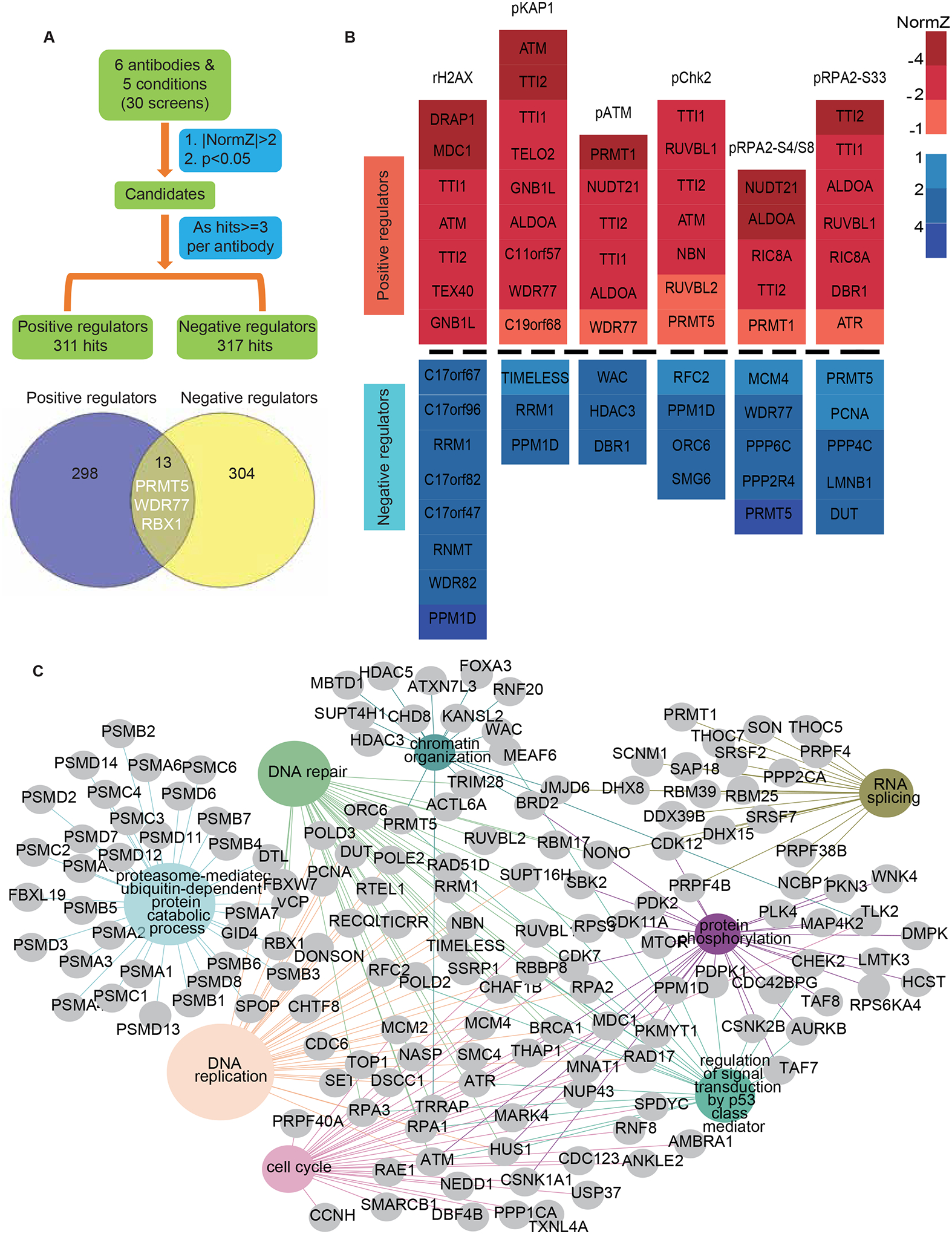
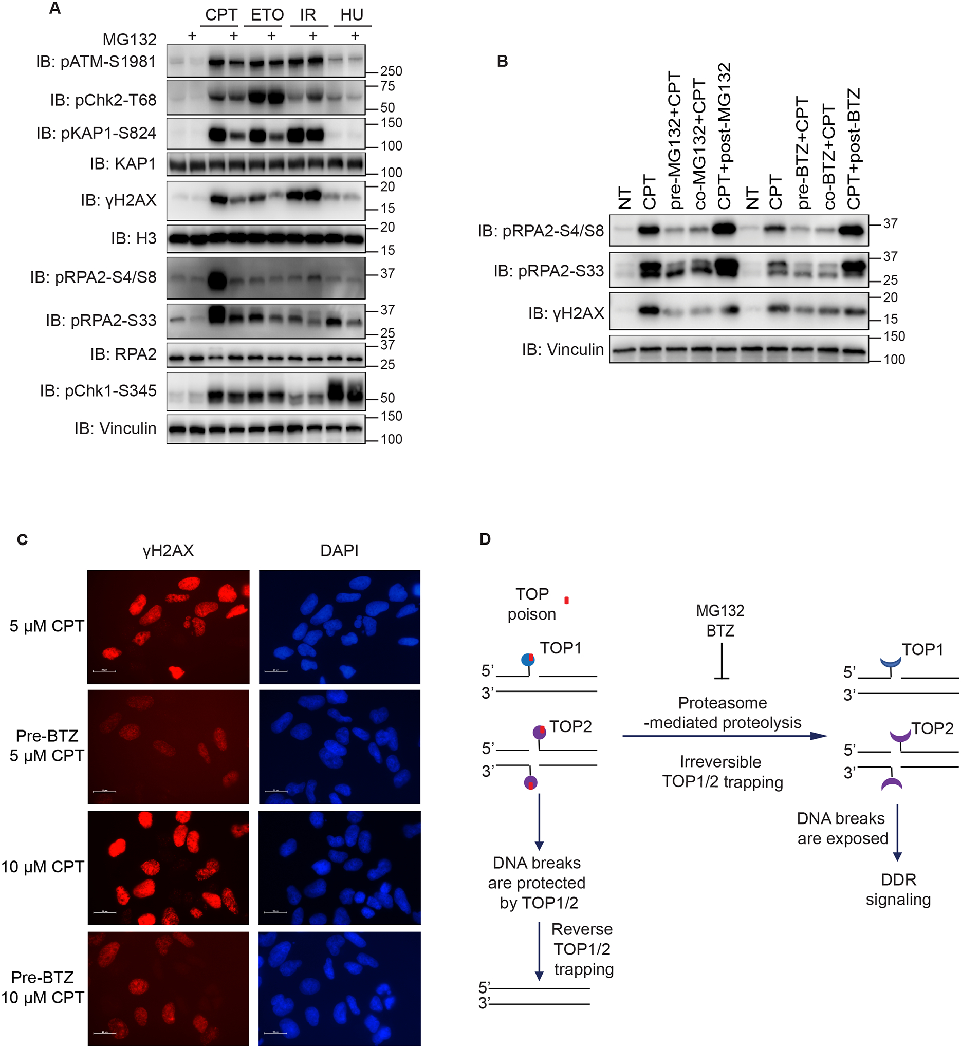
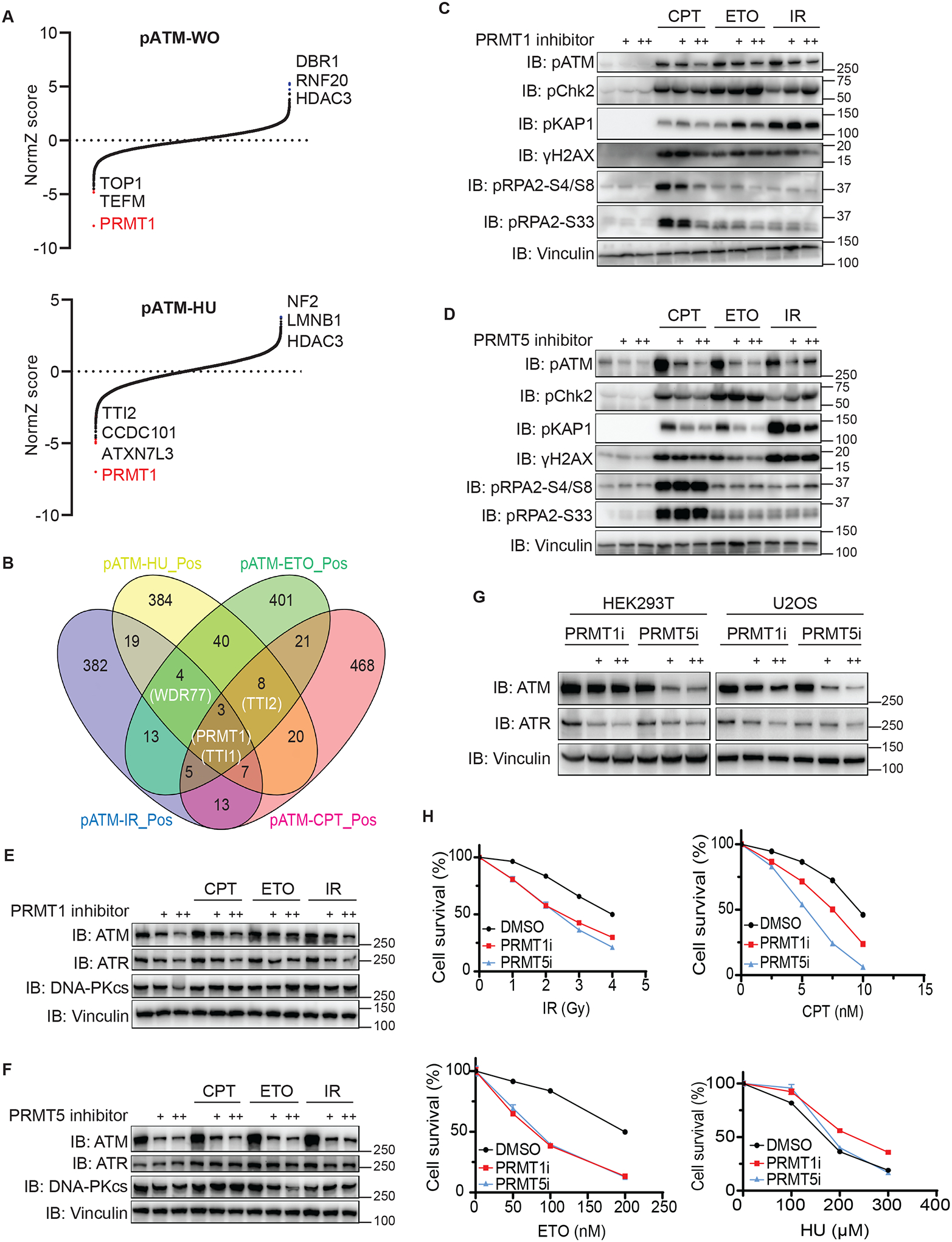
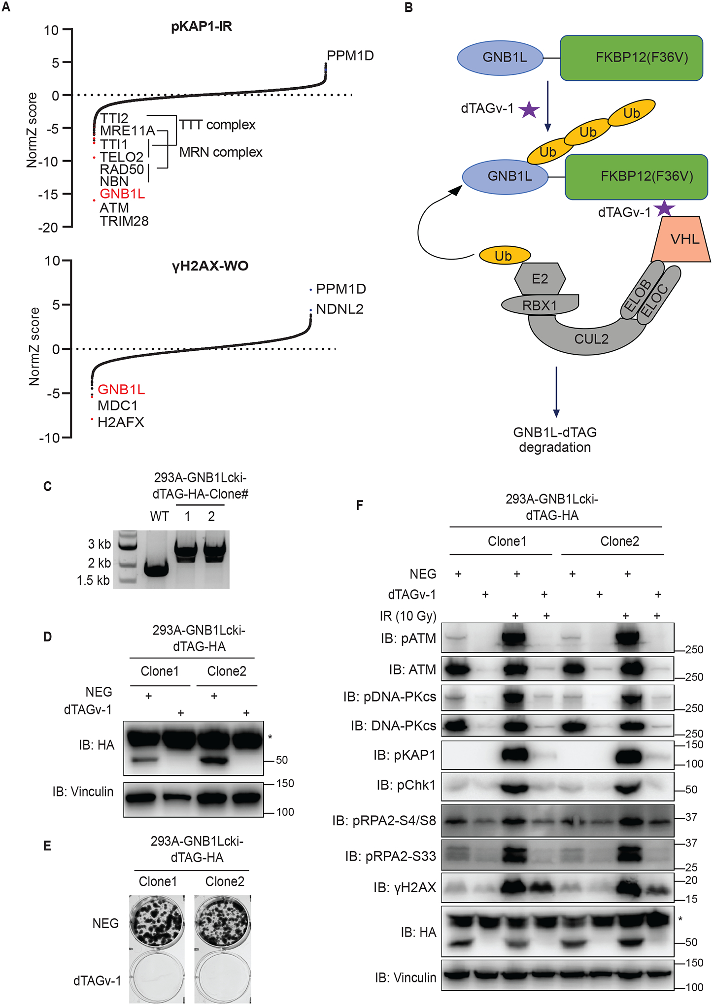
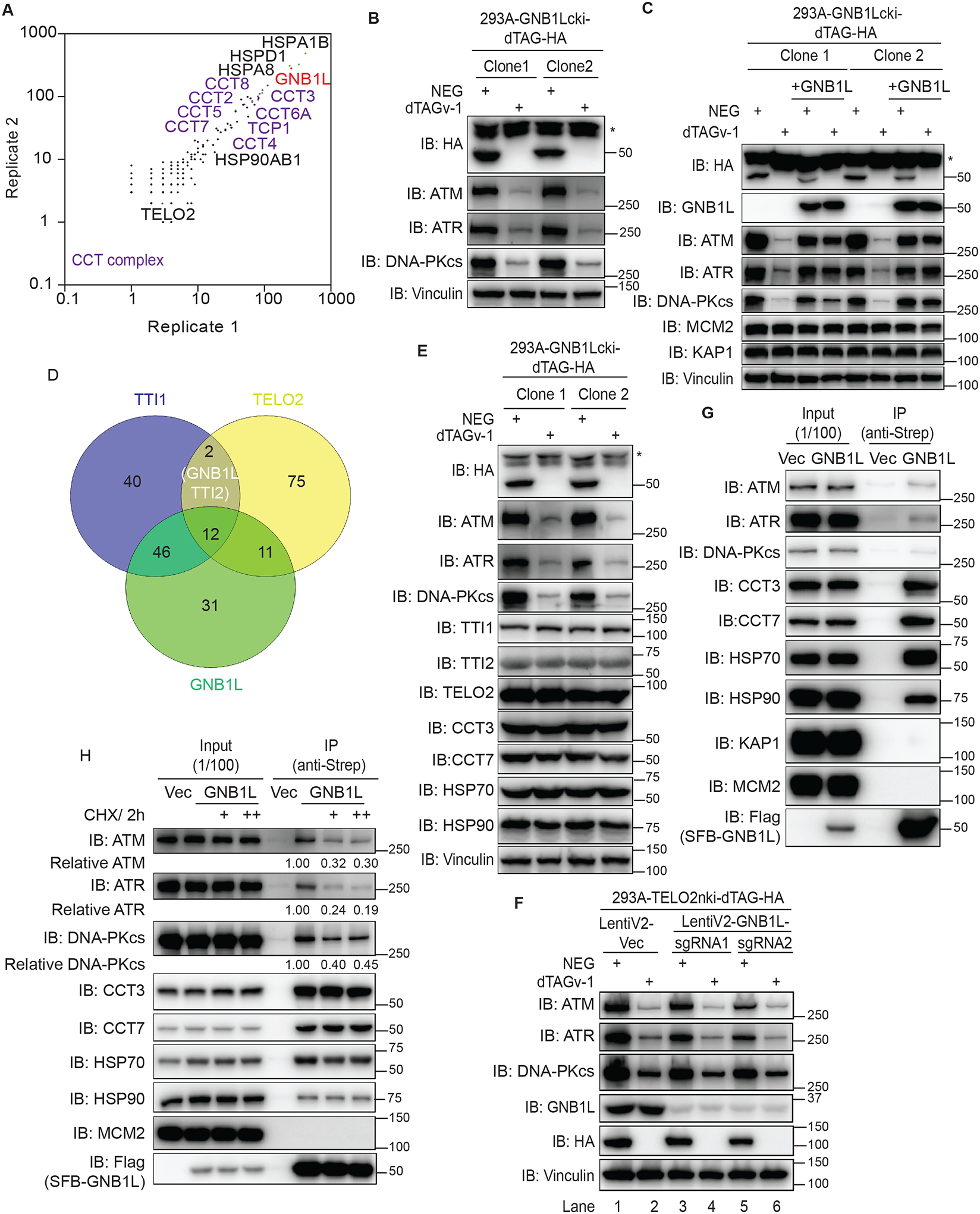
DNA damage-activated signaling pathways are critical for coordinating multiple cellular processes, which must be tightly regulated to maintain genome stability. To provide a comprehensive and unbiased perspective of DNA damage response (DDR) signaling pathways, we performed 30 fluorescence-activated cell sorting (FACS)-based genome-wide CRISPR screens in human cell lines with antibodies recognizing distinct endogenous DNA damage signaling proteins to identify critical regulators involved in DDR. We discovered that proteasome-mediated processing is an early and prerequisite event for cells to trigger camptothecin- and etoposide-induced DDR signaling. Furthermore, we identified PRMT1 and PRMT5 as modulators that regulate ATM protein level. Moreover, we discovered that GNB1L is a key regulator of DDR signaling via its role as a co-chaperone specifically regulating PIKK proteins. Collectively, these screens offer a rich resource for further investigation of DDR, which may provide insight into strategies of targeting these DDR pathways to improve therapeutic outcomes.

Keywords: DDR signaling; FACS-based CRISPR screen; GNB1L; PRMT5; antibody; proteasome.


Conflict of interest statement

Declaration of interests The authors declare no competing interests.

Figures

Figure 1.
Figure 1.. Workflow for FACS-based CRISPR screens
(A) Workflow of the FACS-based CRISPR screening strategy. (B) Heat map shows the correlation of 30 screens based on NormZ scores. Also see Table S1. (C) The criteria for analyzing positive and negative regulators of DDR signaling. (D) Representative GO terms enriched in antibody-based analyses (top panel) and treatment-based analyses (bottom panel) of negative/positive regulators of DDR signaling. The GO term list is provided in Table S3.
Figure 2.
Figure 2.. The regulatory network of DDR signaling revealed by antibody-based analyses
(A) The criteria for analyzing positive and negative regulators of DDR signaling based on antibody. (B) Heatmap plots show the normZ values of hub candidates. Red and blue colors represent positive and negative regulators, respectively. NormZ values were grouped by their absolute values labelled by different colors. (C) Representation of the Reactome pathway network for the proteins in A. The pathways highlighted in color were significantly enriched. The GO term list is provided in Table S4.
Figure 3.
Figure 3.. Proteasome inhibition reduces both CPT- and ETO-induced DDR signaling
(A) Immunoblots of DDR signaling proteins from HEK293A cells. Cells were treated with 10 μM MG132 for 1 h or subjected to mock treatment and then treated with CPT (1 μM, 1 h), ETO (10 μM, 1 h), IR (10 Gy, allowed recovery for 1 h), or HU (10 mM, 1 h). Cells were then directly lysed by SDS loading buffer for Western blotting. Anti-Vinculin, anti-KAP1, anti-RPA2 and anti-H3 are the loading controls for Western blotting. (B) Immunoblots of indicated proteins from HEK293A cells treated with either MG132 or BTZ and CPT. Specifically, cells were treated with 1 μM CPT for 1 h together with 1 h of pretreatment, co-treatment, and posttreatment with 10 μM BTZ or MG132. (C) Immunofluorescent staining of γH2AX in HEK293A cells. Cells were treated with 10 μM BTZ for 1 h and then 5 μM or 10 μM CPT for 1 h. Representative images of γH2AX were shown (scale bar 20 μm). (D) Schematic of the working hypothesis of proteasome-mediated proteolysis and DDR signaling.
Figure 4.
Figure 4.. PRMT1 and PRMT5 regulate ATM protein levels
(A) Results of DrugZ analysis in FACS-based screens performed with an antibody recognizing endogenous pATM. (B) Venn diagram of the overlap of hits in screens performed with the pATM antibody. Courtesy: Oliveros, J.C. (2007–2015) Venny. An interactive tool for comparing lists with Venn’s diagrams. Publicly available at http://bioinfogp.cnb.csic.es/tools/venny/index. html. (C) Immunoblots of DDR signaling proteins in HEK293A cells treated with 1 μM or 10 μM PRMT1 inhibitor EPZ019997 for 3 days, which was followed by DNA-damaging treatment. (D) Immunoblots of DDR signaling proteins in HEK293A cells treated with 1 μM or 10 μM PRMT5 inhibitor EPZ015666 for 3 days, which was followed by DNA-damaging treatment. (E) Immunoblots of the indicated proteins in the samples in C. (F) Immunoblots of the indicated proteins in the samples in D. (G) Immunoblots of the indicated proteins prepared from HEK293T and U2OS cells. Cells were treated with the PRMT1 or PRMT5 inhibitor for 3 days. (H) Treatment of PRMT1 or PRMT5 inhibitor sensitized cells to DNA damaging agent. HEK293A cell survival was determined with the use of CellTiter-Glo luminescence assays. Data are represented as mean ± SEM. n= 6 biological independent replicates.
Figure 5.
Figure 5.. GNB1L depletion significantly reduces both basal and IR-induced DDR signaling
(A) Results of DrugZ analysis in FACS-based screens performed with antibodies recognizing endogenous pKAP1 or γH2AX. (B) Schematic of the dTAG system using VHL-recruiting dTAGv-1 to promote the degradation of GNB1L-dTAG protein. (C) PCR validation of GNB1Lcki-dTAG clones of HEK293A cells. (D) Western blot validation of the GNB1Lcki-dTAG cell lines. (E) Clonogenic survival of GNB1L-dTAG cells in the presence of dTAGv-1 or dTAGv-1-NEG (NEG). (F) Immunoblots of DDR signaling proteins in GNB1L-dTAG cells. Cells were treated with dTAGv-1 or dTAGv-1-NEG for 3 days and followed by exposure to IR (10 Gy, allowed recovery for 1 h) or mock treatment.
Figure 6.
Figure 6.. GNB1L associates with the CCT complex and regulates PIKK proteins
(A) The profile of the GNB1L interactome in HEK293T cells identified using mass spectrometry. (B) Immunoblots of the indicated proteins in lysates prepared from GNB1L-dTAG cells. The cells were treated with dTAGv-1 or dTAGv-1-NEG for 3 days. (C) Immunoblots of the indicated proteins prepared from GNB1L-dTAG cells and the reconstituted GNB1L cell lines generated from GNB1L-dTAG cells. Cells were treated with dTAGv-1 or dTAGv-1-NEG for 3 days. (D) Venn diagram showing the overlapping co-dependency genes for TTI1, TELO2, and GNB1L. The co-dependency gene lists for TTI1, TELO2, and GNB1L are from depmap. (E) Immunoblots of the indicated proteins in the samples in B. (F) HEK293A-TELO2-dTAG cells were treated with 1 μM NEG or dTAGv-1 and further infected with control virus (vector) or virus expressing LentiV2-GNB1L-sgRNA1/2. Cells were lysed directly after 3 days and immunoblotted for the indicated proteins. (G) Co-immunoprecipitation assay identification of the interaction of GNB1L with endogenous PIKKs, CCT3, CCT7, HSP70, and HSP90. (H) Co-immunoprecipitation assay identification of the interaction of GNB1L with endogenous PIKKs, CCT3, or HSP90 in the presence of CHX (100 μg/ml or 200 μg/ml) for 2 h in HEK293T cells as indicated. The quantification was performed with Image J.

Similar articles

Cited by

References

    1. Marechal A, and Zou L (2013). DNA damage sensing by the ATM and ATR kinases. Cold Spring Harb Perspect Biol 5. 10.1101/cshperspect.a012716. - DOI - PMC - PubMed
    1. Lanz MC, Dibitetto D, and Smolka MB (2019). DNA damage kinase signaling: checkpoint and repair at 30 years. EMBO J 38, e101801. 10.15252/embj.2019101801. - DOI - PMC - PubMed
    1. Ciccia A, and Elledge SJ (2010). The DNA damage response: making it safe to play with knives. Mol Cell 40, 179–204. 10.1016/j.molcel.2010.09.019. - DOI - PMC - PubMed
    1. Lindahl T, and Barnes DE (2000). Repair of endogenous DNA damage. Cold Spring Harb Symp Quant Biol 65, 127–133. 10.1101/sqb.2000.65.127. - DOI - PubMed
    1. Ghosal G, and Chen J (2013). DNA damage tolerance: a double-edged sword guarding the genome. Transl Cancer Res 2, 107–129. 10.3978/j.issn.2218-676X.2013.04.01. - DOI - PMC - PubMed

Publication types

MeSH terms

Substances