. 2023 Nov;165(5):1136-1150.
doi: 10.1053/j.gastro.2023.07.017. Epub 2023 Aug 2.

Ceramides Increase Fatty Acid Utilization in Intestinal Progenitors to Enhance Stemness and Increase Tumor Risk

Affiliations

Ceramides Increase Fatty Acid Utilization in Intestinal Progenitors to Enhance Stemness and Increase Tumor Risk

Ying Li et al. Gastroenterology. 2023 Nov.

Abstract

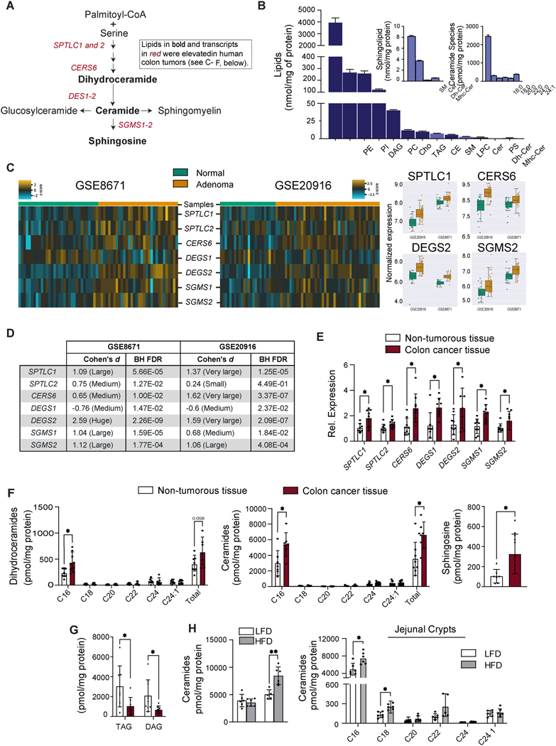
Background & aims: Cancers of the alimentary tract, including esophageal adenocarcinomas, colorectal cancers, and cancers of the gastric cardia, are common comorbidities of obesity. Prolonged, excessive delivery of macronutrients to the cells lining the gut can increase one's risk for these cancers by inducing imbalances in the rate of intestinal stem cell proliferation vs differentiation, which can produce polyps and other aberrant growths. We investigated whether ceramides, which are sphingolipids that serve as a signal of nutritional excess, alter stem cell behaviors to influence cancer risk.

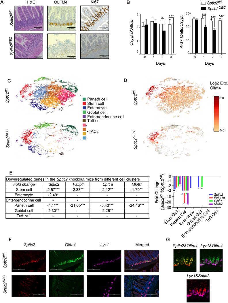
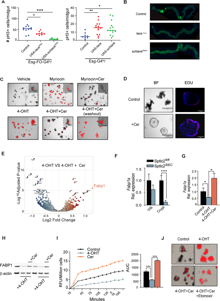
Methods: We profiled sphingolipids and sphingolipid-synthesizing enzymes in human adenomas and tumors. Thereafter, we manipulated expression of sphingolipid-producing enzymes, including serine palmitoyltransferase (SPT), in intestinal progenitors of mice, cultured organoids, and Drosophila to discern whether sphingolipids altered stem cell proliferation and metabolism.

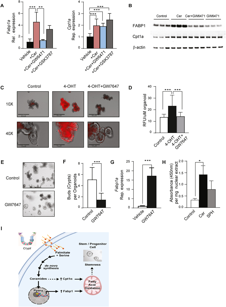
Results: SPT, which diverts dietary fatty acids and amino acids into the biosynthetic pathway that produces ceramides and other sphingolipids, is a critical modulator of intestinal stem cell homeostasis. SPT and other enzymes in the sphingolipid biosynthesis pathway are up-regulated in human intestinal adenomas. They produce ceramides, which serve as prostemness signals that stimulate peroxisome-proliferator activated receptor-α and induce fatty acid binding protein-1. These actions lead to increased lipid utilization and enhanced proliferation of intestinal progenitors.

Conclusions: Ceramides serve as critical links between dietary macronutrients, epithelial regeneration, and cancer risk.

Keywords: Ceramides; Colorectal Cancer; Metabolism; Sphingolipids; Stem Cell.


Conflict of interest statement

Conflict of Interests.

SAS is a founder and shareholder of Centaurus Therapeutics. LPW is a shareholder of Centaurus Therapeutics. None of the other authors have relevant conflicts relevant to this manuscript.

Figures

Figure 1.
Figure 1.. Sphingolipids and sphingolipid-synthesizing genes are upregulated in human colon cancer.
(A) Schematic of the sphingolipid synthesis pathway. Shown in bold font are the lipids, and in red font the transcripts, that were upregulated in human colon cancer (see C-F, below). (B) Major lipid species in the mouse small intestine were detected by LC/MS. Sphingolipids are in light blue. (C) Comparison of sphingolipid gene expression in control tissue vs. non-cancer adenoma tissue. Briefly, microarray expression values were z-score scaled and made into a heat map (left). We also included boxplots (right) for 4 genes that have the most significant changes in both data sets. Samples were clustered by calculating the Euclidean distance between centroids. GSE8671 and GSE20916 samples were assessed against genes probe-set of sphingolipid synthesis. The associated analysis code can be found in this link (https://github.com/j-berg/li_2021). (D) Cohen’s effect sizes were used to describe the significance of the gene regulation between normal and non-cancer adenoma tissue. (E) qPCR analysis of transcripts encoding sphingolipid synthesizing enzymes in colon cancer tissue or adjacent non-tumorous tissue obtained from patients. (n=9) (F) Quantification of sphingolipids in colon cancer tissue and adjacent non-tumorous tissue from patients. (n=9) (G) Triacylglycerol and diacylglycerol in colon cancer tissue and adjacent non-tumorous tissue obtained from patients, (n=9). (H) Quantification of sphingolipids in the intestine of C57BL/6 mice fed with a low fat diet (LFD) versus high fat diet (HFD) for 12 weeks. (n=7). (*p≤0.05, ** p≤0.01 and ***p<0.001). Abbreviations: PE, phosphatidylethanolamine; PI, phosphatidylinositol; DAG, diacylglycerol; PC, phosphatidylcholine; Cho, cholesterol; TAG, triacylglycerol; CE, cholesterol esthers; SM, sphingomyelin; LPC, lysophosphatidylcholine; Cer, ceramide; PS, phosphatidylserine; Dh-Cer, dihydroceramide; Mhc-Cer, monohexosylceramide; Sptlc1, serine palmitoyltransferase long chain base subunit 1; Sptlc2, serine palmitoyltransferase, long chain base subunit 2; Cers6, ceramide synthase-6; Degs1, dihydroceramide desaturase-1; Degs2, dihydroceramide desaturase-2; Sgms1, sphingomyelin synthase-1; Sgms2, sphingomyelin synthase-2.
Fig. 2.
Fig. 2.. Deletion of Sptlc2 from the intestines disrupts the epithelium and depletes intestinal stem cells.
(A) H&E staining, anti-OLFM4 immunostaning, and anti-Ki67 immunostaining of the jejunum 4-days after intraperitoneal (IP) administration of tamoxifen (1mg/mouse) to Sptlc2δIEC and Sptlc2fl/fl mice. (B) Quantification of crypts per villus or Ki67 positive cells per crypt from Sptlc2δIEC and Sptlcfl/fl mice (n=100). (* p≤0.05, ** p≤0.01 and *** p≤0.001) (C) Identification of crypt-enriched intestinal epithelial cell type clusters from Sptlc2δIEC and Sptlcfl/fl mice by single cell RNA sequencing (tSNE). tSNE plot depicting 22,360 cells from Sptlcfl/fl mice and 11,751 cells from Sptlc2δIEC mice. Cell viability from the control mouse was 79% and from the knockout mouse was 74.9%. (D) tSNE plot of the expression pattern of Olfm4 generated with the Loupe Browser (10X Genomics). (E) Downregulated genes in the SptlcδIEC mice from different cell clusters (identified in the single cell RNA sequencing analysis, numbers are the fold change compared to Sptlcfl/fl controls). (F) Sptlc2δISC and Sptlc2fl/fl mice were given tamoxifen by intraperitoneal injection (3mg/day for 5 consecutive days). In situ hybridization (RNAscope) measuring Sptlc2, Olfm4, and Lyz1 mRNA expression was conducted in the intestine on day 5 after tamoxifen injection. Nuclei were stained by DAPI. (G) Samples from the Sptlc2fl/fl mice stained as in F, above, showing combinations of Sptlc2&Olfm4, Lyz1&Olfm4, and Lyz1&Sptlc2 stains. Abbreviations: IEC, intestinal epithelia cell; H&E, Hematoxylin and Eosin staining; TACs, transit amplifying cells; Lyz1, Lysozyme C1; Olfm4, Olfactomedin 4; ISC, intestinal stem cell; serine palmitoyltransferase, long chain base subunit 2; tSNE, t-stochastic neighborhood embedding.
Fig. 3.
Fig. 3.. Ceramides promote stemness and intestinal organoid survival by stimulating FABP1 expression to enhance fatty acid uptake.
Genetic modulation of sphingolipid synthesis genes in the midgut of Drosophila: (A) The graph on the left depicts midgut mitosis (pH3+ cells) in control (w1118, n=12) versus Lace (n=8) or Schlank (n=11) knockdown. Knockdown was controlled by a progenitor-specific and temperature-sensitive Esg-flip out (FO)- Gal4 driver (Esg-FO-G4ts). The graph on the right depicts the quantification of midguts mitosis (pH3+ cells) in control (w1118, n=8) versus Lace (n=14) and Schlank (n=14) gain-of-function conditions, with transgene expression controlled by a progenitor-specific and temperature-sensitive Esg-Gal4 driver (Esg-G4ts) (n=50). *denotes p≤0.05, ** p≤0.01 and *** p≤0.001. (B) Panels showing confocal images of the midguts bearing GFP positive ApcRas tumor clones at 4 weeks (n=8,10, 10 for control, laceKD, and schlankKD). Intestinal organoids were cultured from crypts isolated from the jejunum of SptlcδIEC mice. (C) Propidium iodide (PI) staining of organoids treated with either vehicle, myriocin (10μM), or 4OHT (200ng/ml) for 48 hours. Some samples were supplemented with C2-ceramide (25μM, Cer). In the sample on the lower right, the added C2-ceramide was then removed from the media (washout) for the final 24 hours. Insets show high magnification images of single organoids. (D) Secondary organoids were treated with either vehicle or C2-ceramide (25μM) for 7 days. The images on the left show the organoid morphology, while the ones on the right show staining for the proliferation marker EDU. (E) Volcano plot depicting the RNAseq datasets obtained from 4OHT-treated organoids (200 ng/ml) supplemented with or without C2-ceramide (25μM) for 48 hours. (F) qPCR analysis of Fabp1 expression in the villi or crypts from the jejuna of the Sptlcfl/fl or SptlcδIEC mice, respectively. (G) qPCR analysis of Fabp1 gene expression in organoids treated with vehicle, 4OHT (200 ng/ml), or 4OHT+C2-ceramide (Cer, 25 μM) for 48 hours. (H) Western blot showing FABP1 and actin expression following Sptlc2 deletion with 4-OHT and/or C2-ceramide supplementation for 48 hours. (I) Fatty acid uptake into cells dissociated from organoids that were treated with vehicle, 4OHT, or C2-ceramide for 48 hours (n=4). The bar graph depicts the area under the curve (3 independent experiments). (J) PI staining images of organoids treated with vehicle, 4OHT, 4OHT plus C2-ceramide or 4OHT plus C2 ceramide plus etomoxir (100μM). Abbreviations: Cer, (d18:1/2:0) N-acetoyl-D-erythro-sphingosine; 4-OHT, 4-hydroxytamoxifen; RFU, relative fluorescence unit; IEC, intestinal epithelia cell; FABP1, fatty acid binding protein 1; AUC, area under the curve; Sptlc2, serine palmitoyltransferase; Cpt1a, Carnitine palmitoyltransferase 1A.
Fig. 4.
Fig. 4.. Ceramides increase FABP1 expression through PPARα activation.
(A) qPCR analysis of Fabp1 and Cpt1a expression in organoids treated with vehicle, C2-ceramide (25 μM), and C2-ceramide with GW6471(PPARα antagonist, 1μM) or GSK3787 (PPARδ antagonist, 1μM) for 48 hours. (B) Western blot analysis of FABP1, CPT1a, and actin in organoids treated with C2-ceramide with or without the PPARα antagonist GW6471(GW6471, 1μM) for 48 hours. (C) Propidium iodide (PI) staining of organoids treated with either vehicle, 4OHT (200ng/ml) or 4OHT with the PPARα agonist GW7647 for 48 hours. (D) Quantification of PI staining of 4C (3 independent experiments). (E) Secondary organoids treated with either vehicle or GW7647 (1μM) for 7 days. (F) Quantification of (E), the crypts growing from each organoid. (n=50, *p<0.05, **p<0.01, ***p<0.001). (G) qPCR analysis of Fabp1 expression in organoids treated with either vehicle or GW7647 (1μM). (H) Quantification of PPRE binding activity of PPARα in nuclear extracts from organoids treated with vehicle (Control), C2 ceramide (Cer, 25 μM) and sphingosine (SPH, 25μM) for 48 hours. (n=4, *p≤0.05). (I) Schematic depicting the major conclusion of the paper, including the regulatory events that link exogenous palmitate to the regulation of stemness through a ceramide-PPARα-FABP1 axis. Abbreviations: Cer, (d18:1/2:0) N-acetoyl-D-erythro-sphingosine; 4-OHT, 4-hydroxytamoxifen; RFU, relative fluorescence unit; IEC, intestinal epithelia cell; FABP1, fatty acid binding protein 1; Cpt1a, Carnitine palmitoyltransferase 1A; PPARα, Peroxisome proliferator-activated receptor alpha; AUC, area under the curve; OD, optical density; FAO, fatty acid oxidation; PPRE, peroxisome proliferator hormone response elements.

References

    1. Leblond CP, Stevens CE. The constant renewal of the intestinal epithelium in the albino rat. Anat Rec 1948;100:357–77. - PubMed
    1. Beyaz S, Mana MD, Roper J, et al. High-fat diet enhances stemness and tumorigenicity of intestinal progenitors. Nature 2016;531:53–8. - PMC - PubMed
    1. Maddocks ODK, Athineos D, Cheung EC, et al. Modulating the therapeutic response of tumours to dietary serine and glycine starvation. Nature 2017;544:372–376. - PubMed
    1. Summers SA, Chaurasia B, Holland WL. Metabolic Messengers: Ceramides . Nature Metabolism 2019;1:1051–1058 - PMC - PubMed
    1. Bensard CL, Wisidagama DR, Olson KA, et al. Regulation of Tumor Initiation by the Mitochondrial Pyruvate Carrier. Cell Metab 2019. - PMC - PubMed

Publication types

LinkOut - more resources