. 2023 Aug 4;21(1):253.
doi: 10.1186/s12951-023-02006-x.

Anti-glioma effect of ginseng-derived exosomes-like nanoparticles by active blood-brain-barrier penetration and tumor microenvironment modulation

Affiliations

Anti-glioma effect of ginseng-derived exosomes-like nanoparticles by active blood-brain-barrier penetration and tumor microenvironment modulation

Jisu Kim et al. J Nanobiotechnology. .

Abstract

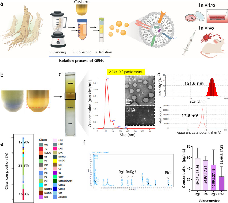
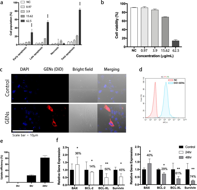
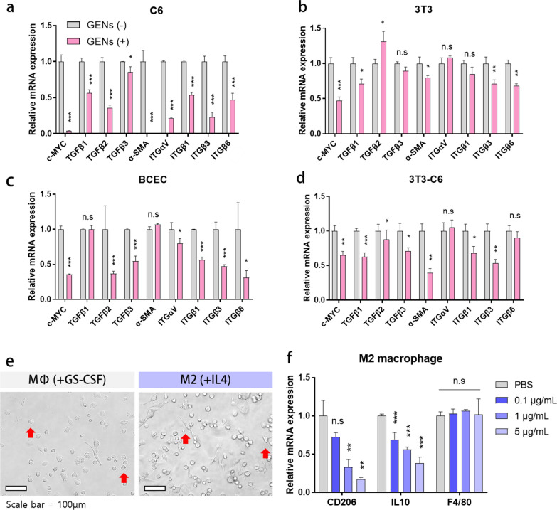
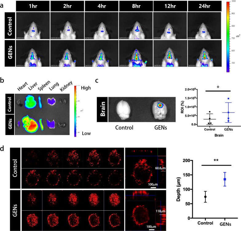
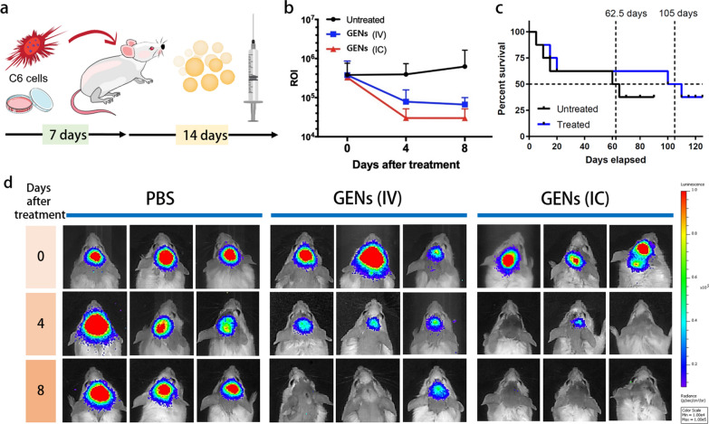
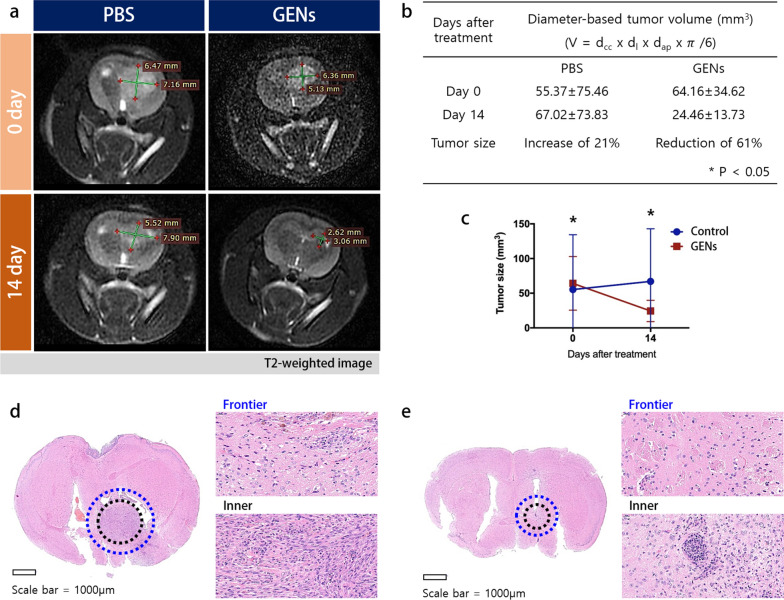
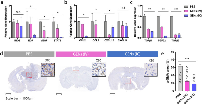
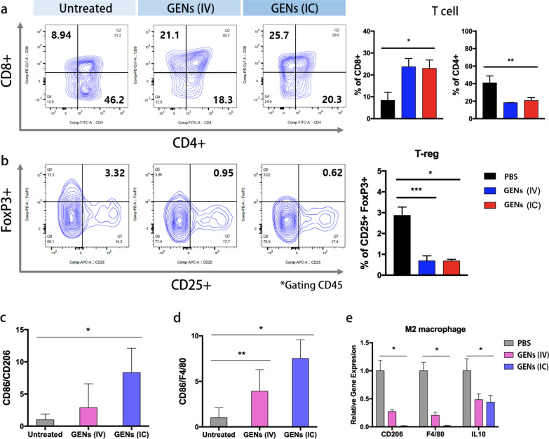
Inhibition of tumor growth and normalization of immune responses in the tumor microenvironment (TME) are critical issues for improving cancer therapy. However, in the treatment of glioma, effective nanomedicine has limited access to the brain because of the blood-brain barrier (BBB). Previously, we demonstrated nano-sized ginseng-derived exosome-like nanoparticles (GENs) consisting of phospholipids including various bioactive components, and evaluated anti-tumor immune responses in T cells and Tregs to inhibit tumor progression. It was found that the enhanced targeting ability of GENs to the BBB and glioma induced a significant therapeutic effect and exhibited strong efficacy in recruiting M1 macrophage expression in the TME. GENs were demonstrated to be successful candidates in glioma therapeutics both in vitro and in vivo, suggesting excellent potential for inhibiting glioma progression and regulating tumor-associated macrophages (TAMs).

Keywords: Anti-glioma; Ginseng; Ginseng-derived exosome-like nanoparticles; Plant-derived exosome-like nanoparticles; Tumor microenvironment.


Conflict of interest statement

The authors declare no competing interests.

Figures

Fig. 1
Fig. 1
Isolation and characterization of ginseng-derived exosome-like nanoparticles (GENs). a A schematic illustration of the isolation process of GENs from fresh ginseng. Ginseng juice was sequentially centrifuged at low and high velocity of speed to isolate GENs. The sucrose cushion method was employed by layering sucrose at concentrations of 68% and 27%. GENs were purified by a buoyant density from 1.13 to 1.19 g/mL using a density gradient method. After the purification of GENs, the cellular toxicity and therapeutic efficiency of GENs were evaluated by in vitro and in vivo analyses. b The sucrose cushion method to prevent disruption of the extracellular vesicles (EVs) and contaminants caused by excessive aggregates of EVs during centrifugation (right). c The specific layer between 8 and 30% of sucrose was achieved and the concentration of 2.24 × 10.13 particles/mL and size distribution were determined with a serial dilution of GENs by nanoparticle tracking analysis (NTA). The round-shaped morphology of GENs was observed by transmission electron microscopy (TEM; inset). d The size and zeta potential of GENs were measured at 37 ℃ by dynamic light scattering (DLS). e A graph of lipidomic analysis was reported as the percentage of the components of lipids of GENs. f The concentration of ginsenosides of GENs was determined by high performance liquid chromatography (HPLC). wax esters (WE), triglycerides (TG), sulfoquinovosyldiacylglycerol (SQDG), sphingosine (So), phosphatidylserine (PS), phosphatidylinositol phosphate (PIP), phosphatidylinositol (PI), phosphatidylgylcerols (PG), phosphatidylethanolamine (PE), phosphatidylcholine (PC), phosphatidic acid (PA), monogalactosylmonoacylglycerol (MGMG), monogalactosylacylglycerols (MGDG), monoacylglycerol (MG), lysophosphatidylglycerol (LPG), lysophosphatidylethanolamine (LPE), lysophophatidylcholine (LPC), lysophosphatidic acid (LPA), digalactosylmonoacylglycerol (dgmg), digalactosyldiacylglycerol (DGDG), diacylglycerol (DG), coenzyme (Co), cardiolipin (CL), ceramide 1-phosphates (CerP), CerG3GNAc1, diglycosylceramide (CerG2), glucosylceramide (CerG1), ceramides (Cer), acylglucosyl-sitosterol esters (AGlcSiE)
Fig. 2
Fig. 2
Apoptosis in C6 glioma cells treated with ginseng-derived exosome like nanoparticles (GENs) and cellular uptake of GENs. a Cell apoptosis assay in C6 glioma cells by Annexin V/PI after treatment with GENs at concentrations ranging from 0.975 to 62.5 μg/mL for 24 h (the concentrations were determined by Bradford assay). b Cell viability after treatment with GENs at the indicated concentrations was expressed as a percentage of cells in the control group (three independent experiments). c Confocal fluorescence imaging showed the internalization of conventional liposomes made from egg yolk and GENs by C6 glioma cells. After incubating C6 glioma cells with DiD-labeled GENs for 6 h, the distribution of GENs was assessed. Nucleic acid and GENs were stained with Hoechst (blue) and DiD staining dye (red), respectively. d Cellular uptake of GENs after 6 h of incubation. e Measurement of uptake efficiency of GENs in cells for 24 h. f Quantitative PCR analysis for the mRNA expression of apoptosis-associated genes in C6 glioma cells treated with GENs. 50 µg/mL of GENs was treated in C6 glioma cells for 24 h and 48 h. The mRNA expression levels were normalized to those of GAPDH (housekeeping gene) and expressed as a ratio of the control group (*, P < 0.05; **, P < 0.01; ***, P < 0.001)
Fig. 3
Fig. 3
Inhibition of overexpressed c-MYC and activation of TGF-β and integrin in glioma cells, fibroblasts, and endothelial cells and the effect of treatment of M2 bone marrow-derived macrophages (BMMs). ad Gene expression levels of c-MYC proto-oncogene, TGFβ1, 2, 3, integrin αVβ1, 3, 6, and α-SMA in C6, 3T3, BCEC, and 3T3-C6 co-culture. e The morphology of BMMs, as observed by microscopy. BMMs were stimulated with GS-CSF for 7 days and with IL4 for 3 days. f The mRNA expression levels in M2 macrophages after treatment with serially diluted GENs (*, P < 0.05; ** P < 0.01; ***, P < 0.001)
Fig. 4
Fig. 4
The targeting ability of GENs to glioma in vivo. a In vivo fluorescence imaging of DiD-labeled GENs. Conventional liposomes based on egg yolk were used as a control. After intravenous (IV) administration of DiD-labeled GENs, fluorescence imaging was performed at different time points. b Ex vivo fluorescence imaging of the dissected organs 24 h after the IV injection of DiD-labeled GENs. Higher fluorescence intensity in the brain was observed in GENs compared to the control group. c Relative region of interest (ROI) of fluorescence intensity in the brain samples. Using ROI as a measure, the graph shows that GENs exhibited an almost 2.26-fold higher fluorescence intensity on average than the control group. d Penetration of GENs and conventional liposomes into 3D tumor spheroids (*P < 0.05; **P < 0.01)
Fig. 5
Fig. 5
In vivo anticancer effect induced by GENs and Kaplan–Meier survival analysis. a Schematic illustration of the orthotopic model using a Balb/C mice. After the implantation of C6 glioma cells into the brain, GENs were injected every other day for 14 days. b A region of interest (ROI) of luminescence intensity compared to the control group after the indicated number of days after the treatment (0 to 8 days). c The survival rate of tumor-bearing mice treated with GENs compared to the control group (N = 8). d After orthotopic treatment, the luminescence intensity of C6 cells (expressing luciferase) was measured by an in vivo imaging system. On day 8 after the treatment, a significant reduction in the luminescence intensity of C6 glioma was observed in the group treated with GENs compared to the control group (N = 8)
Fig. 6
Fig. 6
Orthotopic-induced xenograft brain tumor tissue hematoxylin and eosin (H&E) staining and magnetic resonance imaging (MRI) measurement of the C6 glioma in Wistar rat. a After 14 days of treatment with GENs, the tumor volume was measured by MRI imaging. b, c A table and graph for the diameter-based tumor volume. Tumors decreased in volume by 61% at day 14 after treatment with GENs, whereas tumors increased volume by 21% in the control group. de H&E staining of the brain tissue of an control tumor and treated tumor, respectively (*P < 0.05)
Fig. 7
Fig. 7
Gene expression of CAFs in the tumor microenvironment in tumor-bearing mice. a mRNA expression of CAF-related genes was decreased in the treatment group. b Reduced gene expression of chemokines. c Decreased gene expression of the TGFβ family in the treatment groups. Grey, PBS; pink, intravenous (IV) administration; blue, intracranial (IC) administration. de After treatment with GENs, α-SMA immunohistochemical staining in the brain tissue was determined (brown) (*, P < 0.05; **, P < 0.01; ***, P < 0.001)
Fig. 8
Fig. 8
T cells and regulatory T cells (Tregs) are induced by decreased cytokines in the tumor microenvironment (TME). a Analysis of regulation of T cells. Fluorescently conjugated anti-CD8, CD4, and CD45 monoclonal antibodies were used to analyze T cells. The abundance of CD8 of T cells, gated CD45, was increased in the GENs treatment groups, whereas the abundance of CD4 was decreased compared to the PBS group. b Fluorescently conjugated anti-FoxP3, CD25, and CD45 monoclonal antibodies were used to analyze Tregs. Tregs cell population was evaluated by FACS analysis. The abundance of Tregs was reduced in the GENs treatment groups. ce The ratio of the relative gene expressions of M1 and M2 (*, P < 0.05; **, P < 0.01; ***, P < 0.001)
Fig. 9
Fig. 9
Prediction of miRNA targets and gene silencing effect of miRNAs mediated apoptosis of tumor cells through the regulation of apoptotic c-MYC signaling pathway. a miRNA targets were predicted using miRNA target prediction tool based on sequence-based miRNA target prediction. b The binding site of ptc-miR396f in the mRNA of c-MYC. c Relative gene expression levels of c-MYC and BCL2 with miRNAs. There was a significant gene silencing effect compared to mock-transfected and control groups, which are normalized to non-targeting control (NTC) (*, P < 0.05; ***, P < 0.001)

References

    1. Lin D, Wang M, Chen Y, Gong J, Chen L, Shi X, Lan F, Chen Z, Xiong T, Sun H, Wan S. Trends in Intracranial Glioma Incidence and Mortality in the United States, 1975–2018. Front Oncol. 2021;11:748061. - PMC - PubMed
    1. Koutu V, Rajawat S, Shastri L, Malik MM. Apoptosis and inhibition of human epithelial cancer cells by ZnO nanoparticles synthesized using plant extract. Adv Nano Res. 2019;7(4):233–240.
    1. Arani AG, Farazin A, Mohammadimehr M. The effect of nanoparticles on enhancement of the specific mechanical properties of the composite structures: a review research. Adv Nano Res. 2021;10(4):327–337.
    1. Hong C, Liang J, Xia J, Zhu Y, Guo Y, Wang A, Lu C, Ren H, Chen C, Li S, et al. One stone four birds: a novel liposomal delivery system multi-functionalized with ginsenoside rh2 for tumor targeting therapy. Nanomicro Lett. 2020;12(1):129. - PMC - PubMed
    1. Hong C, Wang D, Liang J, Guo Y, Zhu Y, Xia J, Qin J, Zhan H, Wang J. Novel ginsenoside-based multifunctional liposomal delivery system for combination therapy of gastric cancer. Theranostics. 2019;9(15):4437–4449. - PMC - PubMed

LinkOut - more resources