. 2023 Sep;11(9):1231-1242.
doi: 10.1016/j.jchf.2023.05.031. Epub 2023 Aug 2.

Serum Proteomic Analysis of Peripartum Cardiomyopathy Reveals Distinctive Dysregulation of Inflammatory and Cholesterol Metabolism Pathways

Collaborators, Affiliations

Serum Proteomic Analysis of Peripartum Cardiomyopathy Reveals Distinctive Dysregulation of Inflammatory and Cholesterol Metabolism Pathways

Jana P Lovell et al. JACC Heart Fail. 2023 Sep.

Abstract

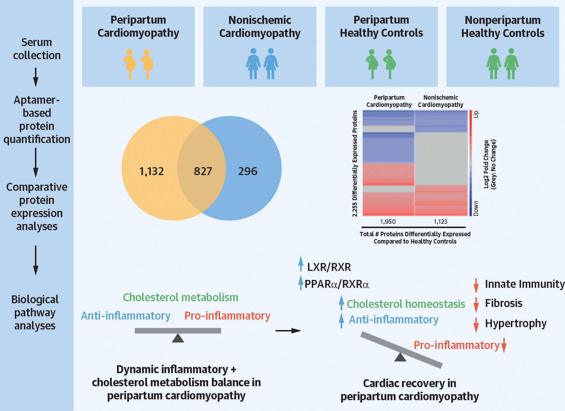
Background: The pathophysiology of peripartum cardiomyopathy (PPCM) and its distinctive biological features remain incompletely understood. High-throughput serum proteomic profiling, a powerful tool to gain insights into the pathophysiology of diseases at a systems biology level, has never been used to investigate PPCM relative to nonischemic cardiomyopathy.

Objectives: The aim of this study was to characterize the pathophysiology of PPCM through serum proteomic analysis.

Methods: Aptamer-based proteomic analysis (SomaScan 7K) was performed on serum samples from women with PPCM (n = 67), women with nonischemic nonperipartum cardiomyopathy (NPCM) (n = 31), and age-matched healthy peripartum and nonperipartum women (n = 10 each). Serum samples were obtained from the IPAC (Investigation of Pregnancy-Associated Cardiomyopathy) and IMAC2 (Intervention in Myocarditis and Acute Cardiomyopathy) studies.

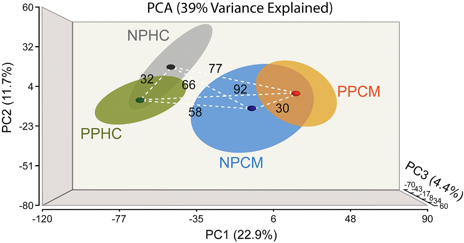
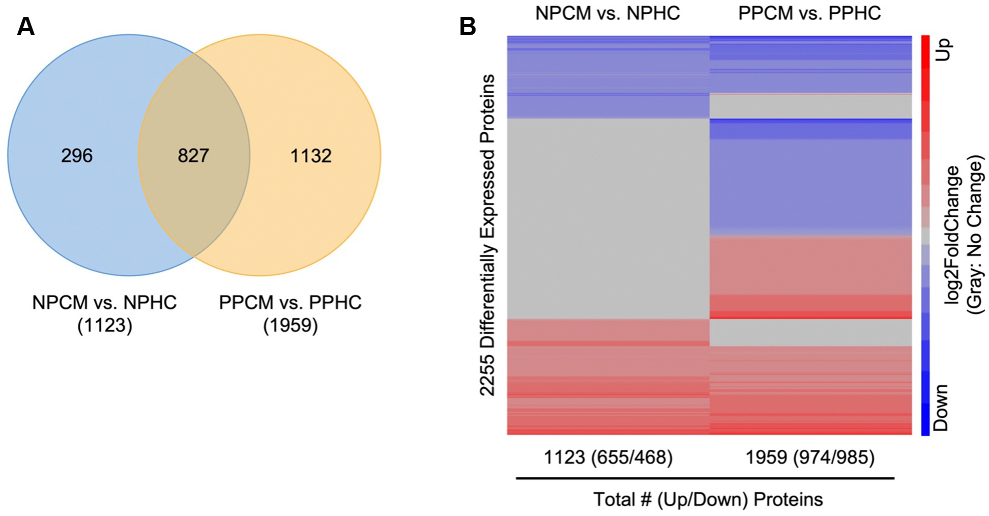
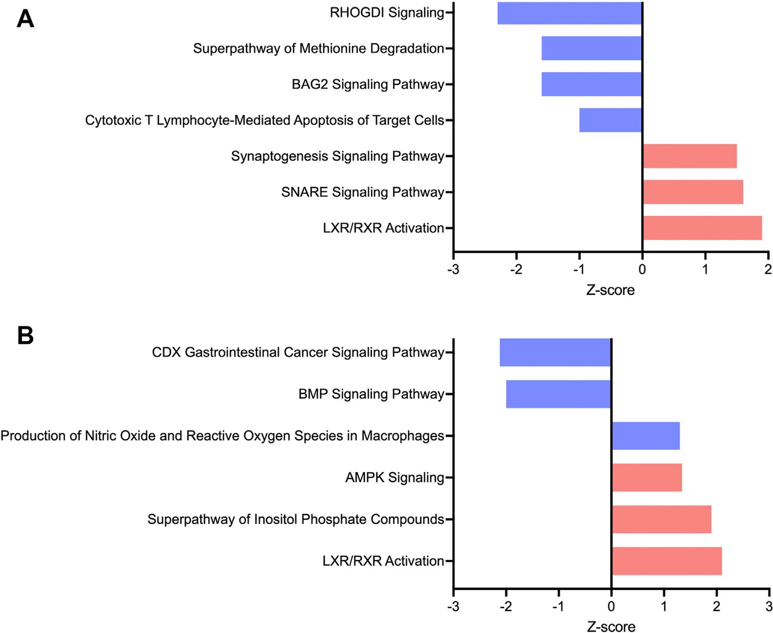
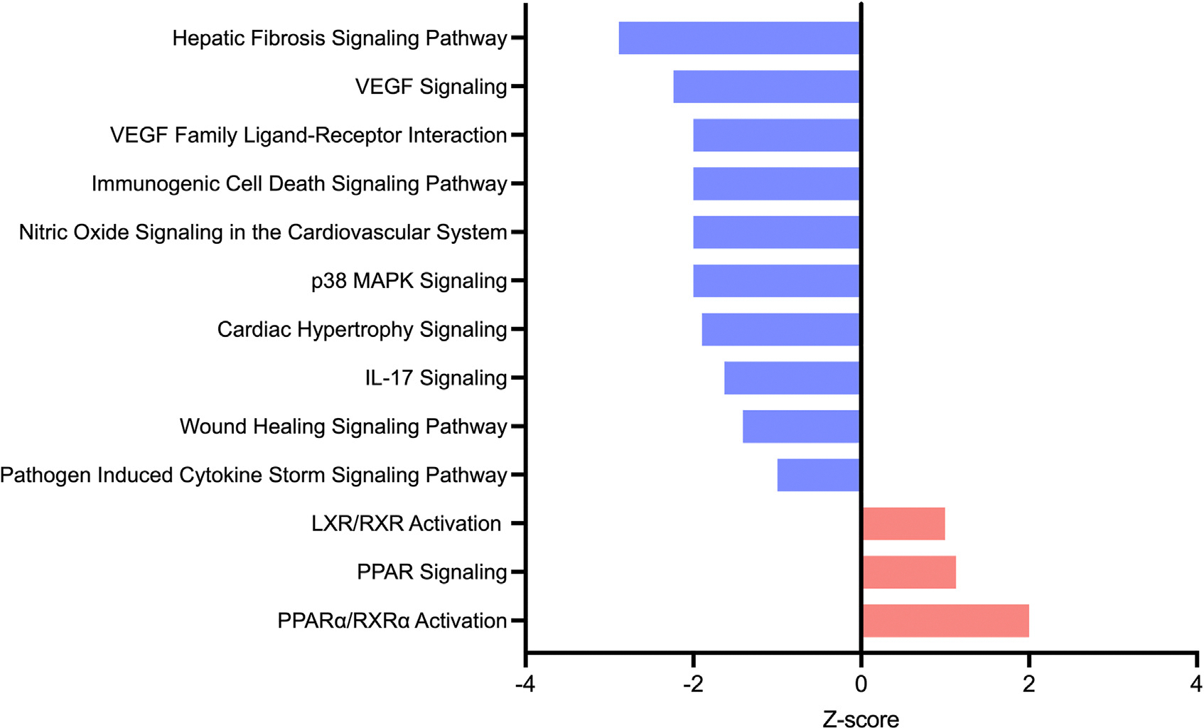
Results: Principal component analysis revealed unique clustering of each patient group (P for difference <0.001). Biological pathway analyses of differentially measured proteins in PPCM relative to NPCM, before and after normalization to pertinent healthy controls, highlighted specific dysregulation of inflammatory pathways in PPCM, including the upregulation of the cholesterol metabolism-related anti-inflammatory pathway liver-X receptor/retinoid-X receptor (LXR/RXR) (P < 0.01, Z-score 1.9-2.1). Cardiac recovery by 12 months in PPCM was associated with the downregulation of pro-inflammatory pathways and the upregulation of LXR/RXR, and an additional RXR-dependent pathway involved in the regulation of inflammation and metabolism, peroxisome proliferator-activated receptor α/RXRα signaling.

Conclusions: Serum proteomic profiling of PPCM relative to NPCM and healthy controls indicated that PPCM is a distinct disease entity characterized by the unique dysregulation of inflammation-related pathways and cholesterol metabolism-related anti-inflammatory pathways. These findings provide insight into the pathophysiology of PPCM and point to novel potential therapeutic targets.

Keywords: heart failure; inflammation; pregnancy; proteomics; serum proteomics.


Conflict of interest statement

Funding Support and Author Disclosures This study has been funded through National Heart, Lung, and Blood Institute grants 5K08HLO145108-03 and 1R01HL160716-01 to Dr Adamo and T32-HL007227 to Dr Lovell, institutional funds from the Johns Hopkins Division of Cardiology awarded to Dr Adamo, and the Blumenthal Scholarship in Preventative Cardiology and American Heart Association 979462 funds awarded to Dr Sharma. Investigation of Pregnancy-Associated Cardiomyopathy was funded by National Institutes of Health grant HL102429 and IMAC2 by HL075038. All other authors have reported that they have no relationships relevant to the contents of this paper to disclose.

Figures

FIGURE 1
FIGURE 1. PCA Plot Demonstrating the Spatial Relationship of Serum Proteomic Profiles of Patient Groups
There is unique clustering of each patient group with partial overlap of healthy control groups and partial overlap of the cardiomyopathy groups. The dashed white lines with numbers indicate the Euclidean distances between centroids (bold small circles) of different patient groups (large ellipsoids at 1× SD of the centroid). The distance between healthy control groups is similar to the distance between cardiomyopathy groups, suggesting there is a significant biological difference between PPCM and nonischemic nonperipartum cardiomyopathy. NPCM = nonperipartum cardiomyopathy; NPHC = nonperipartum healthy controls; PCA = principal components analysis; PPCM = peripartum cardiomyopathy; PPHC = peripartum healthy controls.
FIGURE 2
FIGURE 2. PPCM and NPCM Are Characterized by Dysregulation in Specific Sets of Serum Proteins With Partial Overlap
(A) Venn diagram showing comparative analyses of serum protein levels between each cardiomyopathy group and their pertinent controls (fold-difference ≥1.5, q < 0.05). There is an overlap of 827 proteins dysregulated in NPCM and PPCM with 296 proteins uniquely dysregulated in NPCM and 1,132 proteins uniquely dysregulated in PPCM. (B) Comparative heat maps of proteins differentially expressed in NPCM and PPCM relative to their respective healthy controls indicate that NPCM and PPCM are defined each by dysregulation of a unique set of serum proteins with partial overlap. Proteins without significant differential fold-differences are shaded in gray to highlight differences and similarities between PPCM and NPCM. Red = increased relative level; blue = decreased relative level. Abbreviations as in Figure 1.
FIGURE 3
FIGURE 3. LXR/RXR Activation Is Significantly Upregulated in PPCM
Ingenuity pathway analysis (IPA) of proteins dysregulated in PPCM relative to nonischemic cardiomyopathy with (A) and without (B) normalization to healthy controls (fold-difference ≥1.5, q < 0.05). Canonical pathways dysregulated in PPCM with P < 0.05 are sorted by Z-score. Positive Z-scores (red bars) indicate upregulation. Negative Z-scores (blue bars) indicate downregulation. AMPK = adenosine monophosphate-activated protein kinase; BAG2 = Bcl2-associated anthanogene 2; BMP = bone morphogenetic protein; CDX = caudal type homeobox; LXR/RXR = liver-X receptor/retinoid-X receptor; RHOGDI = rho GDP-dissociation inhibitor; SNARE = soluble N-ethylmaleimide-sensitive factor attachment protein receptor; other abbreviation as in Table 1.
FIGURE 4
FIGURE 4. Pro-Inflammatory and Pathways Involved in Adverse Cardiac Remodeling Are Downregulated in Recovered PPCM While Anti-Inflammatory Pathways Are Upregulated
IPA of proteins dysregulated in PPCM patients who experience recovery of left ventricular (LV) systolic function by 12 months after initial diagnosis (fold-difference ≥1.15, P < 0.05). Canonical pathways dysregulated in PPCM with P < 0.05 are sorted by Z-score. Positive Z-scores (red bars) indicate upregulation. Negative Z-scores (blue bars) indicate downregulation. IL = interleukin; MAPK = mitogen-activated protein kinase; PPAR = peroxisome proliferator-activated receptor; VEGF = vascular endothelial growth factor; other abbreviations as in Figures 1 and 3.
CENTRAL ILLUSTRATION
CENTRAL ILLUSTRATION. Serum Proteomic Analysis of Peripartum Cardiomyopathy
Aptamer-based serum proteomic analysis revealed peripartum cardiomyopathy is a distinct disease entity relative to nonischemic nonperipartum cardiomyopathy, with each group characterized by dysregulation of a unique set of serum proteins that overlap partially. Biological pathway analyses indicate that peripartum cardiomyopathy is characterized by dysregulation of inflammatory and cholesterol metabolism-related pathways. The upregulation of retinoid-X receptor (RXR)-mediated pathways regulating metabolism and inflammation may contribute to cardiac recovery in peripartum cardiomyopathy. LXR = liver-X receptor; PPAR = peroxisome proliferator-activated receptor.

Comment in

Similar articles

Cited by

References

    1. Sliwa K, Hilfiker-Kleiner D, Petrie MC, et al. Current state of knowledge on aetiology, diagnosis, management, and therapy of peripartum cardiomyopathy: a position statement from the Heart Failure Association of the European Society of Cardiology Working Group on peripartum cardiomyopathy. Eur J Heart Fail. 2010:767–778. 10.1093/eurjhf/hfq120 - DOI - PubMed
    1. Bauersachs J, König T, van der Meer P, et al. Pathophysiology, diagnosis and management of peripartum cardiomyopathy: a position statement from the Heart Failure Association of the European Society of Cardiology Study Group on peripartum cardiomyopathy. Eur J Heart Fail. 2019;21(7):827–843. 10.1002/ejhf.1493 - DOI - PubMed
    1. Cooper LT, Mather PJ, Alexis JD, et al. Myocardial recovery in peripartum cardiomyopathy: prospective comparison with recent onset cardiomyopathy in men and nonperipartum women. J Card Fail. 2012;18(1):28–33. 10.1016/j.cardfail.2011.09.009 - DOI - PMC - PubMed
    1. McNamara DM, Elkayam U, Alharethi R, et al. Clinical outcomes for peripartum cardiomyopathy in North America: results of the IPAC Study (Investigations of Pregnancy-Associated Cardiomyopathy). J Am Coll Cardiol. 2015;66(8):905–914. 10.1016/j.jacc.2015.06.1309 - DOI - PMC - PubMed
    1. Goland S, Modi K, Bitar F, et al. Clinical profile and predictors of complications in peripartum cardiomyopathy. J Card Fail. 2009;15(8):645–650. 10.1016/j.cardfail.2009.03.008 - DOI - PubMed

Publication types