Review
. 2023 Sep:69:101814.
doi: 10.1016/j.smim.2023.101814. Epub 2023 Aug 4.

Immune aging - A mechanism in autoimmune disease

Affiliations
Review

Immune aging - A mechanism in autoimmune disease

Yanyan Zheng et al. Semin Immunol. 2023 Sep.

Abstract

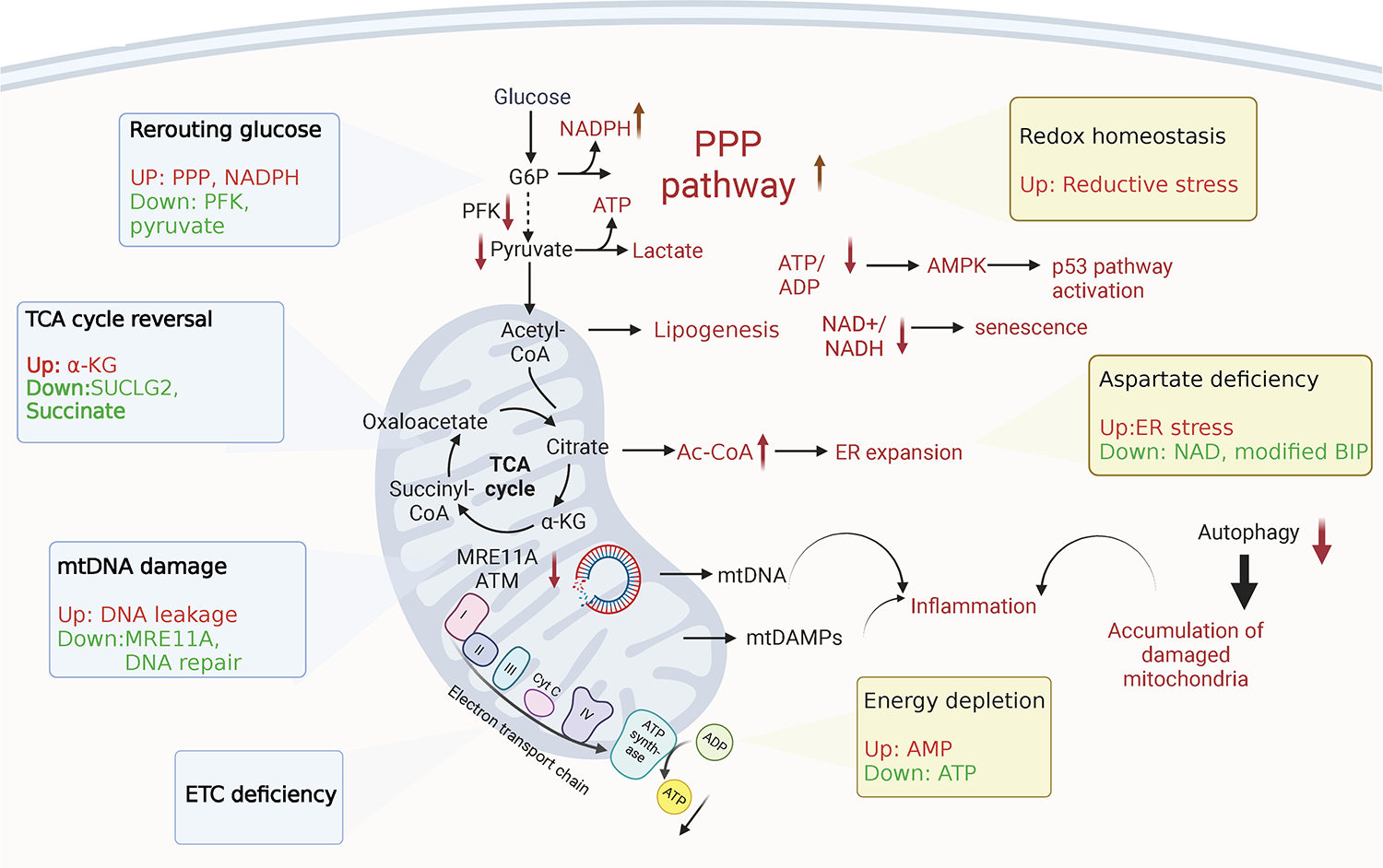
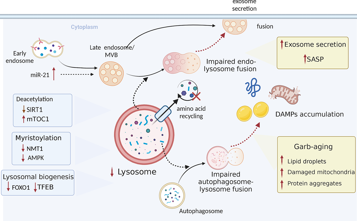
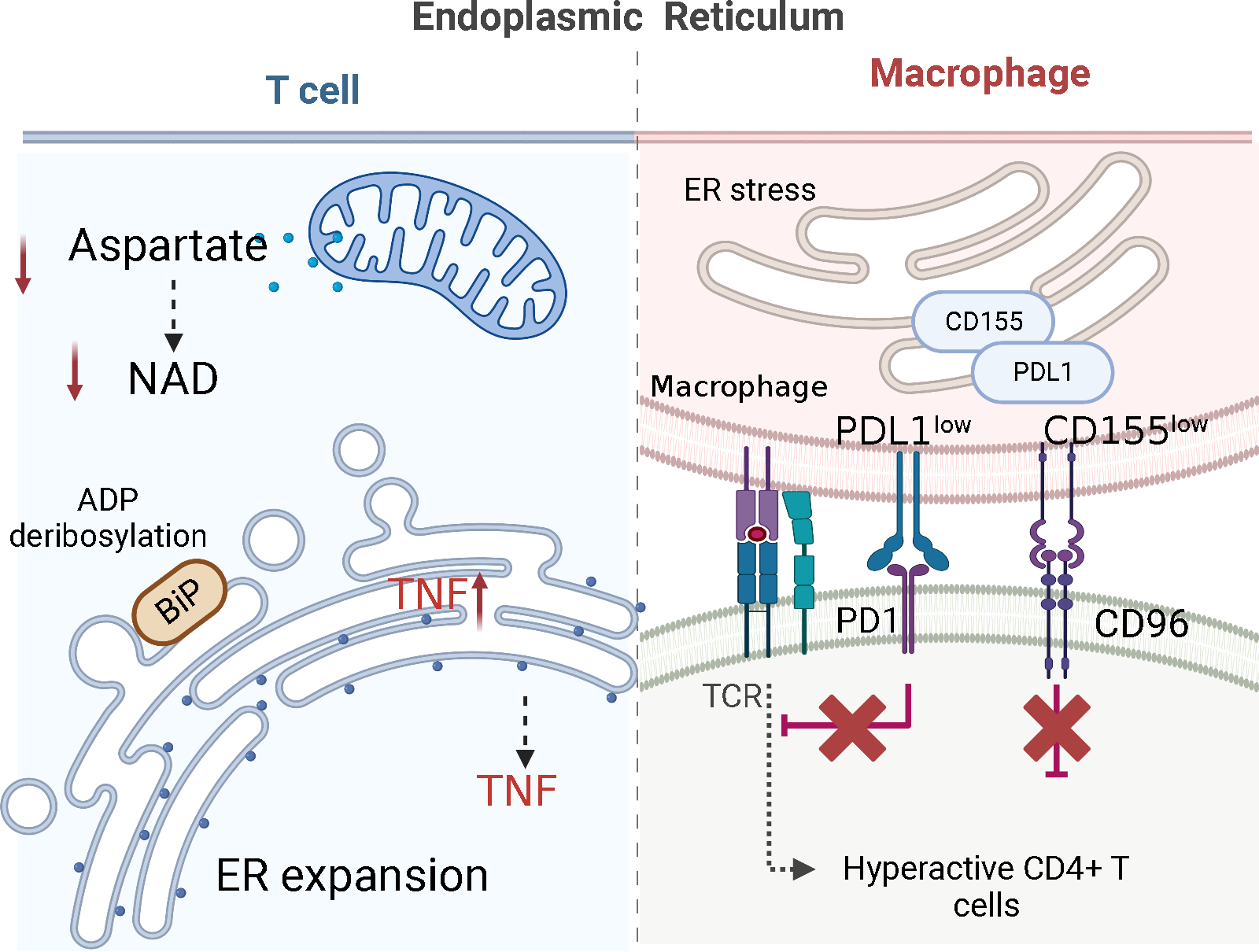
Evidence is emerging that the process of immune aging is a mechanism leading to autoimmunity. Over lifetime, the immune system adapts to profound changes in hematopoiesis and lymphogenesis, and progressively restructures in face of an ever-expanding exposome. Older adults fail to generate adequate immune responses against microbial infections and tumors, but accumulate aged T cells, B cells and myeloid cells. Age-associated B cells are highly efficient in autoantibody production. T-cell aging promotes the accrual of end-differentiated effector T cells with potent cytotoxic and pro-inflammatory abilities and myeloid cell aging supports a low grade, sterile and chronic inflammatory state (inflammaging). In pre-disposed individuals, immune aging can lead to frank autoimmune disease, manifesting with chronic inflammation and irreversible tissue damage. Emerging data support the concept that autoimmunity results from aging-induced failure of fundamental cellular processes in immune effector cells: genomic instability, loss of mitochondrial fitness, failing proteostasis, dwindling lysosomal degradation and inefficient autophagy. Here, we have reviewed the evidence that malfunctional mitochondria, disabled lysosomes and stressed endoplasmic reticula induce pathogenic T cells and macrophages that drive two autoimmune diseases, rheumatoid arthritis (RA) and giant cell arteritis (GCA). Recognizing immune aging as a risk factor for autoimmunity will open new avenues of immunomodulatory therapy, including the repair of malfunctioning mitochondria and lysosomes.

Keywords: Autoimmune disease; Giant cell arteritis; Immune aging; Rheumatoid arthritis; T cell aging.


Conflict of interest statement

Declaration of Competing Interest The authors declare no conflicts of interest.

Figures

Fig. 1
Fig. 1. Mitochondria in immune aging-related autoimmune disease.
Aging mitochondria show defects in various aspects, including glucose rerouting, tricarboxylic acid cycle (TCA) reversal and reduced electron transport chain (ETC) efficiency in metabolism, as well as accumulation of mitochondrial DNA damage. Aging immune effector cells suffer from ATP depletion, metabolite imbalance, loss of redox homeostasis and ER stress. Damaged mitochondria contribute to autoimmune inflammation through multiple mechanisms, specifically, in rheumatoid arthritis.
Fig. 2
Fig. 2. Lysosomes and autophagosomes in immune aging-related autoimmune disease.
Lysosome biogenesis decreases with age. Increased mTOC1 activity inhibits autophagic processes. Impaired endosome or autophagosome-lysosome fusion results in increased exosome secretion and extracellular accumulation of damage-associated molecular patterns, which contributes to senescence-associated secretory phenotype (SASP) and inflammation.
Fig.3
Fig.3. The endoplasmic reticulum (ER) in immune aging-related autoimmune disease.
(1) The size of the ER and the maturation of proteins are controlled by mitochondria-derived aspartate, which regenerates cytoplasmic NAD+ to ADP-ribosylate the ER stress sensor BiP. In the pre-aged T cells of rheumatoid arthritis patients, shortage of mitochondrial aspartate leads to an increase in ER size, accumulation of tumor necrosis factor (TNF) mRNA on ER membranes and excessive production of TNF. (2) Macrophages from patients with giant cell arteritis retain the checkpoint ligand CD155 in the endoplasmic reticulum (ER) and fail to bring it to the cell surface. CD155low antigen-presenting cells induce expansion of CD4+CD96+ T cells, which become hyperactive and tissue invasive.
Fig.4
Fig.4. Inter-organelle contact sites.
Inter-organelle contact sites, areas of close vicinity between the membranes of two organelles, function in calcium and lipid transfer etc. and are emerging as hot spots of cellular regulation. These inter-organelle sites participate in organelle biogenesis, division, apoptosis, and autophagy and are emerging as critical regulators of immune cells function in health and disease.

References

    1. Berner F, Bomze D, Diem S, Ali OH, Fassler M, Ring S et al. Association of Checkpoint Inhibitor-Induced Toxic Effects With Shared Cancer and Tissue Antigens in Non-Small Cell Lung Cancer. JAMA Oncol, 2019;5:1043–7. - PMC - PubMed
    1. Berner F, Bomze D, Lichtensteiger C, Walter V, Niederer R, Hasan Ali O et al. Autoreactive napsin A-specific T cells are enriched in lung tumors and inflammatory lung lesions during immune checkpoint blockade. Sci Immunol, 2022;7:eabn9644. - PubMed
    1. Cancro MP. Age-Associated B Cells. Annu Rev Immunol, 2020;38:315–40. - PubMed
    1. Zhao TV, Sato Y, Goronzy JJ, Weyand CM. T-Cell Aging-Associated Phenotypes in Autoimmune Disease. Front Aging, 2022;3:867950. - PMC - PubMed
    1. Goronzy JJ, Weyand CM. Mechanisms underlying T cell ageing. Nat Rev Immunol, 2019;19:573–83. - PMC - PubMed

Publication types