. 2023 Aug 5;13(1):12720.
doi: 10.1038/s41598-023-37874-8.

PP2A-B55alpha controls keratinocyte adhesion through dephosphorylation of the Desmoplakin C-terminus

Affiliations

PP2A-B55alpha controls keratinocyte adhesion through dephosphorylation of the Desmoplakin C-terminus

Abbey L Perl et al. Sci Rep. .

Abstract

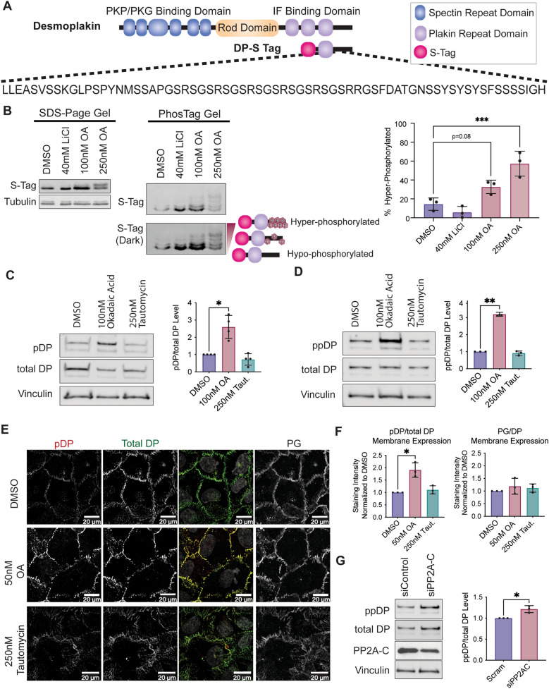
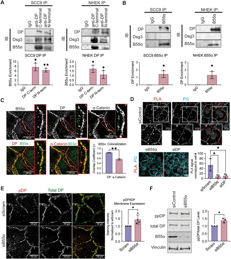
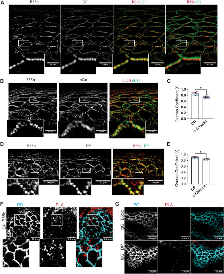
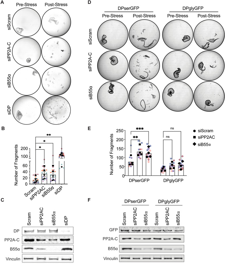
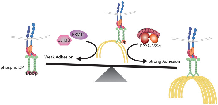
Critical for the maintenance of epidermal integrity and function are attachments between intermediate filaments (IF) and intercellular junctions called desmosomes. The desmosomal cytoplasmic plaque protein desmoplakin (DP) is essential for anchoring IF to the junction. DP-IF interactions are regulated by a phospho-regulatory motif within the DP C-terminus controlling keratinocyte intercellular adhesion. Here we identify the protein phosphatase 2A (PP2A)-B55α holoenzyme as the major serine/threonine phosphatase regulating DP's C-terminus and consequent intercellular adhesion. Using a combination of chemical and genetic approaches, we show that the PP2A-B55α holoenzyme interacts with DP at intercellular membranes in 2D- and 3D- epidermal models and human skin samples. Our experiments demonstrate that PP2A-B55α regulates the phosphorylation status of junctional DP and is required for maintaining strong desmosome-mediated intercellular adhesion. These data identify PP2A-B55α as part of a regulatory module capable of tuning intercellular adhesion strength and a candidate disease target in desmosome-related disorders of the skin and heart.


Figures

Figure 1
Figure 1
PP2A is a phosphatase for the C-terminal domain of DP. (A) Schematic of DP structural domains and the DP-S-Tag construct containing only residues 2628–2871. Highlighted below is the 68 residue-long “GSR” repeat phospho-regulatory motif downstream of the IF binding site capable of regulating DP-IF interactions. (B) A 15% acrylamide gel without (left) and with (right) Phos-Tag molecule capable of separating protein by its number of phosphate groups. Protein lysates are from SCC9 cells transfected with the DP-S Tag C-terminus and treated with either LiCl or OA for 3 h. Phos-Tag gel was analyzed using an anti-S-Tag antibody to visualize the total DP-S-Tag expression. Quantification of the % Phosphorylated DP-Stag is on right. Percent hyper-phosphorylated was calculated by (phosphorylated signal/total signal)*100 to get a final percentage. (CF) SCC9s were treated with inhibitors preferential for either PP2A (50 and 100 nM OA) or PP1 (250 nM Tautomycin) for 3 h. Western blot analysis of S2849 phosphorylated DP (pDP) (C) and the dual S2845/S2849 phosphorylated DP (ppDP) (D). (E) Immunofluorescence staining of S2849 phosphorylated DP (pDP) in drug treated SCC9’s. (F) Staining intensity from [E] was quantified at the membrane using PG stain as a mask. (G) Amount of the dual S2845/S2849 phosphorylated DP (ppDP) was analyzed in total SCC9 cells transfected with siRNA targeting the PP2A-C subunit. Statistical analyses were performed on normalized data using One-way ANOVA with multiple comparisons (BF) or a student t-test (G). * < 0.05; ** < 0.01; *** < 0.001.
Figure 2
Figure 2
DP is found in complex with the PP2A regulatory subunit B55⍺ in SCC9 cells. (A) Immunoprecipitation of endogenous DP using antibodies targeting DP’s C-terminus or N-terminus and blotting back for Dsg3 or B55⍺. SCC9s (left) and NHEKs (right) were grown for 2 days in high-calcium media (HCM). (B) Immunoprecipitation of endogenous B55⍺ and blotting back for DP or Dsg3. SCC9s (left) and NHEKs (right) were grown for 2 days in high-calcium media (HCM). (C) SCC9 immunofluorescence co-stained for B55⍺, DP, and ⍺-Catenin. Overlayed images are shown below. Colocalization analysis as determined by an object-based colocalization analysis tool represented as overlap coefficient measurements. (D) Proximity ligation analysis performed on SCC9 cell transfected with siRNA targeting either a Scramble control, B55α, or DP. A fluorescence-based PLA signal was measured on fixed coverslips incubated with B55α and DP targeting antibodies. (E) Immunofluorescence staining of S2849 phosphorylated DP (pDP) in SCC9 cells transfected with siRNA targeting the B55α subunit. Staining intensity from was quantified at the membrane using PG stain as a mask. (F) Amount of the dual S2845/S2849 phosphorylated DP (ppDP) were analyzed in total SCC9 cells transfected with siRNA targeting the B55α subunit. Statistical analyses were performed using a One-way ANOVA with multiple comparisons (A,D) or a student t-test (BC,EF). * < 0.05; ** < 0.01; *** < 0.001.
Figure 3
Figure 3
B55alpha is found localized to sites of intercellular membranes in the 3D epidermis in vitro and in vivo. (AB) 3D organotypic reconstructed skin cultures grown for 6 days in culture and immunofluorescence staining of B55⍺, DP, and PG (A) and B55⍺, α-Catenin, and PG (B) was performed. (C) Colocalization analysis of (AB) as determined by an object-based colocalization analysis tool represented as overlap coefficient measurements. (D) Human skin samples were stained for B55⍺ and DP using immunofluorescence. (E) Colocalization analysis of (D) and (Supplemental Fig. 4D) as determined by an object-based colocalization analysis tool represented as overlap coefficient measurements. (F–G) Proximity ligation analysis performed on fixed human skin samples. A fluorescence-based PLA signal was measure on fixed slides incubated with either B55α and DP targeting antibodies or B55α and IgG. Statistical analyses were performed using a student t-test. * < 0.05; ** < 0.01.
Figure 4
Figure 4
PP2A-B55alpha regulates epithelial cell adhesion and barrier through DP. (A) A dispase-adhesion assay was performed in SCC9s transfected with siRNA targeting Scramble, B55⍺, or DP and grown in normal HCM for 5 days. Images represent SCC9 monolayers pre- (left) and post- (right) exposure to mechanical stress. (B) Monolayer fragmentation from (A) was quantified and represented as total fragment number from n = 5. (C) Western blot validation of siRNA knockdown in (A). (D) A dispase-adhesion assay was performed in A431 cells expressing either dox-inducible DPserGFP or DPglyGFP and transfected with siRNA targeting Scramble, B55⍺, or DP. Cells were grown in normal HCM with 1 μg/mL Dox for 6 days. Images represent A431 monolayers pre- (left) and post- (right) exposure to mechanical stress. (E) Monolayer fragmentation from (A) was quantified and represented as total fragment number from n = 4. (F) Western blot validation of siRNA knockdown in (C). Statistical analyses were performed using a One-way ANOVA with multiple comparisons. * < 0.05; ** < .01.
Figure 5
Figure 5
A balance between PP2A-B55alpha and GSK3β/PRMT1 activities on DP’s C-terminus controls desmosome-adhesion strength through regulating the DP-IF connection. A schematic representing the regulatory switch controlling desmosome-mediated adhesion through the phospho-regulation of the DP-IF interaction. Albrecht et al. previously identified GSK3β and PRMT1 as cooperating to induce the phosphorylation of DP’s C-terminus that inhibits DP-IF binding and weakens desmosomal adhesion. Our work has uncovered of PP2A-B55α as an opposing regulatory node capable of dephosphorylating DP’s C-terminus, inducing increased cell adhesion potentially through in increased association between DP and IF.

References

    1. Broussard JA, et al. Scaling up single-cell mechanics to multicellular tissues - the role of the intermediate filament-desmosome network. J. Cell. Sci. 2020 doi: 10.1242/jcs.228031. - DOI - PMC - PubMed
    1. Hatzfeld M, Keil R, Magin TM. Desmosomes and intermediate filaments: Their consequences for tissue mechanics. Cold Spring Harb. Perspect. Biol. 2017 doi: 10.1101/cshperspect.a029157. - DOI - PMC - PubMed
    1. Getsios S, Huen AC, Green KJ. Working out the strength and flexibility of desmosomes. Nat. Rev. Mol. Cell Biol. 2004;5:271–281. doi: 10.1038/nrm1356. - DOI - PubMed
    1. Vasioukhin V, Bowers E, Bauer C, Degenstein L, Fuchs E. Desmoplakin is essential in epidermal sheet formation. Nat. Cell Biol. 2001;3:1076–1085. doi: 10.1038/ncb1201-1076. - DOI - PubMed
    1. Gallicano GI, Bauer C, Fuchs E. Rescuing desmoplakin function in extra-embryonic ectoderm reveals the importance of this protein in embryonic heart, neuroepithelium, skin and vasculature. Development. 2001;128:929–941. doi: 10.1242/dev.128.6.929. - DOI - PubMed

Publication types