. 2023 Nov;152(5):1336-1344.e5.
doi: 10.1016/j.jaci.2023.07.013. Epub 2023 Aug 5.

Hemophagocytic lymphohistiocytosis-like hyperinflammation due to a de novo mutation in DPP9

Affiliations

Hemophagocytic lymphohistiocytosis-like hyperinflammation due to a de novo mutation in DPP9

Christine Wolf et al. J Allergy Clin Immunol. 2023 Nov.

Erratum in

  • Corrigendum.
    [No authors listed] [No authors listed] J Allergy Clin Immunol. 2025 Oct;156(4):1124. doi: 10.1016/j.jaci.2025.08.002. Epub 2025 Aug 27. J Allergy Clin Immunol. 2025. PMID: 40864024 No abstract available.

Abstract

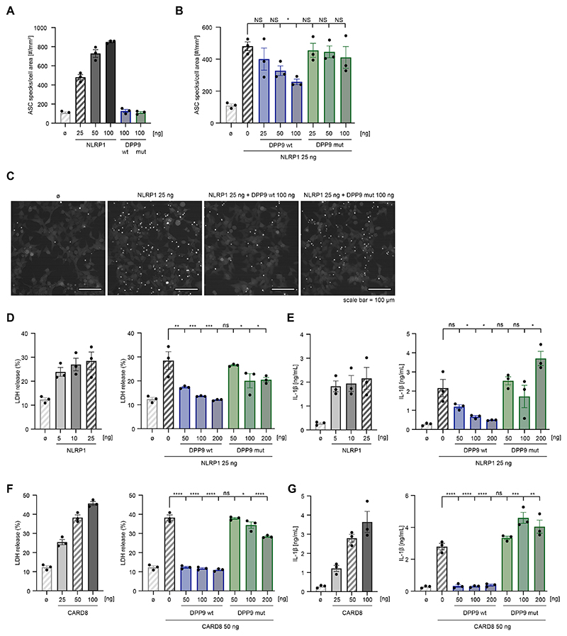
Background: Genetic defects in components of inflammasomes can cause autoinflammation. Biallelic loss-of-function mutations in dipeptidyl peptidase 9 (DPP9), a negative regulator of the NLRP1 and CARD8 inflammasomes, have recently been shown to cause an inborn error of immunity characterized by pancytopenia, skin manifestations, and increased susceptibility to infections.

Objective: We sought to study the molecular basis of autoinflammation in a patient with severe infancy-onset hyperinflammation associated with signs of fulminant hemophagocytic lymphohistiocytosis.

Methods: Using heterologous cell models as well as patient cells, we performed genetic, immunologic, and molecular investigations to identify the genetic cause and to assess the impact of the identified mutation on inflammasome activation.

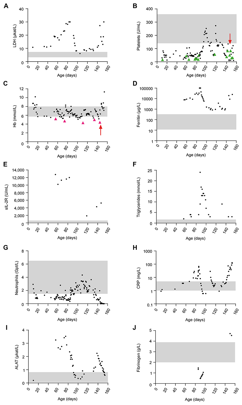
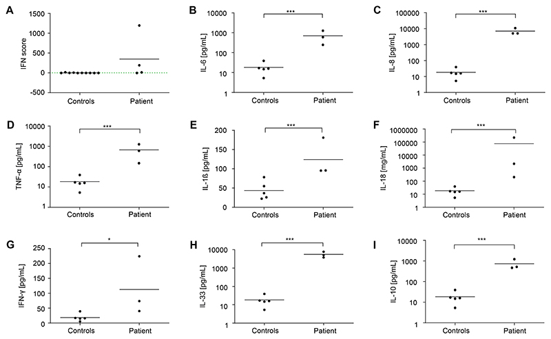
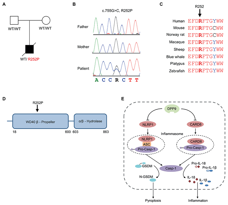
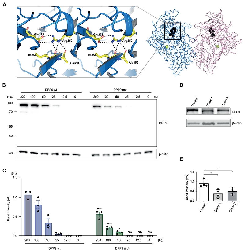
Results: The patient exhibited pancytopenia with decreased neutrophils and T, B, and natural killer cells, and markedly elevated levels of lactate dehydrogenase, ferritin, soluble IL-2 receptor, and triglycerides. In addition, serum levels of IL-1β and IL-18 were massively increased, consistent with inflammasome activation. Genetic analysis revealed a previously undescribed de novo mutation in DPP9 (c.755G>C, p.Arg252Pro) affecting a highly conserved amino acid residue. The mutation led to destabilization of the DPP9 protein as shown in transiently transfected HEK293T cells and in patient-derived induced pluripotent stem cells. Using functional inflammasome assays in HEK293T cells, we demonstrated that mutant DPP9 failed to restrain the NLRP1 and CARD8 inflammasomes, resulting in constitutive inflammasome activation. These findings suggest that the Arg252Pro DPP9 mutation acts in a dominant-negative manner.

Conclusions: A de novo mutation in DPP9 leads to severe infancy-onset autoinflammation because of unleashed inflammasome activation.

Keywords: CARD8; DPP9; IL-18; IL-1β; Inborn error of immunity; NLRP1; autoinflammation; hemophagocytic lymphohistiocytosis; inflammasome; proinflammatory cytokines.


Conflict of interest statement

Disclosure of Potential Conflict of Interest

The authors declare that they have no relevant conflicts of interest.

Figures

Figure 1
Figure 1. Laboratory findings.
A-J, Blood parameters of the patient taken at the indicated day of life. The gray shaded areas mark the normal range for each parameter. B and C, Green and purple arrowheads indicate platelet and erythrocyte transfusions, respectively, and the red arrows mark allogeneic hematopoietic stem cell transplantation. ALAT, Alanine-aminotransferase; CRP, C-reactive protein; Hb, hemoglobin; sIL-2R, soluble IL-2 receptor.
Figure 2
Figure 2. Interferon signature and serum cytokines.
A, IFN signature in patient PBMCs measured at different time points. An IFN score of 12.49 (dashed green line) indicates the median IFN score of 10 healthy controls + 2 SD. B-I, Cytokine levels in patient sera collected at different time points, compared with 5 healthy controls. Data are presented as mean of 3 technical replicates. The Mann-Whitney U test was used. ***P < .001; *P < .05.
Figure 3
Figure 3. Identification of a de novo DPP9 mutation.
A, Pedigree of the family. B, Electropherograms showing a heterozygous DPP9 mutation in the patient, which is absent in his healthy parents. C, Multiple sequence alignment showing high conservation of R252 within the DPP9 protein. D, Schematic of domain architecture of DPP9 with the R252P mutation indicated. E, Model depicting the negative regulatory function of DPP9 on the NRLP1 and CARD8 inflammasomes.
Figure 4
Figure 4. Impact of the DPP9 mutation R252P on protein structure and stability.
A, Model of DPP9 focusing on the R252 pocket (PDB: 6EOR, 6EOQ, 6QZV7). Left, The plot displays the Arg252 pocket in stereo. Right, A chain trace of the DPP9 dimer is shown with Arg252 (black). The position of the active site Ser730 is indicated (yellow sphere). B, HEK293T cells were transiently transfected with plasmids encoding for wild-type DPP9 (wt) or the DPP9 mutant (R252P) at the indicated concentrations. Expression levels were determined 24 hours later via immunoblotting. One representative immunoblot of 3 independent experiments is depicted. C, The band intensities of 3 independent experiments were quantified and summarized as mean ± SEM. A 2-way ANOVA was performed followed by the Šidák multiple comparisons test. ****P < .0001; *P < .05. D, One representative immunoblot of 4 independent experiments showing DPP9 expression in patient-derived iPSCs compared with a control iPSC line. β-actin served as loading control. E, The band intensities, normalized to β-actin, of 4 independent experiments were quantified and summarized as mean values. The Mann-Whitney U test was used. *P < .05. AU, Arbitrary unit; NS, not significant.
Figure 5
Figure 5. DPP9 R252P fails to inhibit NLRP1- and CARD8-dependent inflammasome activation.
A-C, HEK293T cells expressing fluorescently tagged ASC were transfected with the indicated plasmids or left untreated (B). After 24 hours, cells were imaged to quantify ASC speck formation. A and B, The number of ASC specks was quantified per cell area. Data are presented as mean ± SEM of 3 independent experiments. A 1-way ANOVA was performed followed by the Dunnett multiple comparisons test: *P < .05. Note that certain control conditions are shown twice for each panel, and the corresponding bars are shaded. C, Representative fields of view of 1 representative experiment of 3 independent experiments conducted as in Fig 5B. Scale bar, 100 mm. D-G, HEK293T cells were transfected with inflammasome components and indicated amounts of NLRP1 or CARD8 and/ or wild-type or mutant (R252P) DPP9 for 24 hours and treated with 4 mM Val-boroPro for 4 hours. Cytotoxicity was determined by LDH assay and IL-1β release was measured by ELISA. Data are presented as mean ± SEM of 3 independent experiments. A 1-way ANOVA was performed followed by the Dunnett multiple comparisons test. ****P < .0001; ***P < .001; **P < .01; *P < .05. Note that certain control conditions are shown twice for each panel, and the corresponding bars are shaded. NS, Not significant.

References

    1. Broz P, Dixit VM. Inflammasomes: mechanism of assembly, regulation and signalling. Nat Rev Immunol. 2016;16:407–20. - PubMed
    1. Guo H, Callaway JB, Ting JPY. Inflammasomes: mechanism of action, role in disease, and therapeutics. Nat Med. 2015;21:677–87. - PMC - PubMed
    1. Taabazuing CY, Griswold AR, Bachovchin DA. The NLRP1 and CARD8 inflammasomes. Immunol Rev. 2020;297:13–25. - PMC - PubMed
    1. Harapas CR, Steiner A, Davidson S, Masters SL. An update on autoinflammatory diseases: inflammasomopathies. Curr Rheumatol Rep. 2018;20:40. - PubMed
    1. Romberg N, Vogel TP, Canna SW. NLRC4 inflammasomopathies. Curr Opin Allergy Clin Immunol. 2017;17:398–404. - PMC - PubMed

Publication types

MeSH terms

Substances