. 2023 Sep;10(27):e2300759.
doi: 10.1002/advs.202300759. Epub 2023 Aug 6.

eIF3f Mediates SGOC Pathway Reprogramming by Enhancing Deubiquitinating Activity in Colorectal Cancer

Affiliations

eIF3f Mediates SGOC Pathway Reprogramming by Enhancing Deubiquitinating Activity in Colorectal Cancer

Qihao Pan et al. Adv Sci (Weinh). 2023 Sep.

Abstract

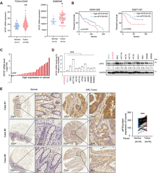
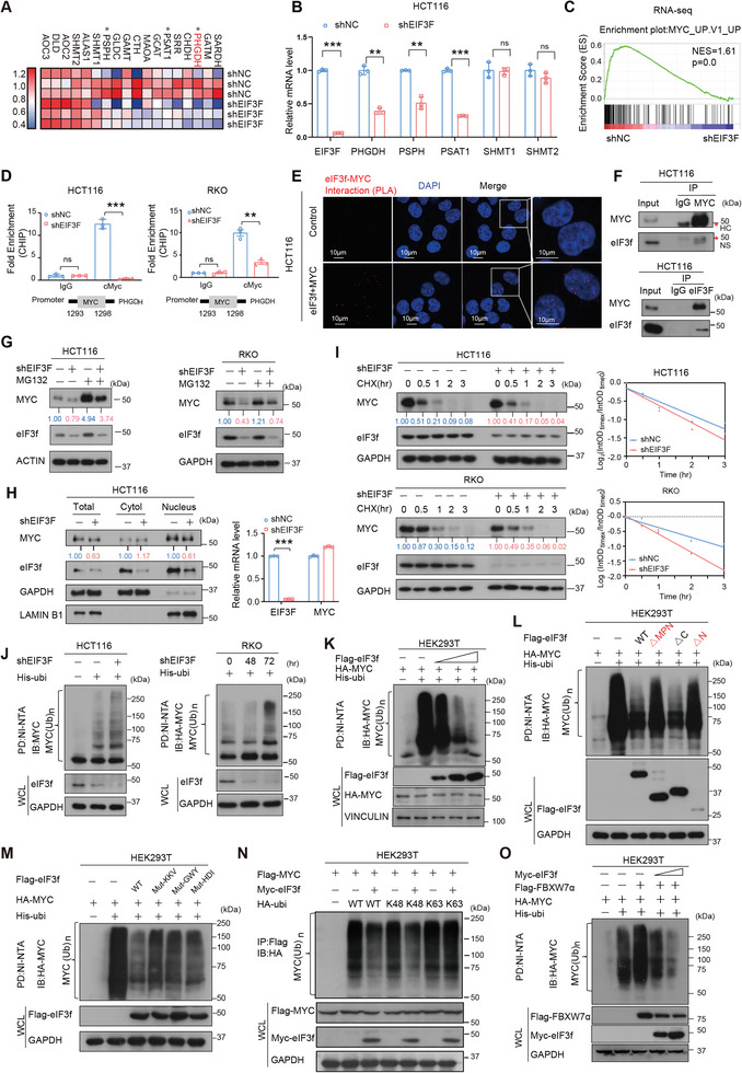
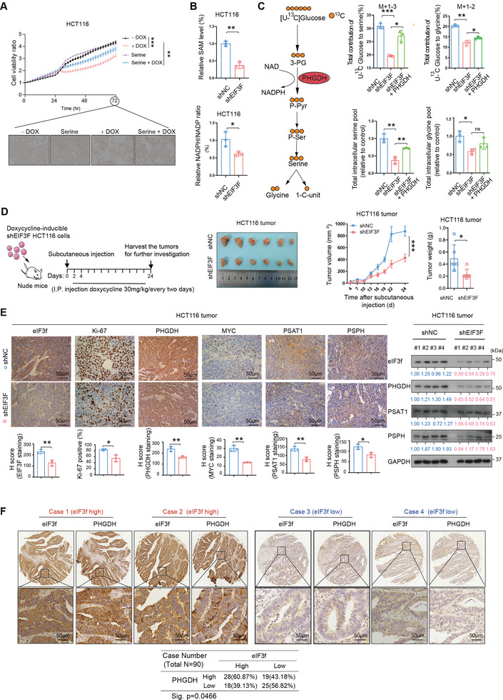
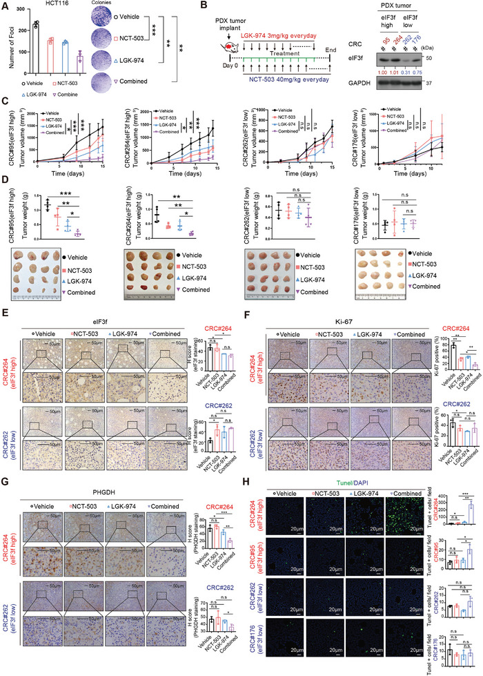
Numerous studies have demonstrated that individual proteins can moonlight. Eukaryotic Initiation translation factor 3, f subunit (eIF3f) is involved in critical biological functions; however, its role independent of protein translation in regulating colorectal cancer (CRC) is not characterized. Here, it is demonstrated that eIF3f is upregulated in CRC tumor tissues and that both Wnt and EGF signaling pathways are participating in eIF3f's oncogenic impact on targeting phosphoglycerate dehydrogenase (PHGDH) during CRC development. Mechanistically, EGF blocks FBXW7β-mediated PHGDH ubiquitination through GSK3β deactivation, and eIF3f antagonizes FBXW7β-mediated PHGDH ubiquitination through its deubiquitinating activity. Additionally, Wnt signals transcriptionally activate the expression of eIF3f, which also exerts its deubiquitinating activity toward MYC, thereby increasing MYC-mediated PHGDH transcription. Thereby, both impacts allow eIF3f to elevate the expression of PHGDH, enhancing Serine-Glycine-One-Carbon (SGOC) signaling pathway to facilitate CRC development. In summary, the study uncovers the intrinsic role and underlying molecular mechanism of eIF3f in SGOC signaling, providing novel insight into the strategies to target eIF3f-PHGDH axis in CRC.

Keywords: FBXW7; MYC; PHGDH; Wnt; colorectal cancer; eIF3f; ubiquitination.


Conflict of interest statement

The authors declare no conflict of interest.

Figures

Figure 1
Figure 1
eIF3f is overexpressed in CRC. A) Expression level of eIF3f in colorectal tumor and normal tissues (TCGA‐COAD and GSE9348). Unpaired student's t test was performed. Solid lines denote the medians, the 5th and 95th percentiles. **p < 0.01, *p < 0.05. B) Kaplan‐Meier plot of overall survival time and log‐rank test based on EIF3F expression in GSE41258 and GSE71187 datasets. C) Waterfall plot of relative EIF3F mRNA levels of 26 paired samples of CRC and the adjacent normal tissue measured by qRT‐PCR. D) qRT‐PCR and immunoblotting of eIF3f expression in CRC and normal colonic cells (NCM460) and gastric epithelial cells (GES‐1). ***P<0.001. E) eIF3f is overexpressed in tumor than adjacent normal tissue. Analysis of eIF3f protein expression level in CRC and normal tissues by IHC staining of colorectal cancer tissue microarray (TMA). Representative different eIF3f staining images. The staining intensity and percentage were analyzed by Halo pathology software, paired t‐test were used, ***p < 0.001.
Figure 2
Figure 2
eIF3f deubiquitinates PHGDH. A) PHGDH mRNA expression was assessed from TCGA‐COAD and GSE9348 database. ***p < 0.001, ****p < 0.0001. B) Representative images of proximity ligation assay (PLA) results revealed that eIF3f interacted with PHGDH. The red signals demonstrate eIF3f‐PHGDH interaction. The nuclei of the cells were stained with DAPI (Blue signals). C) Endogenous co‐IP results indicated that EIF3F interacted with PHGDH. D) DOX‐inducible KD of eIF3f leads to downregulation of PHGDH. E) MG132 reversed EIF3F KD‐mediated PHGDH downregulation. F) eIF3F KD increased the turnover rate of PHGDH. G) Immunoblot analysis of poly‐ubiquitinated PHGDH in poly‐ubiquitination assays of indicated cells expressing DOX inducible shEIF3F and treated with 20 × 10−6 m MG132 for 6 h. The cell lysates were pulled down by nickel beads and immunoblotted with an anti‐ PHGDH antibody. H) eIF3F increased the steady expression of PHGDH. I) Overexpression of eIF3f reduced the ubiquitination level of PHGDH in a dose‐dependent manner. J) eIF3f deubiquitinated K48‐ubiquitin linkage of PHGDH. K) Schematic representation of vectors expressing WT or serial deletion mutants of Flag‐eIF3f (Upper panel). Deubiquitinating activity of different eIF3f mutants toward PHGDH ubiquitination. L) MPN mutations affects the deubiquitinating activity of eIF3f toward PHGDH ubiquitination. Schematic representation of the species alignment and the mutation sites (Upper panel). HEK293T cells co‐transfected with HA‐PHGDH and EIF3F wild‐type or mutants construct were treated with 50 × 10−6 m MG132 for 6 h before harvesting. Cells were lysed in guanidine‐HCl containing buffer and cell lysates were then pull down (PD) with nickel beads (NI‐NTA) and immunoblotted with HA‐PHGDH (lower panel).
Figure 3
Figure 3
PHGDH is a substrate of FBXW7β. A) Screening of potential E3 ligases for PHGDH in HCT‐116 cells by transient transfection of indicated overexpression plasmids. B) FBXW7β decreases the steady state expression of PHGDH. C) Endogenous co‐IP result showed the interaction between FBXW7 β and PHGDH. D) MG132 reversed FBXW7β‐mediated PHGDH downregulation. E) FBXW7 β increases the poly‐ubiquitinated level of PHGDH. F) Gel‐filtration chromatography fraction analysis of cell lysates from shNC or shEIF3F HCT‐116 cells. Molecular size of eluted fraction is indicated above. G) eIF3f expression level affects FBXW7β‐mediated PHGDH polyubiquitination. H) Amino acid sequence of the putative FBXW7β binding motifs in PHGDH. I) 497A/501A mutant of PHGDH is not vulnerable to FBXW7β‐mediated downregulation. J) 497A/501A mutant of PHGDH is resistant to FBXW7β‐mediated ubiquitination. K) 497A/501A mutant of PHGDH is resistant to FBXW7β‐mediated acceleration of protein turn‐over. L) CHIR (GSK3β inhibitor) treatment increased PHGDH protein level. M) CHIR reversed FBXW7β ‐mediated PHGDH downregulation. N) Endogenous co‐IP results indicated that GSK3β interacted with PHGDH. O) GSK3β increases the steady‐state level of PHGDH. P) GSK3β kinase dead mutant (K85A) failed to enhance PHGDH phosphorylation. Q) GSK3β kinase dead mutant (K85A) failed to mediate PHGDH ubiquitination.
Figure 4
Figure 4
eIF3f regulated PHGDH transcription via stabilizing MYC. A) Heatmap of the expression of genes that mediate serine/one‐carbon metabolism in cells expressing shNC or shEIF3F in HCT‐116 cells. B) qRT‐PCR results showed the mRNA levels of genes related to serine synthesis pathway after eIF3f was knocked down. **P < 0.01, *** P <0.001, ns = no significant. C) GSEA analysis of RNA‐seq data revealed that eIF3f is associated with the expression of MYC‐targeted genes. D) ChIP‐PCR analysis revealed binding of transcription factor MYC on PHGDH promoter. MYC binds on the promoter region (1293bp‐1298 bp) of PHGDH. **P < 0.01, *** P <0.001. E) Representative images of proximity ligation assay (PLA) results revealed that eIF3f interacted with MYC. The red signals demonstrate eIF3f‐MYC interaction. The nuclei of the cells were stained with DAPI (Blue signals). F) HCT116 cell lysates were immunoprecipitated with either eIF3f or MYC antibody and immunoblotted with the indicated antibodies. IgG was used as a control. “* HC” indicated heavy chain. G) eIF3f KD leads to MYC downregulation. eIF3f was knocked down after DOX treatment. H) eIF3f KD leads to MYC downregulation in the nucleus. Cell lysates were harvested and separated into total lysates, cytoplasmic lysates and nucleus lysates via nucleus fraction followed by immunoblotting with indicated antibodies. mRNA expression of MYC is not changed after eIF3f was knocked down. ***P <0.001. I) Immunoblot analysis of the MYC protein turnover rate in indicated cells with eIF3f KD. Cycloheximide (CHX). eIF3f was knocked down after DOX treatment. J) Immunoblot analysis of poly‐ubiquitinated MYC in poly‐ubiquitination assays of indicated cells expressing DOX inducible shEIF3F and treated with 20 × 10−6 m MG132 for 6 h. The cell lysates were pulled down by nickel beads and immunoblotted with an anti‐MYC antibody. K‐L) HEK293T cells were transfected with increasing doses of Flag‐EIF3F, or EIF3F deletion constructs and His‐ubiquitin. The cell lysates were pulled down by nickel beads and immunoblotted with an anti‐MYC antibody. M) MPN mutations affects the deubiquitinating activity of eIF3f toward MYC ubiquitination. N) eIF3f deubiquitinates K48‐linked poly‐ubiquitination of MYC. O) eIF3f antagonizes FBXW7α‐mediated MYC ubiquitination.
Figure 5
Figure 5
eIF3f regulates SGOC pathway in CRC. A) Serine could partially rescue the growth inhibition of CRC cells induced by EIF3F knockdown in HCT‐116 cells. IncuCyte was used to measure the confluence of the cells. Each time point was relative to Time 0 h. TWO WAY‐ANOVA test was used to test the significance. ** P <0.01, ***P < 0.001. B) Measurement of SAM and NADH/NAD+ levels in HCT‐116 cells transduced with EIF3F shRNA. *P < 0.05, **P < 0.01. C) Scheme of metabolite tracing of [U‐13C]‐labeled glucose to serine and glycine metabolism (Left). Incorporation of [U‐13C] glucose into the indicated metabolites at 24 h in HCT‐116 cells expressing indicated plasmids (Right). *P < 0.05, **P < 0.01, *** P <0.001. D) Tumor growth curves of HCT‐116 (1×10⁶) colon cancer cells with or without EIF3F knockdown. EIF3F knockdown was induced by doxycycline(30 mg kg−1) treatment. Cells were subcutaneously injected into nude mice (n = 6). The tumors were isolated at the end of the experiments. Tumor volume and tumor weight were measured. E) Representative IHC images of EIF3F, Ki67, PHGDH, MYC, PSAT1, and PSPH staining in the subcutaneous tumor tissues generated in (H). Staining intensity were quantitated. Unpaired student's t test was used to test the significance, *P < 0.05, **P < 0.01. Scale bars represent 50 µm. Immunoblot analysis of indicated protein levels in the subcutaneous tumor tissues generated in (H). F) Representative EIF3F and PHGDH IHC staining in the tissue microarray (TMA). Case 1 and 2 are representatives of a patient with EIF3F high‐expressed colon cancer. Case 3 and 4 are representatives of a patient with EIF3F low‐expressed colon cancer. Chi‐square analysis shows the correlation of eIF3f and PHGDH expression in human CRC tissue microarray specimens (n = 90).
Figure 6
Figure 6
Wnt signaling promoted EIF3F transcription. A) Immunoblotting of EIF3F in HCT‐116 (left panel) and HEK293T (right panel) cells treated with the indicated concentrations of WNT inhibitor (NCB0846) for 48 h. MYC is measured as a positive control of NCB0846 treatment. B) qRT‐PCR analysis of EIF3F in indicated cells treated with the indicated concentrations of WNT inhibitor (NCB0846) for 48 h. Student's t‐test was used. *** P < 0.001. C) qRT‐PCR analysis of EIF3F in HCT‐116 cells treated with Wnt‐3a containing conditioned medium for 12 h. Student's t‐test was used. D) qRT‐PCR analysis of EIF3F in HCT‐116 cells treated with indicated treatment. Student's t‐test was used. *** P <0.001. E) Immunoblotting results and qRT‐PCR analysis of EIF3F expression in HCT116 with β‐catenin overexpression. *** P < 0.001, ns = no significant. F) qRT‐PCR analysis of EIF3F expression in HCT116 cells expressing TCF4. **P < 0.01, ns = no significant. G) Four potential TCF4 binding sites in the promoter of EIF3F predicted by JASPAR website. H) Luciferase activity was detected by dual luciferase reporter assay after HEK‐293T cells transfected with the indicated reporter plasmids (Basic or EIF3F promoter) and TCF4 expression plasmids. *P < 0.05, ns = no significant. I) Chromatin immunoprecipitation of TCF4 and IgG in HCT116 cells, followed by qPCR for the indicated loci on EIF3F promoter. Data were presented as mean ± SD of three independent experiments. **P < 0.01, ns = no significant. J) TCF4 and β‐catenin binds to the same site of EIF3F promoter. ChIP analysis of EIF3F promoter in HCT‐116 cells using antibodies against TCF4 (left panel) and β‐catenin (right panel). Bars represent means ± SD, n = 3, student's t test. *** P <0.001.
Figure 7
Figure 7
Combined treatment of NCT‐503 and LGK‐974 reduced EIF3F high CRC PDX tumors growth with better efficacy in vivo. A) Quantitation and representative images showed the inhibitory effect on HCT‐116 colony formation by different treatment. B) The scheme of combined treatment on CRC PDX model (Left). Treatment schedule of PHGDH inhibitor NCT‐503 and Wnt inhibitor LGK‐974 were indicated. The mice were treated with indicated treatment via intraperitoneal injection. Immunoblotting of the expression level of EIF3F in indicated CRC PDX tumors was shown (Right). C,D) Tumor volume and tumor weight of EIF3F high CRC PDX tumor or EIF3F low CRC PDX tumor with indicated treatment. Xenograft PDX tumor volume was measured twice a week. E–G) Representative images of immunohistochemical staining were used to determine the expression of Ki‐67, EIF3F, and PHGDH in indicated CRC PDX tumors following indicated treatment. H) Combined treatment of NCT‐503 and LGK‐974 significantly induced apoptosis in eIF3f high CRC PDX tumor. Representative immunofluorescent images and quantitation of apoptotic TUNEL+ tumor cells in all CRC PDXs after indicated treatment were shown. *P < 0.05, **P < 0.01, *** P <0.001, ns = no significant.
Figure 8
Figure 8
Schematic summary of eIF3f's deubiquitinating role in regulating PHGDH expression. Model depicts that upon activation of the EGFR‐GSK3β axis, PHGDH is not vulnerable to FBXW7β‐mediated ubiquitination, and is thus stabilized. On the other hand, high eIF3f has a positive impact on PHGDH stability as eIF3f antagonizes Fbxw7β‐mediated PHGDH ubiquitination through its deubiquitinating activity even when EGF is not present. In response to Wnt signaling, β‐catenin/TCF4 directly binds to the promoter of EIF3F to enhance eIF3f transcription. Further, Wnt‐elevated MYC, which is deubiquitinated by eIF3f, can in turn facilitates transcriptional expression of PHGDH to enhance the SGOC pathway, thereby facilitating tumorigenesis.

References

    1. a) Siegel R. L., Miller K. D., Fuchs H. E., Jemal A., CA ‐ Cancer J. Clin. 2021, 71, 7; - PubMed
    2. b) Kocarnik J. M., Shiovitz S., Phipps A. I., Gastroenterol. Rep. 2015, 3, gov046. - PMC - PubMed
    1. Reimers M. S., Zeestraten E. C., Kuppen P. J., Liefers G. J., van de Velde C. J., Gastroenterol. Rep. 2013, 1, 166. - PMC - PubMed
    1. a) Zou S., Fang L., Lee M. H., Gastroenterol. Rep. 2018, 6, 1; - PMC - PubMed
    2. b) Sears C. L., Garrett W. S., Cell Host Microbe 2014, 15, 317; - PMC - PubMed
    3. c) Lee M. H., Cancer Commun. 2021, 41, 937. - PMC - PubMed
    1. a) Crosby D., Bhatia S., Brindle K. M., Coussens L. M., Dive C., Emberton M., Esener S., Fitzgerald R. C., Gambhir S. S., Kuhn P., Rebbeck T. R., Balasubramanian S., Science 2022, 375, eaay9040; - PubMed
    2. b) Akimoto N., Ugai T., Zhong R., Hamada T., Fujiyoshi K., Giannakis M., Wu K., Cao Y., Ng K., Ogino S., Nat. Rev. Clin. Oncol. 2021, 18, 230. - PMC - PubMed
    1. Herrmannová A., Daujotyte D., Yang J. C., Cuchalová L., Gorrec F., Wagner S., Dányi I., Lukavsky P. J., Valásek L. S., Nucleic Acids Res. 2012, 40, 2294. - PMC - PubMed

Publication types

LinkOut - more resources