. 2023 Aug 3;33(3):11553.
doi: 10.4081/ejtm.2023.11553.

Mass spectrometry-based proteomic characterization of the middle-aged mouse brain for animal model research of neuromuscular diseases

Affiliations

Mass spectrometry-based proteomic characterization of the middle-aged mouse brain for animal model research of neuromuscular diseases

Paul Dowling et al. Eur J Transl Myol. .

Abstract

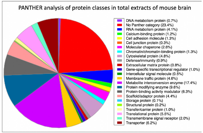
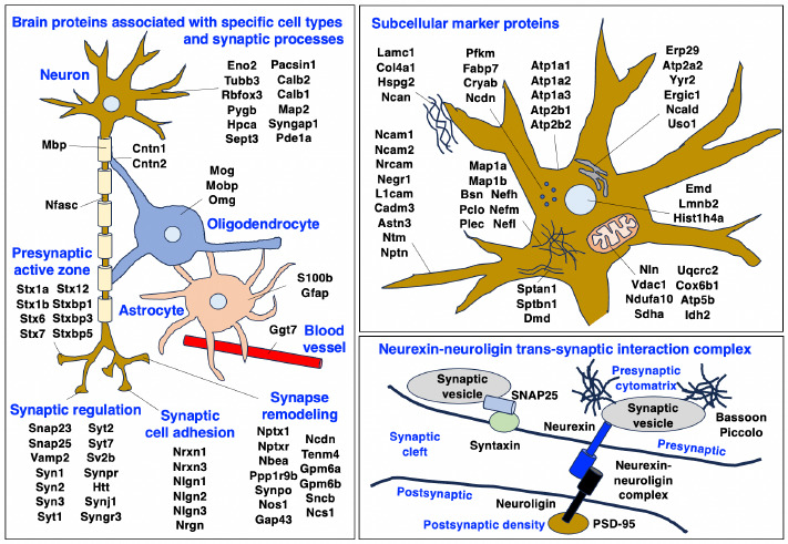
Neuromuscular diseases with primary muscle wasting symptoms may also display multi-systemic changes in the body and exhibit secondary pathophysiological alterations in various non-muscle tissues. In some cases, this includes proteome-wide alterations and/or adaptations in the central nervous system. Thus, in order to provide an improved bioanalytical basis for the comprehensive evaluation of animal models that are routinely used in muscle research, this report describes the mass spectrometry-based proteomic characterization of the mouse brain. Crude tissue extracts were examined by bottom-up proteomics and detected 4558 distinct protein species. The detailed analysis of the brain proteome revealed the presence of abundant cellular proteoforms in the neuronal cytoskeleton, as well as various brain region enriched proteins, including markers of the cerebral cortex, cerebellum, hippocampus and the olfactory bulb. Neuroproteomic markers of specific cell types in the brain were identified in association with various types of neurons and glia cells. Markers of subcellular structures were established for the plasmalemma, nucleus, endoplasmic reticulum, mitochondria and other crucial organelles, as well as synaptic components that are involved in presynaptic vesicle docking, neurotransmitter release and synapse remodelling.


Conflict of interest statement

We confirm that we have read the Journal’s position on issues involved in ethical publication and affirm that this report is consistent with those guidelines.

The mouse is one of the most widely used animal models in neuroscience, including studies of neuromuscular disorders such as muscular dystrophy, or motor neuron disease. Many genetic or acquired muscle diseases exhibit besides characteristic symptoms of fiber degeneration and/or contractile dysfunction also body-wide changes that are illustrated by pathophysiological alterations in non-muscular tissues. For example, the highly heterogeneous group of motor neuron diseases displays severe dysfunction of the motor system, but also a variety of extra-motor abnormalities. The most frequently observed adult-onset form of motor neuron disease is amyotrophic lateral sclerosis, which is characterized by progressive muscular weakness. Primary abnormalities in an extremely large number of genes have been associated with this disorder. Established animal models of amyotrophic lateral sclerosis include the SOD1 mouse and the wobbler mouse. In both patients and animal models of amyotrophic lateral sclerosis, abnormalities are observed in the flow of excitatory signals from neurons in the cortex, brain stem, spinal cord and the neuromuscular junction. Thus, in order to better understand the pathobiochemical mechanisms that underlie muscular atrophy due to the degeneration of lower and upper motor neurons, a comprehensive analysis of the peripheral and central nervous system is essential. Proteomics suggests itself as the methodological approach of choice for such a large-scale and high-throughput analysis due to its swift, unbiased and technology-driven nature. However, the mass spectrometric survey of the mouse model brain and its pathophysiological involvement in motor neuron disease is ideally based on previously established catalogues of the normal brain proteome. This should preferably include the biochemical detection and characterization of brain region enriched markers, cell type specific proteins and the identification of subcellular markers with a distinct function in key organelles of the diverse types of neurons and glial cells.

Another example of a neuromuscular disorder with complex body-wide alterations including abnormal brain functions is Duchenne muscular dystrophy (DMD). This X-linked inherited disorder can be clearly defined as a primary muscle wasting disorder but can also be categorized as a multi-systemic disease. Dystrophinopathies are due to mutations in the DMD gene that results in the almost complete loss of the membrane cytoskeletal protein dystrophin and a reduced expression of its associated glycoprotein complex in voluntary contractile fibers. This weakens the muscle fiber periphery and renders the sarcolemma more susceptible to contraction-induced rupturing of the surface membrane. Hence, the main pathophysiological hallmarks of X-linked muscular dystrophy are muscle membrane leakage and calcium-related myonecrosis, combined with fat substitution, reactive myofibrosis and chronic inflammation of skeletal muscles. Many of these symptoms are also seen in the mdx-type mouse models of dystrophinopathy. However, since the extremely large DMD gene has several promoter regions that produce 8 different tissue-specific dystrophin isoforms, mutation-specific alterations in combination with secondary changes can occur in many organ systems besides the skeletal musculature. This may include the liver, kidney, spleen, gastrointestinal tract and the nervous system. In this context, it is encouraging that the proteomic survey of the dystrophin-deficient brain of the mdx model of Duchenne muscular dystrophy has identified a large number of altered proteins involved in the maintenance of intermediate filaments, membrane repair mechanisms and calcium handling.

In order to establish the scientific basis for comparative proteomic surveys of the central nervous system in relation to mouse model research of neuromuscular diseases, this report outlines the mass spectrometry-based proteomic characterization of the normal middle-aged mouse brain. The proteomic data of markers of brain regions such as the olfactory bulb, hippocampus, cerebellum, hypothalamus and cerebral cortex, various brain cell types and subcellular structures found in neurons, synapsis and glia cells should be helpful as a reference guide for the comprehensive evaluation of the central nervous system in genetic mouse models that are routinely used in basic and applied myology research.

Figures

Fig 1.
Fig 1.
Bioinformatic analysis of the distribution of protein classes in total extracts of mouse brain as determined by mass spectrometric analysis. The pie chart was generated with the help of the PANTHER analysis tool.
Fig 2.
Fig 2.
Overview of the distribution of proteomic markers in specific cell types, synaptic processes and subcellular structures in the middle-aged mouse brain. The figure summarizes identified brain proteins that are specifically associated with neurons, neuron-glia contacts, myelin sheets, Nodes of Ranvier, oligodendrocytes, astrocytes, blood vessels, synaptic regulation, synaptic cell adhesion and synapse remodeling, as well as subcellular structures including the plasmalemma, mitochondria, endoplasmic reticulum, nucleus, cytosol, extracellular matrix and the neurexin-neuroligin synapse complex. The names of the abbreviated brain proteins are listed in Supplemental Tables 1-3.

Similar articles

Cited by

References

    1. Navabpour S, Kwapis JL, Jarome TJ. A neuroscientist's guide to transgenic mice and other genetic tools. Neurosci Biobehav Rev. 2020. Jan;108:732-748. doi: 10.1016/j.neubiorev.2019.12.013. Epub 2019 Dec 13. PMID: 31843544; PMCID: PMC8049509. doi: 10.1016/j.neubiorev.2019.12.013. - PMC - PubMed
    1. Partridge TA. The mdx mouse model as a surrogate for Duchenne muscular dystrophy. FEBS J. 2013. Sep;280(17):4177-86. doi: 10.1111/febs.12267. Epub 2013 Apr 22. PMID: 23551987; PMCID: PMC4147949. - PMC - PubMed
    1. Schmitt-John T. VPS54 and the wobbler mouse. Front Neurosci. 2015. Oct 21;9:381. doi: 10.3389/fnins.2015.00381. PMID: 26539077; PMCID: PMC4612502. - PMC - PubMed
    1. Brown RH, Al-Chalabi A. Amyotrophic Lateral Sclerosis. N Engl J Med. 2017. Jul 13;377(2):162-172. doi: 10.1056/NEJMra1603471. PMID: 28700839. - PubMed
    1. Akçimen F, Lopez ER, Landers JE, Nath A, Chiò A, Chia R, Traynor BJ. Amyotrophic lateral sclerosis: translating genetic discoveries into therapies. Nat Rev Genet. 2023. Apr 6. doi: 10.1038/s41576-023-00592-y. Epub ahead of print. PMID: 37024676. - PMC - PubMed