Review
. 2023 Jul 4;26(2):354.
doi: 10.3892/ol.2023.13940. eCollection 2023 Aug.

Intracorporeal linear‑stapled gastroduodenostomy in totally laparoscopic distal gastrectomy for gastric cancer: Consideration of the intraoperative management of the duodenal wall between the transecting staple line and anastomotic staple line (Review)

Affiliations
Review

Intracorporeal linear‑stapled gastroduodenostomy in totally laparoscopic distal gastrectomy for gastric cancer: Consideration of the intraoperative management of the duodenal wall between the transecting staple line and anastomotic staple line (Review)

Takaya Tokuhara et al. Oncol Lett. .

Abstract

The first part of the duodenum consists of the intraperitoneal segment, called the duodenal bulb, and the retroperitoneal segment. Regarding the blood supplying the duodenal bulb, which is the portion utilized in anastomosing the duodenum and remnant stomach following distal gastrectomy, the arterial pedicles branching off from the gastroduodenal artery are reported to reach the posterior wall first and then spread over the anterior wall, where they anastomose. When performing intracorporeal linear-stapled gastroduodenostomy following totally laparoscopic distal gastrectomy, the blood supply of the duodenal wall between the transecting staple line and anastomotic staple line needs to be considered because both transection of the duodenal bulb and the gastroduodenostomy are performed using an endoscopic linear stapler and the duodenal wall between the staple lines can be ischemic after the anastomosis. Since it needs to be decided intraoperatively whether this duodenal site is preserved or removed, the present review discusses the technical differences among several procedures for intracorporeal linear-stapled gastroduodenostomy, classifying them into two groups on the basis of the intraoperative management of this duodenal site. When this site is preserved, the blood supply of the duodenal wall needs to be retained with certainty. On the other hand, when this site is removed, the ischemic portion of the duodenal wall needs to be identified and removed. Furthermore, in both groups, an adequate anastomotic area needs to be secured. In conclusion, surgeons need to be familiar with the anatomical features of the duodenal bulb, including its blood perfusion and shape, when carrying out intracorporeal linear-stapled gastroduodenostomy.

Keywords: B-I reconstruction; TLDG; blood supply; intracorporeal linear-stapled gastroduodenostomy.


Conflict of interest statement

The authors declare that they have no competing interests.

Figures
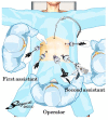
Figure 1.
Figure 1.
Port placements and positions of the operator and assistants. The operator inserted the stapler through the left lower port, and all staples, including transection of the duodenal bulb and gastroduodenostomy, were performed by the operator using their right hand. Photoshop2023 (version 24.5; Adobe Systems, Inc.) was used to generate this figure.
Figure 2.
Figure 2.
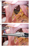
Transection of the duodenal bulb in a posteroanterior direction by the operator positioned between the legs of the patient. (A) The operator retracted the pyloric ring externally with the left hand (white arrow), while the first assistant elevated the posterior wall of the stomach ventrally and the second assistant elevated the liver cranially. (B) The operator transected the duodenal bulb in a posteroanterior direction with the right hand using one endoscopic linear stapler. Photoshop2023 (version 24.5; Adobe Systems, Inc.) was used to generate this figure.
Figure 3.
Figure 3.
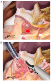
Creating the space required for the fork of the ELS around the cranial side of the duodenal wall. (A) The posterior wall of the duodenal bulb is the mesenteric side, in which the GDA (white arrow) is the marginal artery and the superior duodenal arteries branching from the GDA (red arrow) are the vasae rectae. (B) The operator dissected several cranial supraduodenal vessels branching off the GDA and created the space required for insertion of the ELS fork around the cranial wall of the duodenal bulb. Photoshop2023 (version 24.5; Adobe Systems, Inc.) was used to generate this figure. ELS, endoscopic linear stapler; GDA, gastroduodenal artery.
Figure 4.
Figure 4.
Linear stapling of the cranial wall of the duodenal bulb and the posterior wall of the remnant stomach. (A) The operator inserted the fork of the ELS into the duodenal bulb as far as possible with the right hand, while pulling the staple line of the duodenum externally with the left hand (white arrow). (B) The operator and first assistant changed the position of the duodenal bulb and remnant stomach without creating a gap. (C) The operator fired the ELS with the right hand so that the duodenal and gastric walls between the transecting staple lines and anastomotic staple became as wide as possible. Photoshop2023 (version 24.5; Adobe Systems, Inc.) was used to generate this figure. ELS, endoscopic linear stapler.
Figure 5.
Figure 5.
Modified delta-shaped gastroduodenostomy with linear stapling and single-layer hand suturing was completed. Photoshop2023 (version 24.5; Adobe Systems, Inc.) was used to generate this figure.

References

    1. Jayaraman MV, Mayo-Smith WW, Movson JS, Dupuy DE, Wallach MT. CT of the duodenum: An overlooked segment gets its due. Radiographics. 2001;21:S147–S160. doi: 10.1148/radiographics.21.suppl_1.g01oc01s147. - DOI - PubMed
    1. Reghunath A, Kabilan K, Mittal MK. Exploring the neglected segment of the intestine: The duodenum and its pathologies. Pol J Radiol. 2020;85:e230–e244. doi: 10.5114/pjr.2020.95477. - DOI - PMC - PubMed
    1. Terra C, Ramos-Andrade D, Sá-Marques I, Brito J, Caseiro-Alves F, Curvo-Semedo L. Duodenal imaging on the spotlight: From A to Z. Insights Imaging. 2021;12:94. doi: 10.1186/s13244-021-01045-y. - DOI - PMC - PubMed
    1. Hentati N, Fournier HD, Papon X, Aube C, Vialle R, Mercier P. Arterial supply of the duodenal bulb: An anatomoclinical study. Surg Radiol Anat. 1999;21:159–164. doi: 10.1007/BF01630893. - DOI - PubMed
    1. von Trotha KT, Butz N, Grommes J, Binnebösel M, Charalambakis N, Mühlenbruch G, Schumpelick V, Klinge U, Neumann UP, Prescher A, Krones CJ. Vascular anatomy of the small intestine-a comparative anatomic study on humans and pigs. Int J Colorectal Dis. 2015;30:683–690. doi: 10.1007/s00384-015-2163-4. - DOI - PubMed