Primary leiomyosarcoma of the uterine cervix: an unusual case and critical appraisal
- PMID: 37545785
- PMCID: PMC10401315
- DOI: 10.1093/jscr/rjad439
Primary leiomyosarcoma of the uterine cervix: an unusual case and critical appraisal
Abstract
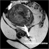
Leiomyosarcomas of the uterine cervix are rare, mostly occurring in perimenopausal women. Diagnosis is based on pathology and immunohistochemistry. Surgery with a total abdominal hysterectomy and bilateral salpingo-oophorectomy remains the standard. A female patient in her 60s presented with heavy postmenopausal bleeding. Vaginal ultrasound scan and magnetic resonance imaging showed a large strongly vascularized cervical mass with features suspicious of sarcomatous degeneration. Positron Emission Tomography-Computed Tomography (PET-CT) did not reveal any evidence of metastases nor lymphadenopathy, but presence of right hydronephrosis. An abdominal hysterectomy with bilateral salpingo-oophorectomy, and end-to-end anastomosis of the right ureter, was performed. Pathology showed an International Federation of Gynecology and Obstetrics (FIGO)-stage 1B leiomyosarcoma of the uterine cervix. No adjuvant treatment was given. Adjuvant radiotherapy reduces the risk of recurrence but no survival impact. The benefit of adjuvant chemotherapy is questionable given the lack of randomized trials. Multidisciplinary research concerning molecular alterations of the disease is required to determine optimal management strategies with potential novel molecular therapies.
Keywords: Cervix; cervical cancer; cervical sarcoma; leiomyosarcoma; sarcoma; uterine sarcoma; uterus.
Published by Oxford University Press and JSCR Publishing Ltd. © The Author(s) 2023.
Conflict of interest statement
None declared.
Figures


References
-
- Irvin W, Presley A, Andersen W, Taylor P, Rice L. Leiomyosarcoma of the cervix. Gynecol Oncol 2003;91:636–42. - PubMed
-
- Thomassin-Naggara I, Siles P, Balvay D, Cuenod CA, Carette MF, Bazot M. MR perfusion for pelvic female imaging. Diagn Interv Imaging 2013;94:1291–8. - PubMed
-
- Exacoustos C, Romanini ME, Amadio A, Amoroso C, Szabolcs B, Zupi E, et al. Can gray-scale and color Doppler sonography differentiate between uterine leiomyosarcoma and leiomyoma? J Clin Ultrasound 2007;35:449–57. - PubMed
Publication types
LinkOut - more resources
Full Text Sources

