[Preprint]. 2023 Jul 24:rs.3.rs-3097806.
doi: 10.21203/rs.3.rs-3097806/v1.

Native top-down proteomics reveals EGFR-ERα signaling crosstalk in breast cancer cells dissociates NUTF2 dimers to modulate ERα signaling and cell growth

Affiliations

Native top-down proteomics reveals EGFR-ERα signaling crosstalk in breast cancer cells dissociates NUTF2 dimers to modulate ERα signaling and cell growth

John Yates 3rd et al. Res Sq. .

Update in

Abstract

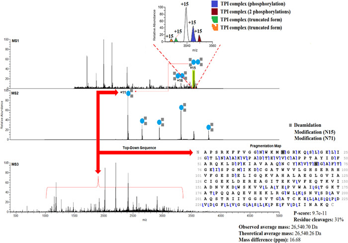
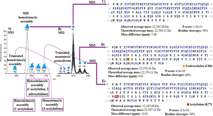
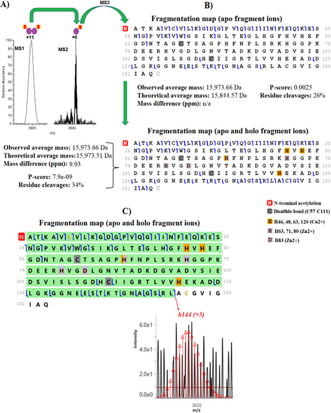
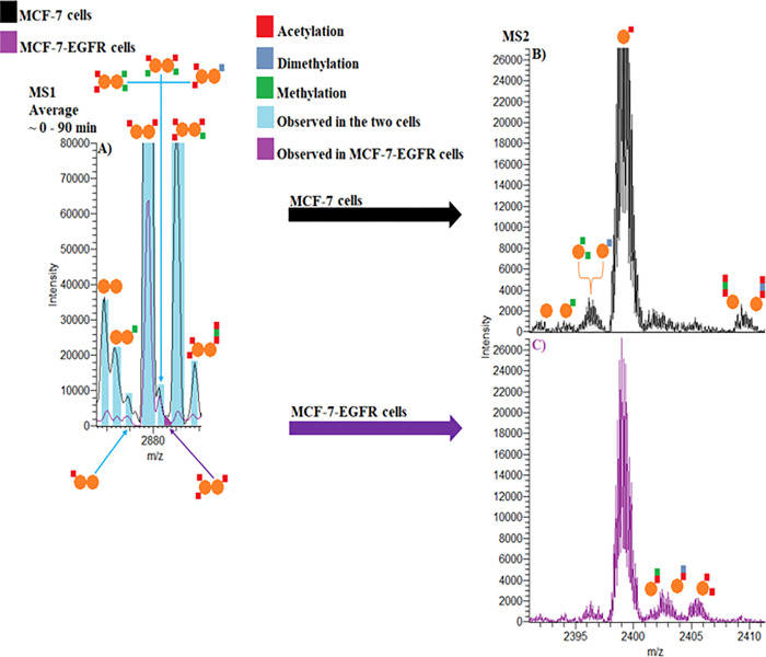
Oligomerization of proteins and their modified forms (proteoforms) produces functional protein complexes 1,2. Complexoforms are complexes that consist of the same set of proteins with different proteoforms 3. The ability to characterize these assemblies within cells is critical to understanding the molecular mechanisms involved in disease and to designing effective drugs. An outstanding biological question is how proteoforms drive function and oligomerization of complexoforms. However, tools to define endogenous proteoform-proteoform/ligand interactions are scarce 4. Here, we present a native top-down proteomics (nTDP) strategy that combines size-exclusion chromatography, nano liquid-chromatography in direct infusion mode, field asymmetric ion mobility spectrometry, and multistage mass spectrometry to identify protein assemblies (≤70 kDa) in breast cancer cells and in cells that overexpress EGFR, a resistance model of estrogen receptor-α (ER-α) targeted therapies. By identifying ~104 complexoforms from 17 protein complexes, our nTDP approach revealed several molecular features of the breast cancer proteome, including EGFR-induced dissociation of nuclear transport factor 2 (NUTF2) assemblies that modulate ER activity. Our findings show that the K4 and K55 posttranslational modification sites discovered with nTDP differentially impact the effects of NUTF2 on the inhibition of the ER signaling pathway. By characterizing endogenous proteoform-proteoform/ligand interactions, we reveal the molecular diversity of complexoforms, which allows us to propose a model for ER drug discovery in the context of designing effective inhibitors to selectively bind and disrupt the actions of targeted ER complexoforms.


Conflict of interest statement

Competing Interests: KRD and NLK are involved in the commercialization of top-down proteomics software including ProSight Native. The other authors have no competing interests to disclose.

Figures

Figure 1
Figure 1
nTDP spectra of TPI complexoforms (dimeric structures) in MCF-7 cells and the fragmentation map with 2 deamidations.
Figure 2
Figure 2
nTDP spectra of the MIF complexoforms (trimeric structures) in MCF-7 cells and its fragmentation maps (unmodified map, nitrosylated map, and acetylated map).
Figure 3
Figure 3. nTDP spectra of the complexoform SOD1 (homodimeric structure) in MCF-7 cells.
(A) MS1 and MS2 spectra of SOD1. (B) Fragmentation maps of SOD1 with apo and holo/apo fragment ions that were generated using the ProSight Lite software. (C) Fragmentation map of SOD1 monomeric proteoform covering regions of the protein where the metal ions are bound and isotopic distribution of the diagnostic ion b144 that were generated using TDValidator software.
Figure 4
Figure 4. nTDP spectra of NUTF2 in MCF-7 and MCF-7-EGFR cells.
(A) Representative native MS spectrum of NUTF2 in MCF-7 (black) and MCF-7-EGFR (purple) cells. (B) MS2 spectrum of NUTF2 monomeric proteoforms in MCF-7 cells. (C) MS2 spectrum of NUTF2 monomeric proteoforms in MCF-7-EGFR cells.
Figure 5
Figure 5
NUTF2 Modulates ERα signaling. (A–C) Crystal structure of one subunit of the NUTF2 dimer interacting with RanGDP and a model of the FxFG nucleoporin peptide, based on PDBs 1GYB and 5BXQ. (D) NUTF2 inhibits ERα-mediated transcription. 3xERE-Luc reporter activity in MCF-7 cells co-transfected with ERα, and empty or NUTF2 expression vectors, and treated in complete medium with vehicle or 1 μM 4OHT for 24 h. N = 7. (E-H) use same legend as D. (E) NUTF2 promotes DNA binding by ERα. 3xERE-Luc reporter activity in HEK293T cells co-transfected with ERα-VP16 fusion protein, and empty vector or NUTF2 expression vectors, and treated in complete medium with vehicle or 1 μM 4OHT for 24 h. N = 5. (F) NUTF2 suppresses ERα-mediated cell proliferation. Stable MCF-7 cells were cultured in complete medium supplemented with vehicle or 1 μM 4OHT for 5 days. N = 4. (G) NUTF2 downregulates GREB1. Stable MCF-7 cells in complete medium were treated for 24 h with vehicle or 1 μM 4OHT. GREB1 mRNA levels were compared by qPCR. N = 2.(H) NUTF2 occupies an ERα-binding site in the GREB1 promoter. Stable MCF-7 cells treated for 1 h with vehicle or 1 μM 4OH, were compared by qChIP assay using ERα, HA-tag and Ran antibodies. N = 3. (I) Confocal microscopy of NUTF2 expressing MCF-7 cells. Cells were fixed, permeabilized and stained with DAPI or antibodies against the HA-tagged NUTF2 plasmid or ERa. See also Figure S14. (J) Volcano plot of RNA-seq data showing the effect of NUTF2 overexpression on gene expression in MCF-7 cells, n = 3. (K) RNA-seq data showing expression profiles of select growth regulatory genes. Mean + SEM, n = 3. (L) Potential upstream regulators of NUTF2 target genes were identified based on published ChIP-seq data sets using LISA (http://www.lisa.cistrome.org).

References

    1. Li H., Nguyen H. H., Ogorzalek Loo R. R., Campuzano I. D. G. & Loo J. A. An integrated native mass spectrometry and top-down proteomics method that connects sequence to structure and function of macromolecular complexes. Nature chemistry 10, 139–148, doi: 10.1038/nchem.2908 (2018). - DOI - PMC - PubMed
    1. Smith L. M. & Kelleher N. L. Proteoform: a single term describing protein complexity. Nature methods 10, 186–187, doi: 10.1038/nmeth.2369 (2013). - DOI - PMC - PubMed
    1. Jensen M. H., Morris E. J., Tran H., Nash M. A. & Tan C. Stochastic ordering of complexoform protein assembly by genetic circuits. PLoS Comput Biol 16, e1007997, doi: 10.1371/journal.pcbi.1007997 (2020). - DOI - PMC - PubMed
    1. Rogawski R. & Sharon M. Characterizing Endogenous Protein Complexes with Biological Mass Spectrometry. Chemical Reviews 122, 7386–7414, doi: 10.1021/acs.chemrev.1c00217 (2022). - DOI - PMC - PubMed
    1. Ryan C. J., Kennedy S., Bajrami I., Matallanas D. & Lord C. J. A Compendium of Co-regulated Protein Complexes in Breast Cancer Reveals Collateral Loss Events. Cell Systems 5, 399–409.e395, doi: 10.1016/j.cels.2017.09.011 (2017). - DOI - PMC - PubMed

Publication types