[Preprint]. 2023 Jul 28:2023.07.28.550765.
doi: 10.1101/2023.07.28.550765.

Prolonged exposure to lung-derived cytokines is associated with inflammatory activation of microglia in patients with COVID-19

Affiliations

Prolonged exposure to lung-derived cytokines is associated with inflammatory activation of microglia in patients with COVID-19

Rogan A Grant et al. bioRxiv. .

Update in

Abstract

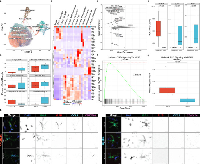
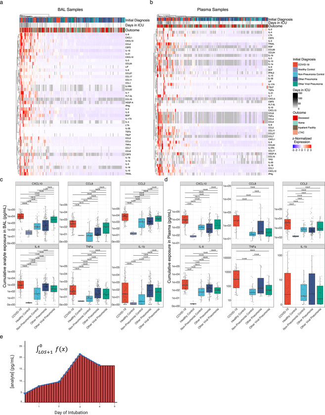
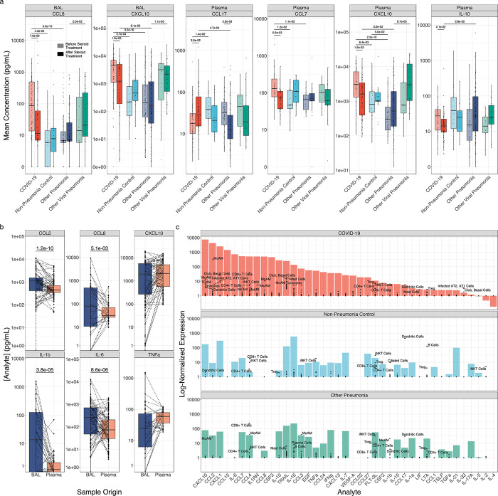
Neurological impairment is the most common finding in patients with post-acute sequelae of COVID-19. Furthermore, survivors of pneumonia from any cause have an elevated risk of dementia1-4. Dysfunction in microglia, the primary immune cell in the brain, has been linked to cognitive impairment in murine models of dementia and in humans5. Here, we report a transcriptional response in human microglia collected from patients who died following COVID-19 suggestive of their activation by TNF-α and other circulating pro-inflammatory cytokines. Consistent with these findings, the levels of 55 alveolar and plasma cytokines were elevated in a cohort of 341 patients with respiratory failure, including 93 unvaccinated patients with COVID-19 and 203 patients with other causes of pneumonia. While peak levels of pro-inflammatory cytokines were similar in patients with pneumonia irrespective of etiology, cumulative cytokine exposure was higher in patients with COVID-19. Corticosteroid treatment, which has been shown to be beneficial in patients with COVID-196, was associated with lower levels of CXCL10, CCL8, and CCL2-molecules that sustain inflammatory circuits between alveolar macrophages harboring SARS-CoV-2 and activated T cells7. These findings suggest that corticosteroids may break this cycle and decrease systemic exposure to lung-derived cytokines and inflammatory activation of microglia in patients with COVID-19.


Conflict of interest statement

Conflict of Interest Statement B.D.S. holds US patent 10,905,706, “Compositions and methods to accelerate resolution of acute lung inflammation,” and serves on the scientific advisory board of Zoe Biosciences, in which he holds stock options. The other authors declare no conflicts of interest.

Figures

Figure 1.
Figure 1.. Microglia exhibit distinct transcriptional responses in patients with COVID-19.
(a) UMAP of 65,767 cells isolated from the frontal lobes of 8 patients postmortem. (b) Relative abundance of microglial cell states as a percentage of total microglia. No significant differences are observed by diagnosis (q ≤ 0.05, Mann-Whitney). (c) Hierarchical clustering of mean marker gene expression by cell type and cell state by diagnosis. (d) MA plot of differentially expressed genes in total microglia in COVID-19 vs controls by pseudo-bulk differential expression analysis. Significantly upregulated genes are shown in red, and significantly downregulated genes are shown in blue (q < 0.05, Wald test). Genes shown in gray are not significantly differentially expressed. (e) Callouts of key markers of cell division and cell-cycle arrest from 1d. All genes shown are significantly differentially expressed (q < 0.05, Wald test). (f) Gene-set enrichment of Hallmark TNF-α Signaling Via NF-κB (M5890) from pseudo-bulk differential expression analysis (q = 5.96×10⁻¹⁶, multilevel splitting Monte Carlo). (g) Median modular expression of Hallmark TNF-α Signaling Via NF-κB (M5890) by diagnosis. Points represent median expression in total microglia from each patient (q = 2.6×10⁻², Mann-Whitney). (h) Representative images of combined immunofluorescence and smFISH (RNAScope) from human frontal lobe tissue sections showing cell cycle-arrested, pro-inflammatory microglia in patients with COVID-19 relative to controls. Images are pseudo-colored by channel as follows. DAPI: blue, IBA1: green, IL1B: red, CCL2: cyan, CDKN1A: magenta.
Figure 2.
Figure 2.. COVID-19 is distinguished from pneumonias of similar severity by expression of T cell chemokines.
(a) Hierarchical clustering of 41 cytokines showing significant variability by diagnosis (q < 0.05, Kruskal–Wallis) from 187 BAL samples collected in the first 48 hours of intubation from 183 patients with an early BAL. (b) Hierarchical clustering of 25 cytokines showing significant variability by diagnosis (q < 0.05, Kruskal–Wallis) from 137 early plasma samples from 134 patients. (c) Expression of COVID-19-defining T lymphocyte and monocyte chemokines and key pro-inflammatory cytokines from 479 BAL samples collected throughout the duration of mechanical ventilation from 332 patients. (d) Expression of COVID-19-defining T lymphocyte and monocyte chemokines and key pro-inflammatory cytokines from 396 plasma samples collected throughout the duration of mechanical ventilation from 262 patients.
Figure 3.
Figure 3.. Cumulative but not peak exposure to pro-inflammatory cytokines is higher in patients with severe SARS-CoV-2 pneumonia compared to patients with severe pneumonia secondary to other pathogens.
(a) Hierarchical clustering of cumulative exposure to 44 BAL cytokines showing significant variability by diagnosis (q < 0.05, Kruskal–Wallis) from 327 patients estimated by geometric integration of the levels of 479 BAL samples collected throughout the duration of mechanical ventilation. (b) Hierarchical clustering of cumulative exposure to 51 plasma cytokines showing significant variability by diagnosis (q < 0.05, Kruskal–Wallis) from 258 patients estimated by geometric integration of the levels of 396 plasma samples collected throughout the duration of mechanical ventilation. (c) Cumulative expression of selected pro-inflammatory cytokines in BAL fluid from (a). (d) Cumulative expression of selected pro-inflammatory cytokines in plasma from (b). (e) Schematic for calculation of cumulative exposure for each cytokine assayed for each patient throughout the course of mechanical ventilation by geometric integration. BAL samples from 3 patients and plasma samples from 2 patients receiving long-term mechanical ventilation were excluded from these analyses.
Figure 4.
Figure 4.. Corticosteroid treatment is associated with reductions in T cell chemokine expression in monocyte-derived alveolar macrophages.
(a) Boxplots of cytokine expression for all BAL fluid and plasma cytokines exhibiting significantly altered expression (q < 0.05, Mann-Whitney) following corticosteroid treatment. Lightly shaded boxes represent cytokine expression values prior to corticosteroid treatment, and darkly shaded boxes represent expression values after corticosteroid treatment. (b) Paired comparisons of cytokine expression in BAL and plasma for all paired samples (paired Mann-Whitney). (c) Deconvolution of “bulk” cytokine expression in BAL fluid by scRNA-seq of cells isolated from BAL fluid. Mean cytokine gene expression for each cell type detected in scRNA-seq data from (black points) is overlaid on bulk cytokine expression by multiplexed cytokine array (filled bars) to identify cell type contributors to cytokine expression.

References

    1. Davydow D. S., Hough C. L., Levine D. A., Langa K. M. & Iwashyna T. J. Functional Disability, Cognitive Impairment, and Depression Following Hospitalization for Pneumonia. Am J Med 126, 615–624.e5 (2013). - PMC - PubMed
    1. Shah F. A. et al. Bidirectional Relationship between Cognitive Function and Pneumonia. Am J Respir Crit Care Med 188, 586–592 (2013). - PMC - PubMed
    1. Tate J. A. et al. Infection hospitalization increases risk of dementia in the elderly. Crit Care Med 42, 1037–1046 (2014). - PMC - PubMed
    1. Taquet M. et al. Neurological and psychiatric risk trajectories after SARS-CoV-2 infection: an analysis of 2-year retrospective cohort studies including 1 284 437 patients. The Lancet Psychiatry 9, 815–827 (2022). - PMC - PubMed
    1. Paolicelli R. C. et al. Microglia states and nomenclature: A field at its crossroads. Neuron 110, 3458–3483 (2022). - PMC - PubMed

Publication types