[Preprint]. 2023 Jul 26:2023.07.25.550509.
doi: 10.1101/2023.07.25.550509.

Pathogenic CANVAS (AAGGG)n repeats stall DNA replication due to the formation of alternative DNA structures

Affiliations

Pathogenic CANVAS (AAGGG)n repeats stall DNA replication due to the formation of alternative DNA structures

Julia A Hisey et al. bioRxiv. .

Update in

Abstract

CANVAS is a recently characterized repeat expansion disease, most commonly caused by homozygous expansions of an intronic (A2G3)n repeat in the RFC1 gene. There are a multitude of repeat motifs found in the human population at this locus, some of which are pathogenic and others benign. In this study, we conducted structure-functional analyses of the main pathogenic (A2G3)n and the main nonpathogenic (A4G)n repeats. We found that the pathogenic, but not the nonpathogenic, repeat presents a potent, orientation-dependent impediment to DNA polymerization in vitro. The pattern of the polymerization blockage is consistent with triplex or quadruplex formation in the presence of magnesium or potassium ions, respectively. Chemical probing of both repeats in supercoiled DNA reveals triplex H-DNA formation by the pathogenic repeat. Consistently, bioinformatic analysis of the S1-END-seq data from human cell lines shows preferential H-DNA formation genome-wide by (A2G3)n motifs over (A4G)n motifs in vivo. Finally, the pathogenic, but not the non-pathogenic, repeat stalls replication fork progression in yeast and human cells. We hypothesize that CANVAS-causing (A2G3)n repeat represents a challenge to genome stability by folding into alternative DNA structures that stall DNA replication.


Figures

Figure 1.
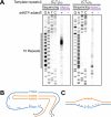
Figure 1.. In vitro polymerization through pathogenic and nonpathogenic repeats.
(A) Polyacrylamide gel electrophoresis separation of ThermoSequenase sequencing reactions performed as described in Materials and Methods. Briefly, 5µg of each plasmid and 0.5pmol of primer pre-annealed and the USBio Thermo Sequenase Cycle Sequencing Kit’s 3’-dNTP internal label cycling sequencing instructions were followed with several modifications detailed in Materials and Methods. (B) Schematic of denatured double-stranded plasmid with primers annealed to allow for ThermoSequenase polymerization through the purine- or pyrimidine-rich strand of (A2G3)10 or (A4G)10 in the template strand. Primers are 98 base pairs or 75 base pairs away from repeats for the purine-rich or pyrimidine-rich template, respectively. Created with BioRender. (C) Model for triplex formation as polymerase progresses through the repeats with the purine-rich strand as the template. Created with BioRender. (D) Polyacrylamide gel electrophoresis separation of in vitro T7 DNA polymerase primer extension reaction. 3.125µM single-stranded oligonucleotides bearing either (A2G3)10 (JH267), (T2C3)10 (JH268), or (A4G)10 (JH269) repeats were pre-annealed with a 2.5 µM 5’-32P labeled primer (JH270) in 10 mM Tris-HCl, pH 7.5, 0.1 mM EDTA by incubation at 95°C for 5 minutes, followed by gradual return to room temperature. 50mM K+ and 10mM Mg2+ were added to the annealing buffer in the left panel. Primer extension reactions consisted of 10nM primer/template and 1µM T7 DNA polymerase in 40 mM Tris-HCl, pH 7.5, 5mM dithiothreitol (DTT), and 55mM K+ (left panel). Primer extension was initiated by addition of 10mM MgCl2, followed by incubation at room temperature for 1 minute, and quenching with formamide/EDTA loading dye. Primer extension products were resolved by electrophoresis on a denaturing 10% polyacrylamide gel. NT=no template (primer added without oligonucleotide template). (E) The same general protocol was followed as in (D), with the following changes: 50mM LiCl, KCl, or NaCl were added to the annealing buffer and 55 mM of the indicated monovalent metal chloride salt was added to the primer extension buffer. Sequencing alongside primer extension reactions were conducted with the templates and primers used for the primer extension reactions and followed the protocol detailed in the Materials and Methods for the sequencing reactions in Figure 1A. NR=no reaction (no addition of MgCl2). (F) Model for G-quadruplex formation as T7 DNA polymerase progresses along the purine-rich template in the presence of K+ in the annealing and primer extension buffer.
Figure 2.
Figure 2.. Potassium permanganate probing of pathogenic and nonpathogenic repeats.
(A) Polyacrylamide gel electrophoresis separation of sequencing reactions and primer extension reactions on potassium permanganate or water treated repeat-containing plasmids using the pyrimidine-rich template. Repeat-containing supercoiled DNA was incubated with potassium permanganate or water, the DNA was precipitated, and used for a primer extension reaction as described in Materials and Methods. (B) H-r3 triplex predicted from chemical probing for (A2G3)10 repeats. (C) DNA unwinding element (DUE) predicted from chemical probing for (A4G)10 repeats. Purple stars in (B) and (C) represent possible KMnO4 modification sites.
Figure 3.
Figure 3.. Bioinformatic analysis of S1-END-seq peaks to determine the triplex-forming potential of various repeats using data from .
For each graph: Top: Graph depicting the percentage of repeats found within 100 nucleotides of S1-END-seq peaks as the repeat length increases. Bottom: Graph depicting the number of repeats as the repeat length increases. (A) Comparison of pathogenic (A2G3)n and (A4G)n motifs genome-wide. (B) Comparison of pentanucleotide motifs with increasing guanine:adenine ratio genome-wide.
Figure 4.
Figure 4.. Analysis of yeast replication intermediates using two-dimensional gel electrophoresis.
(A) Representative gels of the no repeat control, (A2G3)60, (C3T2)60, and (A4G)60 in the lagging strand template of replication in yeast from the yeast 2µ origin of replication. The red arrow indicates replication fork stalling. (B) Densitometry profiles along the arc starting at the 1.5n spot to the 2n spot. These profiles were used for quantification, which was determined as described in ,. (C) Quantification of replication fork slowing via area analysis with the fold change increased normalized to the no repeat control arc. Error bars represent standard error of the mean and non-overlapping error bars were used to determine significance. Created with BioRender and prism.
Figure 5.
Figure 5.. Analysis of human cell replication intermediates using two-dimensional gel electrophoresis.
(A) Representative gels of the no repeat control, (A2G3)60, (C3T2)60, and (A4G)60 in the lagging strand template of replication from the SV40 origin of replication. The red arrow indicates replication fork stalling. (B) Densitometry profiles along the arc starting at the 1n spot to the 1.5n spot. These profiles were used for quantification, which was determined as described in ,. (C) Quantification of replication fork slowing via area analysis with the fold change increased normalized to the no repeat control arc. Error bars represent standard error of the mean and non-overlapping error bars were used to determine significance. Created with BioRender and prism.

References

    1. Cortese A. et al. Biallelic expansion of an intronic repeat in RFC1 is a common cause of late-onset ataxia. Nat Genet 51, 649–658 (2019). - PMC - PubMed
    1. Rafehi H. et al. Bioinformatics-Based Identification of Expanded Repeats: A Non-reference Intronic Pentamer Expansion in RFC1 Causes CANVAS. Am J Hum Genet 105, 151–165 (2019). - PMC - PubMed
    1. Arteche-López A. et al. New Cerebellar Ataxia, Neuropathy, Vestibular Areflexia Syndrome cases are caused by the presence of a nonsense variant in compound heterozygosity with the pathogenic repeat expansion in the RFC1 gene. Clin Genet 103, 236–241 (2023). - PubMed
    1. Cortese A. et al. Cerebellar ataxia, neuropathy and vestibular areflexia syndrome (CANVAS): genetic and clinical aspects. Pract Neurol 22, 14–18 (2022). - PubMed
    1. Cortese A. et al. Cerebellar ataxia, neuropathy, vestibular areflexia syndrome due to RFC1 repeat expansion. Brain 143, 480–490 (2020). - PMC - PubMed

Publication types