

This is a preprint.
Addressing persistent challenges in digital image analysis of cancerous tissues
- PMID: 37547011
- PMCID: PMC10401923
- DOI: 10.1101/2023.07.21.548450
Addressing persistent challenges in digital image analysis of cancerous tissues
Update in
-
Addressing persistent challenges in digital image analysis of cancer tissue: resources developed from a hackathon.Mol Oncol. 2025 Jun;19(6):1565-1581. doi: 10.1002/1878-0261.13783. Epub 2025 Feb 10. Mol Oncol. 2025. PMID: 39927650 Free PMC article. Review.
Abstract
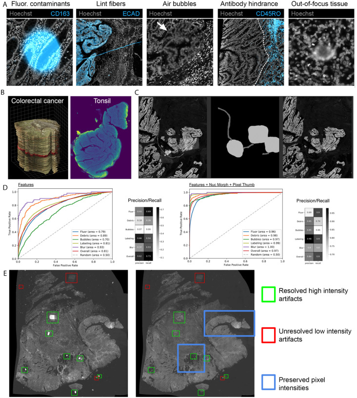
The National Cancer Institute (NCI) supports many research programs and consortia, many of which use imaging as a major modality for characterizing cancerous tissue. A trans-consortia Image Analysis Working Group (IAWG) was established in 2019 with a mission to disseminate imaging-related work and foster collaborations. In 2022, the IAWG held a virtual hackathon focused on addressing challenges of analyzing high dimensional datasets from fixed cancerous tissues. Standard image processing techniques have automated feature extraction, but the next generation of imaging data requires more advanced methods to fully utilize the available information. In this perspective, we discuss current limitations of the automated analysis of multiplexed tissue images, the first steps toward deeper understanding of these limitations, what possible solutions have been developed, any new or refined approaches that were developed during the Image Analysis Hackathon 2022, and where further effort is required. The outstanding problems addressed in the hackathon fell into three main themes: 1) challenges to cell type classification and assessment, 2) translation and visual representation of spatial aspects of high dimensional data, and 3) scaling digital image analyses to large (multi-TB) datasets. We describe the rationale for each specific challenge and the progress made toward addressing it during the hackathon. We also suggest areas that would benefit from more focus and offer insight into broader challenges that the community will need to address as new technologies are developed and integrated into the broad range of image-based modalities and analytical resources already in use within the cancer research community.
Keywords: Multiplexed images; artifact removal; cancer; domain representation; image analysis; scalability; thumbnail generation.
Figures


References
-
- Hajdu SI. A note from history: landmarks in history of cancer, part 4. Cancer. 2012;118(20):4914–4928. - PubMed
-
- Smith JM, Conroy RM. The NIH common fund Human Biomolecular Atlas Program (HuBMAP): Building a framework for mapping the human body. The FASEB Journal. 2018;32:818–2.
Publication types
Grants and funding
LinkOut - more resources
Full Text Sources