. 2023 Jul 26;5(3):100387.
doi: 10.1016/j.ocarto.2023.100387. eCollection 2023 Sep.

Anabolic phenotype in cartilage-specific mitogen-inducible gene-6 knockout mice is independent of transforming growth factor-α

Affiliations

Anabolic phenotype in cartilage-specific mitogen-inducible gene-6 knockout mice is independent of transforming growth factor-α

Ermina Hadzic et al. Osteoarthr Cartil Open. .

Abstract

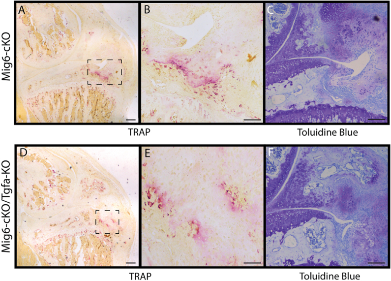
Background/objective: Osteoarthritis (OA) is a whole joint disorder with no disease modifying treatment currently available. The Epidermal Growth Factor Receptor (EGFR) signaling pathway plays an important role in cartilage/bone development and its ligand transforming growth factor-α (TGFα) is upregulated in OA. In contrast, Mitogen-inducible gene 6 (Mig6) is a negative regulator of EGFR, and cartilage-specific Mig-6 deletion results in anabolic effects on cartilage and formation of chondro-osseus nodules (CON). We aimed to attenuate EGFR signaling by inhibiting TGFα production in cartilage-specific Mig6 deficient mice, to test whether this would prevent the formation of CONs.

Methods: We generated double knockout mice by crossing cartilage-specific Mig-6fl/flCol2a1-Cre+/- and whole-body Tgfa± mice to generate experimental and control wild-type mice. Knee and elbow sections were used to examine articular cartilage thickness, cell density, and osteoclast presence. Additionally, immunohistochemistry was completed to analyze phospho-EGFR and SOX9.

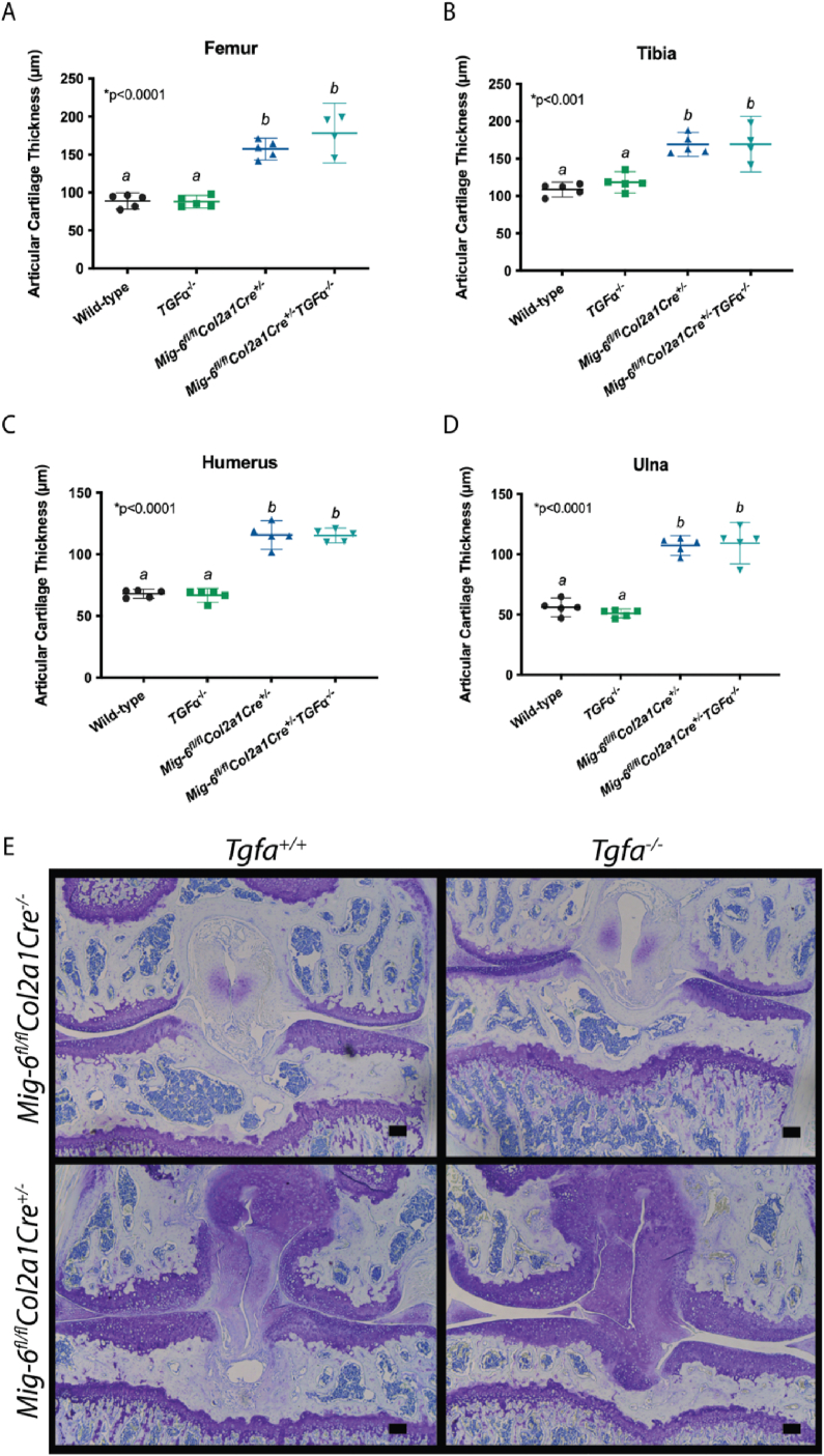
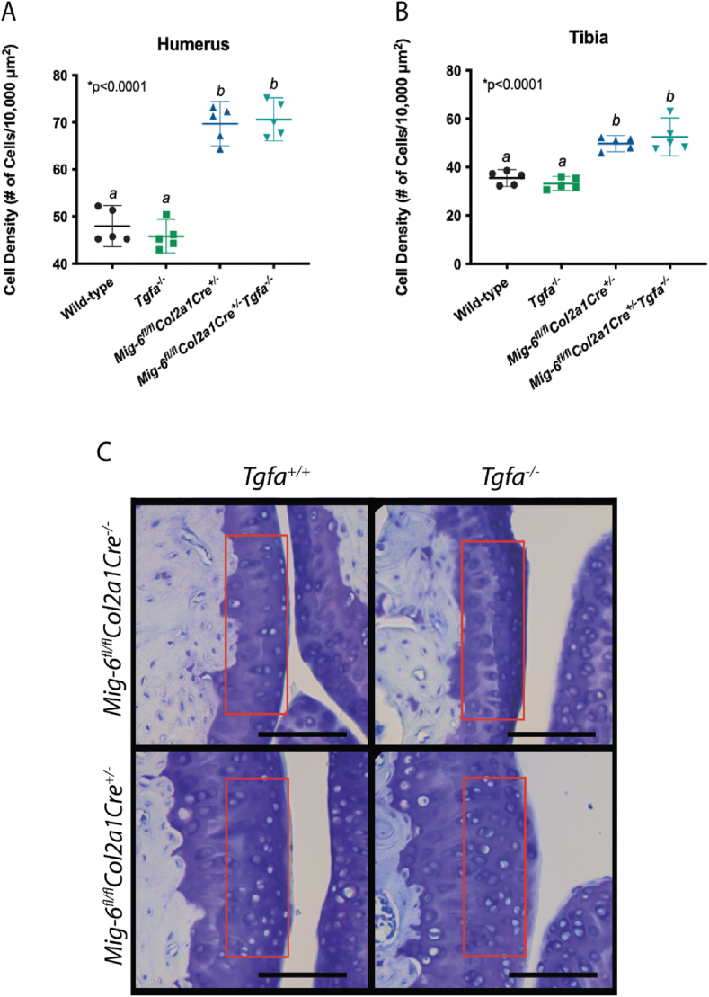
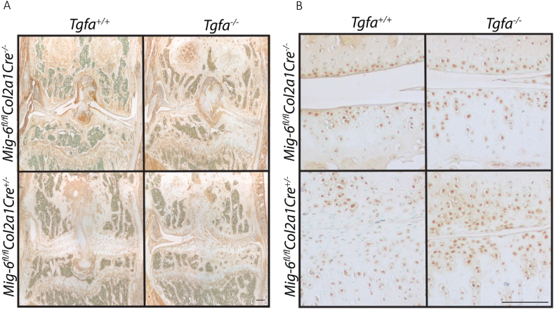
Results: Mig-6 deficient mice display cartilage thickening and CONs at 12 weeks in both the elbow and knee joints, which is independent of TGFα ligand presence. Similarly, articular cartilage cell density is increased in Mig6-cKO/Tgfa-KO and Mig6-cKOmice, but not Tgfa-KO mice, and displays increased SOX9 and phospho-EGFR staining.

Conclusion: The articular cartilage displays increased thickness/cell density and CON formation independent of the presence of TGFα, suggesting the anabolic phenotype in the Mig6-deficient mice is independent of TGFα/EGFR binding. The anabolic phenotype may be due to an alternative EGFR ligand activation, or other non-EGFR specific mechanism. More research is required to elucidate the exact pathway responsible for the anabolic effects.

Keywords: Animal model; Chondrocyte; Epidermal growth factor receptor; Mitogen inducible gene 6; Transforming growth factor-alpha; Transgenic mice.


Conflict of interest statement

The authors declare no conflict of interest.

Figures

Fig. 1
Fig. 1
Phospho-EGFR activity is upregulated in Mig6 deficient mice. Brown staining indicates positive phospho-EGFR staining, which appears more intense and present in more chondrocytes in Mig6 deficient mice, with opposite effects in Tgfa-KO mice. The Mig6-cKO/Tgfa-KO mice seem to have a similar amount of staining to the Mig6-cKO mice. Images represent coronal knee sections, counterstained in methyl green at (A) 4x and (B) 20x. Scale bars ​= ​100 ​μm. N ​= ​5.
Fig. 2
Fig. 2
Mig6 deficient mice display articular cartilage thickening at 12 weeks, regardless of TGFα. Average articular cartilage thickness was calculated using three measurements at different locations on the articular cartilage using ImageJ software on coronal sections. Average articular cartilage thickness was significantly increased in Mig6 deficient mice, regardless of TGFα presence in the (A, E) femur, (B, E) tibia, (C) humerus, and (D) ulna. Graphed data represents a non-parametric one-way ANOVA with Tukey's multiple comparison test, where the individual mean and 95% CI is presented, p values are presented on each graph. (E) Representative images of toluidine blue stained knee sections. Scale bars ​= ​100 ​μm. N ​= ​5.
Fig. 3
Fig. 3
Mig6 deficient mice have significantly more uncalcified cartilage. Average uncalcified and calcified cartilage thickness was calculated on Osteomeasure across three sections, with the medial femoral condyle results presented. (A) The uncalcified cartilage thickness was significantly greater in the Mig6-cKO and Mig6-cKO/Tgfa-KO compared to wild-type and Tgfa-KO, and (C) made up a greater percentage of total cartilage. (B) The thickness of the calcified cartilage was not significantly different between groups, but when looking at the percentage of total calcified cartilage in (D), the Mig6-cKO and Mig6-cKO/Tgfa-KO mice are significantly decreased compared to the wild type and Tgfa-KO mice. The breakdown of uncalcified vs. calcified cartilage can be seen in (F). Subchondral plate thickness was measured (E), with the Mig6-cKO and Mig6-cKO/Tgfa-KO groups demonstrating significantly less thickness compared to the wild-type and Tgfa-KO groups. Graphed data represents a one-way ANOVA with Tukey's multiple comparison test, where the individual mean and 95% CI is presented, p values are presented on each graph. N ​= ​5.
Fig. 4
Fig. 4
Cell density is not affected by TGFα deficiency. Cell density was calculated within the articular cartilage of toluidine blue stained elbow and knee joints. Lacunae with nuclear staining was counted within a 70 ​μm ​× ​200 ​μm region, indicated by the red box (C), representative images of the sagittal elbow is presented. Scale bars ​= ​100 ​μm. Cell density is significantly increased in Mig6 deficient mice, regardless of TGFα presence in the (A) humerus and (B) tibia. Graphed data represents a non-parametric one-way ANOVA with Tukey's multiple comparison test, where the individual mean and 95% CI is presented, p values are presented on each graph. N ​= ​5.
Fig. 5
Fig. 5
SOX9 staining is increased in Mig6 deficient groups. Brown staining indicates positive SRY-box transcription factor 9 (SOX9) nuclear staining, which is present in all groups. The Mig6-cKO/Tgfa-KO mice and Mig6-cKO mice appear to have more positively stained chondrocytes than the Tgfa-KO and wild-type mice. All sections are of the coronal knee, counterstained with methyl green at (A) 4x and (B) 20x. Scale bars ​= ​100 ​μm. N ​= ​5.
Fig. 6
Fig. 6
CONs develop in Mig6 deficient mice independent of TGFα ligand presence. Toluidine blue stained sagittal knee sections of 12-week-old mice demonstrate CON development in Mig6 deficient mice, with or without TGFα. CON development does not appear in wild type or Tgfa-KO mice. Images are representative of N ​= ​2 for the Mig6-cKO/Tgfa-KO mice and N ​= ​3 for the remaining groups. All sections are of the sagittal knee at 10x magnification. Scale bar ​= ​1000 ​μm.
Fig. 7
Fig. 7
TRAP staining is increased within CONs. TRAP staining reveals an increase in osteoclasts within CONs, which appears similar between Mig6-cKO and Mig6-cKO/Tgfa-KO mice. Images (A, D) represent TRAP staining of sagittal knee sections at 4x magnification and (B, E) 20x magnification. (C, F) There is also an increase in purple proteoglycan staining in sagittal knee sections within CONs. Images (B) and (E) are magnified from the dotted box within images (A) and (D), respectively. (A, D) Scale bars ​= ​200 ​μm and (B, C, E, F) scale bars ​= ​100 ​μm.

References

    1. Osteoarthritis cartilage, standardization of osteoarthritis definitions. 2015. https://oarsi.org/research/standardization-osteoarthritis-definitions
    1. Badley E.M., Wilfong J.M., Zahid S., Perruccio A. Arthritis community research and evaluation unit. The status of arthritis in Canada: national report. Arthritis Society. 2019:1–34.
    1. Bannuru R.R., Osani M.C., Vaysbrot E.E., Arden N.K., Bennel K., Bierma-Zeinstra S.M.A., et al. OARSI guidelines for the non-surgical management of knee, hip, and polyarticular osteoarthritis. Osteoarthritis Cartilage. 2019;27(11):1578–1589. doi: 10.1016/j.joca.2019.06.011. - DOI - PubMed
    1. Citri A., Yarden Y. EGF-ERBB signalling: towards the systems level. Nat. Rev. Mol. Cell Biol. 2006;7:505–516. doi: 10.1038/nrm1962. - DOI - PubMed
    1. Olayioye M.A., Neve R.M., Lane H.A., Hynes N.E. The ErbB signaling network: receptor heterodimerization in development and cancer. EMBO J. 2000;19(13):3159–3167. doi: 10.1093/emboj/19.13.3159. - DOI - PMC - PubMed