. 2023 Jul 17:33:404-412.
doi: 10.1016/j.omtn.2023.07.011. eCollection 2023 Sep 12.

Branchpoints as potential targets of exon-skipping therapies for genetic disorders

Affiliations

Branchpoints as potential targets of exon-skipping therapies for genetic disorders

Hiroaki Ohara et al. Mol Ther Nucleic Acids. .

Abstract

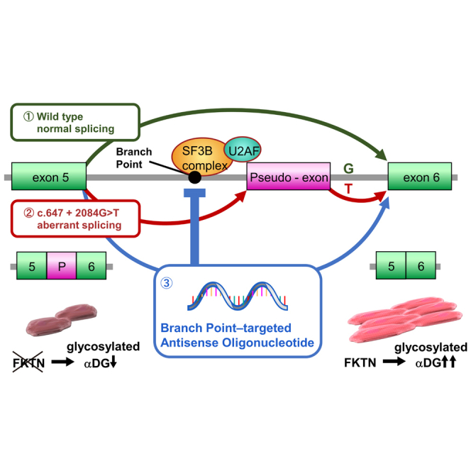
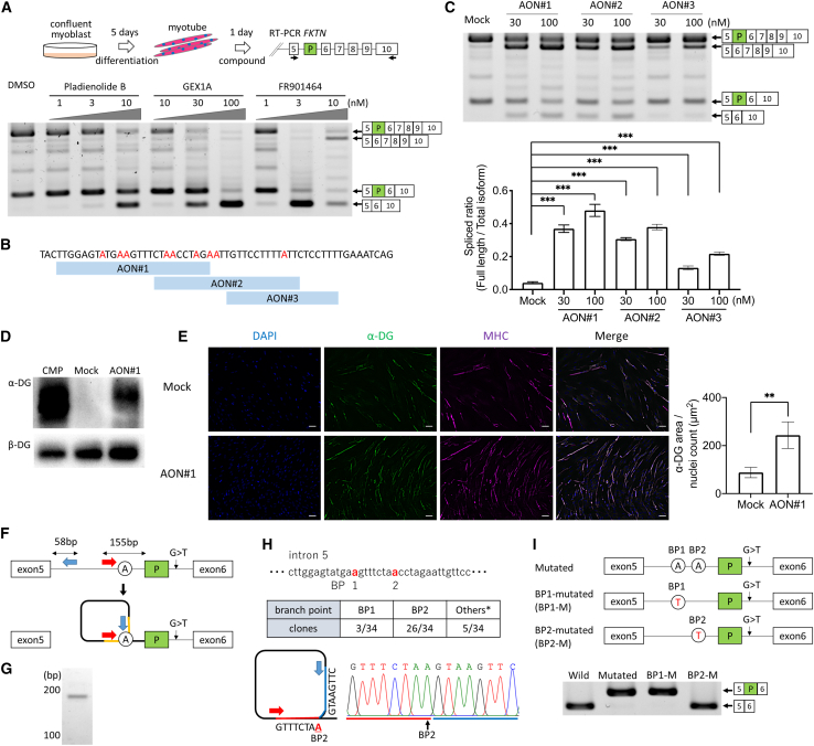
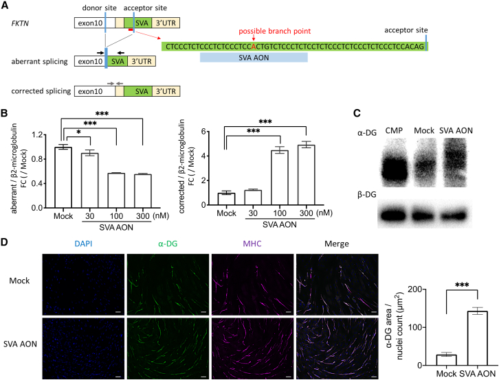
Fukutin (FKTN) c.647+2084G>T creates a pseudo-exon with a premature stop codon, which causes Fukuyama congenital muscular dystrophy (FCMD). We aimed to ameliorate aberrant splicing of FKTN caused by this variant. We screened compounds focusing on splicing regulation using the c.647+2084G>T splicing reporter and discovered that the branchpoint, which is essential for splicing reactions, could be a potential therapeutic target. To confirm the effectiveness of branchpoints as targets for exon skipping, we designed branchpoint-targeted antisense oligonucleotides (BP-AONs). This restored normal FKTN mRNA and protein production in FCMD patient myotubes. We identified a functional BP by detecting splicing intermediates and creating BP mutations in the FKTN reporter gene; this BP was non-redundant and sufficiently blocked by BP-AONs. Next, a BP-AON was designed for a different FCMD-causing variant, which induces pathogenic exon trapping by a common SINE-VNTR-Alu-type retrotransposon. Notably, this BP-AON also restored normal FKTN mRNA and protein production in FCMD patient myotubes. Our findings suggest that BPs could be potential targets in exon-skipping therapeutic strategies for genetic disorders.

Keywords: MT: Oligonucleotides: Therapies and Applications; antisense oligonucleotide; branchpoint; exon-skipping therapy; muscular dystrophy; splice-switching; splicing regulatory elements.


Conflict of interest statement

The authors declare no competing interests.

Figures

None
Graphical abstract
Figure 1
Figure 1
Identification of SF3B1 inhibitors as potent agents for suppressing the c.647+2084G>T-induced pseudo-exon of FKTN (A) Diagram of the FKTN splicing reporter, FKTN minigene of exon 5–6 with intron 5 (wild type), or exon 5–6 with c.647+2084G>T-harboring intron 5 (mutated type) with a HiBiT tag. Filled arrows indicate primers for RT-PCR. (B and C) HEK293 cells were transfected with the wild-type or mutated reporter for 24 h. The luminescence value was quantified by relative luminescence units (RLUs) generated by the C-terminal HiBiT tag using the Nano-Glo HiBiT Lytic Detection System (B). Pseudo-exon expression was confirmed using RT-PCR (C). The larger band corresponds to the 64-bp pseudo-exon inclusion between exons 5 and 6. (D and E) HEK293 cells were transfected with the wild-type or mutated reporter for 4 h and treated with TG003 (30 μM) or DMSO for 24 h. DMSO was used as a negative control because it was used as a solvent for the compounds. The effect of TG003 was confirmed by HiBiT assay (D) and RT-PCR (E). The NanoLuc luciferase activity of TG003 was 1.7-fold higher than that of DMSO. (F) Scatterplot of the fold change of RLUs using the FKTN splicing reporter in focused library screening. (G) RT-PCR analysis of the FKTN splicing reporter with SF3B1 inhibitors (10 μM) and DMSO in HEK293 cells after 24 h. (H) HiBiT assay using the FKTN splicing reporter with pladienolide B (1–10 nM), GEX1A (1–100 nM), and FR901464 (1–10 nM). (I) SF3B1 inhibitor effect in patient-derived myotubes with the FKTN c.647+2084G>T mutation. RT-PCR analysis was performed using pladienolide B (1, 3, or 10 nM), GEX1A (10, 30, or 100 nM), FR901464 (1, 3, or 10 nM), or DMSO. Filled arrows indicate primers for RT-PCR. Data are shown as mean ± SD (n = 4). ∗∗∗p < 0.001, calculated by t test using Welch’s two-sample t test.
Figure 2
Figure 2
BP-AON induced pseudo-exon skipping of FKTN mRNA (A) RT-PCR analysis for FKTN exons 5–10 in patient-derived myotubes with a c.647+2084G>T in the presence of SF3B1 inhibitors. Filled arrows indicate primers for RT-PCR. (B) AON design targeting BPs upstream of the pseudo-exon. Adenines located within the 18- 50-nt window from the pseudo-exon are colored red. (C) RT-PCR analysis was performed with AON#1, AON#2, AON#3 (30 or 100 nM), or mock. The full length (FL)/total splice ratio was quantified using intensity analysis. (D) Western blotting of glycosylated α-dystroglycan (α-DG) in patient-derived myotubes treated with AON#1 (100 nM) or mock. β-Dystroglycan (β-DG) was used as a loading control. (E) Patient-derived myotubes showing α-DG glycosylation (clone IIH6, green) and myosin heavy chain (clone MF20, magenta) after treatment with AON#1 (100 nM) or mock. Scale bars, 50 μm. (F) Schematic of divergent primers for lariat introns in the FKTN splicing reporter upstream of the pseudo-exon, where “A” denotes the BP. RT-PCR detected branched structures of introns (yellow). (G) RT-PCR results for the lariat intron. (H) BPs were detected using TA cloning and Sanger sequencing. The main BP, BP2, the adenine located 38 bp from the pseudo-exon, is shown selectively. (I) Diagram showing FKTN-mutated, BP1-mutated (BP1-M), and BP2-mutated (BP2-M) reporters. RT-PCR analysis revealed skipping of the pseudo-exon in BP2-M. Data represent the mean ± SD (n = 4). ∗∗p < 0.01 and ∗∗∗p < 0.001, calculated using Welch’s two-sample t test or one-way ANOVA with independent post hoc Tukey’s multiple-comparisons test.
Figure 3
Figure 3
BP-AON effect in myotubes derived from a patient with FCMD with homozygous SVA retrotransposon insertions (A) Representation of abnormal splicing in FKTN with SVA retrotransposon insertions and AON design targeting one possible non-redundant BP (SVA-AON). Shown is a schematic of primers for abnormal RT-PCR products (black arrow) and corrected RT-PCR products (gray arrow). (B) qRT-PCR of aberrant splicing and corrected splicing in myotubes treated with SVA-AON (30, 100, or 300 nM) or mock. β2-Microglobulin was used as an internal control. Data are shown as mean ± SD (n = 3). (C) Western blot of glycosylated α-DG treated with SVA-AON (100 nM) or mock. β-DG was used as a loading control. (D) Patient-derived myotubes showing α-DG glycosylation (clone IIH6, green) and myosin heavy chain (clone MF20, magenta) after treatment with SVA-AON (100 nM) or mock. Scale bars, 50 μm. Data represent the mean ± SD (n = 4). ∗p < 0.05, ∗∗p < 0.01, and ∗∗∗p < 0.001, calculated using Welch’s two-sample t test or one-way ANOVA with independent post hoc Tukey’s multiple-comparisons test.

References

    1. Vaz-Drago R., Custódio N., Carmo-Fonseca M. Deep intronic mutations and human disease. Hum. Genet. 2017;136:1093–1111. - PubMed
    1. Okubo M., Noguchi S., Awaya T., Hosokawa M., Tsukui N., Ogawa M., Hayashi S., Komaki H., Mori-Yoshimura M., Oya Y., et al. RNA-seq analysis, targeted long-read sequencing and in silico prediction to unravel pathogenic intronic events and complicated splicing abnormalities in dystrophinopathy. Hum. Genet. 2023;142:59–71. - PubMed
    1. Fukuyama Y., Osawa M., Suzuki H. Congenital progressive muscular dystrophy of the Fukuyama type - clinical, genetic and pathological considerations. Brain Dev. 1981;3:1–29. - PubMed
    1. Kanagawa M., Kobayashi K., Tajiri M., Manya H., Kuga A., Yamaguchi Y., Akasaka-Manya K., Furukawa J.I., Mizuno M., Kawakami H., et al. Identification of a Post-translational Modification with Ribitol-Phosphate and Its Defect in Muscular Dystrophy. Cell Rep. 2016;14:2209–2223. - PubMed
    1. Muntoni F., Brockington M., Blake D.J., Torelli S., Brown S.C. Defective glycosylation in muscular dystrophy. Lancet. 2002;360:1419–1421. - PubMed