Observational Study
. 2023 Dec;53(12):e14074.
doi: 10.1111/eci.14074. Epub 2023 Aug 7.

Phenylacetylglutamine and trimethylamine N-oxide: Two uremic players, different actions

Affiliations
Observational Study

Phenylacetylglutamine and trimethylamine N-oxide: Two uremic players, different actions

Sam Hobson et al. Eur J Clin Invest. 2023 Dec.

Abstract

Background: Chronic kidney disease (CKD) patients exhibit a heightened cardiovascular (CV) risk which may be partially explained by increased medial vascular calcification. Although gut-derived uremic toxin trimethylamine N-oxide (TMAO) is associated with calcium-phosphate deposition, studies investigating phenylacetylglutamine's (PAG) pro-calcifying potential are missing.

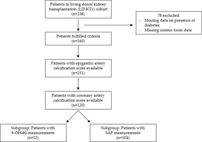
Methods: The effect of TMAO and PAG in vascular calcification was investigated using 120 kidney failure patients undergoing living-donor kidney transplantation (LD-KTx), in an observational, cross-sectional manner. Uremic toxin concentrations were related to coronary artery calcification (CAC) score, epigastric artery calcification score, and markers of established non-traditional risk factors that constitute to the 'perfect storm' that drives early vascular aging in this patient population. Vascular smooth muscle cells were incubated with TMAO or PAG to determine their calcifying effects in vitro and analyse associated pathways by which these toxins may promote vascular calcification.

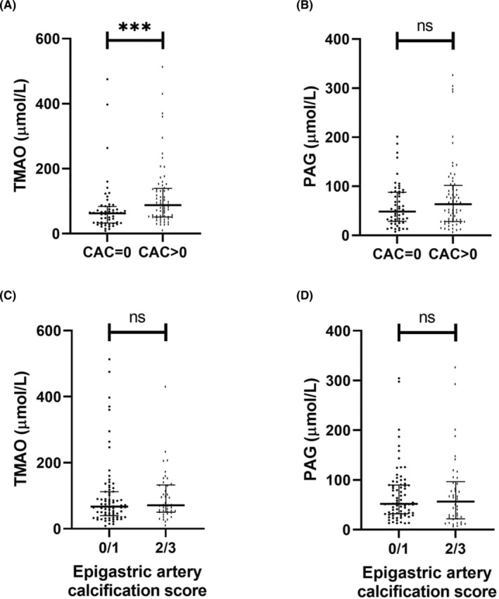
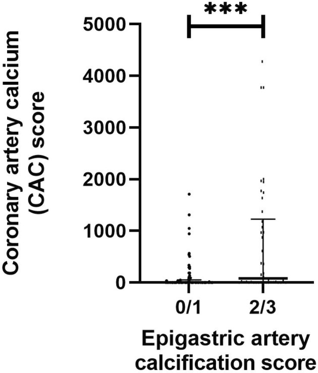
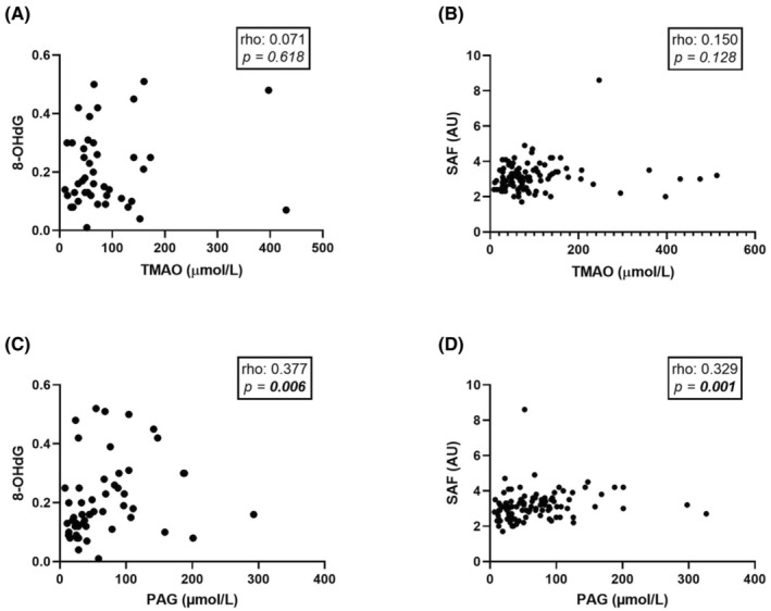
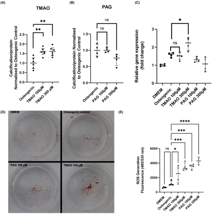
Results: TMAO, but not PAG, was independently associated with CAC score after adjustment for CKD-related risk factors in kidney failure patients. Neither toxin was associated with epigastric artery calcification score; however, PAG was independently, positively associated with 8-hydroxydeoxyguanosine. Similarly, TMAO, but not PAG, promoted calcium-phosphate deposition in vitro, while both uremic solutes induced oxidative stress.

Conclusions: In conclusion, our translational data confirm TMAO's pro-calcifying effects, but both toxins induced free radical production detrimental to vascular maintenance. Our findings suggest these gut-derived uremic toxins have different actions on the vessel wall and therapeutically targeting TMAO may help reduce CV-related mortality in CKD.

Keywords: TMAO; kidney failure; oxidative stress; phenylacetylglutamine; vascular calcification.


Conflict of interest statement

P.S. reports serving in advisory or leadership roles for Astellas, AstraZeneca, Baxter, Fresenius Medical Care (FMC), GlaxoSmithKline, Reata and Vifor; receiving honoraria from Astellas, AstraZeneca, Baxter, FMC, Novo Nordisk, Boerhinger Ingelheim and Pfizer/Bristol Square Myers (BSM); receiving research funding from AstraZeneca and Bayer; serving on advisory boards for AstraZeneca, Baxter, FMC, GlaxoSmithKline, Reata and Vifor. All other authors of this manuscript have nothing to declare.

Figures

FIGURE 1
FIGURE 1
Flowchart depicting study design. 8‐OHdG, 8‐hydroxydeoxyguanosine; SAF, skin autofluorescence.
FIGURE 2
FIGURE 2
Comparisons between TMAO and PAG versus coronary artery calcification or epigastric artery calcification in a living‐donor kidney transplantation patient cohort (n = 120). (A) Serum TMAO concentration in study population stratified by presence of CAC. (B) Serum PAG concentration in study population stratified by presence of CAC. For (A and B), CAC score was quantified in Agatston units using non‐contrast multi‐detector cardiac CT scan. n = 55 and n = 65 for CAC = 0 and CAC >0 groups, respectively. (C) Serum TMAO concentration in relation with epigastric artery calcification score. (D) Serum PAG concentration in relation with epigastric artery calcification score. For (C and D), epigastric arteries were obtained at living‐donor kidney transplantation and scored for calcification by a trained pathologist on tissue sections 1–2 μm thick. Vessels were stained with von Kossa (silver nitrate plus nuclear fast red) and the extent of medial calcification was graded from 0 (no calcification) to 3 (severely calcified). Groups were formed based on the degree of calcification, that is, 0/1 corresponds to non‐calcified and mildly calcified vessels (n = 78), and 2/3 corresponds to moderately and severely calcified vessels (n = 42). Data are presented as mean ± standard error of mean. Differences between groups were assessed by non‐parametric Mann–Whitney U test. ***p < .001. CAC, coronary artery calcium; PAG, phenylacetylglutamine; TMAO, trimethylamine N‐oxide.
FIGURE 3
FIGURE 3
Representative epigastric artery calcification score images. Arteries were isolated at living‐donor kidney transplantation from kidney failure patients and scored by a trained pathologist. Photos taken by Magnus Söderberg.
FIGURE 4
FIGURE 4
Comparisons between CAC score and epigastric artery calcification in the living‐donor kidney transplantation patient cohort (n = 120). CAC score was quantified in Agatston units using non‐contrast multi‐detector cardiac CT scan. Epigastric arteries were obtained at living‐donor kidney transplantation and scored for calcification by a trained pathologist on tissue sections 1–2 μm thick. Vessels were stained with von Kossa (silver nitrate plus nuclear fast red) before the extent of medial calcification was graded from 0 (no calcification) to 3 (severely calcified). Groups were formed based on degree of calcification, that is, 0/1 (n = 78) corresponds to non‐calcified and mildly calcified vessels, and 2/3 (n = 42) corresponds to moderately and severely calcified vessels. Data are presented as mean ± standard error of mean. Differences between groups were assessed by non‐parametric Mann–Whitney U test. ***p < .001.
FIGURE 5
FIGURE 5
Correlations between TMAO and PAG versus 8‐OHdG and SAF in the living‐donor kidney transplantation cohort (n = 120). (A) TMAO correlated with 8‐OHdG. (B) TMAO correlated with SAF. (C) PAG correlated with 8‐OHdG. (D) PAG correlated with SAF. Univariate correlations were assessed using Spearman's rank correlation. Significant associations (p < .05) are depicted in bold. 8‐OHdG, 8‐hydroxydeoxyguanosine; PAG, phenylacetylglutamine; SAF, skin autofluorescence; TMAO, trimethylamine N‐oxide.
FIGURE 6
FIGURE 6
In vitro experiments investigating the effect of TMAO and PAG in VSMC calcification and oxidative stress. (A) TMAO‐induced calcification assay using BoneTag Optical Dye (n = 6 per group). (B) PAG‐induced calcification assay using BoneTag Optical Dye. Aortic VSMCs were incubated with uremic toxins for 7 days in the background of high phosphate (2.5 mM phosphate). For both experiments, readouts were normalised for protein content using BCA assay (n = 4 per group). (C) mRNA expression analysis of osteogenic marker Runx2. Relative gene expression was normalised to DMEM control group (n = 4 per group). (D) Representative images of alizarin red staining for specified conditions. (E) Reactive oxygen species assay for specified conditions using DCFDA/H2DCFDA assay (n = 4 per group). Readout taken 4 h after addition of toxins. Statistics: data presented as mean ± SEM. Groups were compared using one‐way ANOVA followed by group‐wise comparisons using Tukey's post hoc test. p‐values <.05 were deemed statistically significant. *p < .05, **p < .01, ***p < .001, ****p < .0001. PAG, phenylacetylglutamine; ROS, reactive oxygen species; TMAO, trimethylamine N‐oxide.

References

    1. Hill NR, Fatoba ST, Oke JL, et al. Global prevalence of chronic kidney disease – a systematic review and meta‐analysis. PLoS One. 2016;11:e0158765. - PMC - PubMed
    1. Kovesdy CP. Epidemiology of chronic kidney disease: an update 2022. Kidney Int Suppl (2011). 2022;12:7‐11. - PMC - PubMed
    1. Bikbov B, Purcell CA, Levey AS, et al. Global, regional, and national burden of chronic kidney disease, 1990–2017: a systematic analysis for the global burden of disease study 2017. Lancet. 2020;395:709‐733. - PMC - PubMed
    1. Jankowski J, Floege J, Fliser D, Böhm M, Marx N. Cardiovascular disease in chronic kidney disease: pathophysiological insights and therapeutic options. Circulation. 2021;143:1157‐1172. - PMC - PubMed
    1. Kooman JP, Kotanko P, Schols AMWJ, Shiels PG, Stenvinkel P. Chronic kidney disease and premature ageing. Nat Rev Nephrol. 2014;10:732‐742. - PubMed

Publication types