

Experimenting with ASL-based arterialized cerebral blood volume as a novel imaging biomarker in grading glial neoplasms
- PMID: 37548164
- PMCID: PMC10649543
- DOI: 10.1177/19714009231193163
Experimenting with ASL-based arterialized cerebral blood volume as a novel imaging biomarker in grading glial neoplasms
Abstract
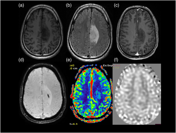
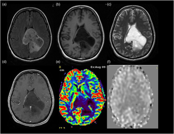
Background: Perfusion imaging is one of the methods used to grade glial neoplasms, and in this study we evaluated the role of ASL perfusion in grading brain glioma.
Purpose: The aim is to evaluate the role of arterialized cerebral blood volume (aCBV) of multi-delay ASL perfusion for grading glial neoplasm.
Materials and methods: This study is a prospective observational study of 56 patients with glial neoplasms of the brain who underwent surgery, and only cases with positive diagnosis of glioma are included to evaluate the novel diagnostic parameter.
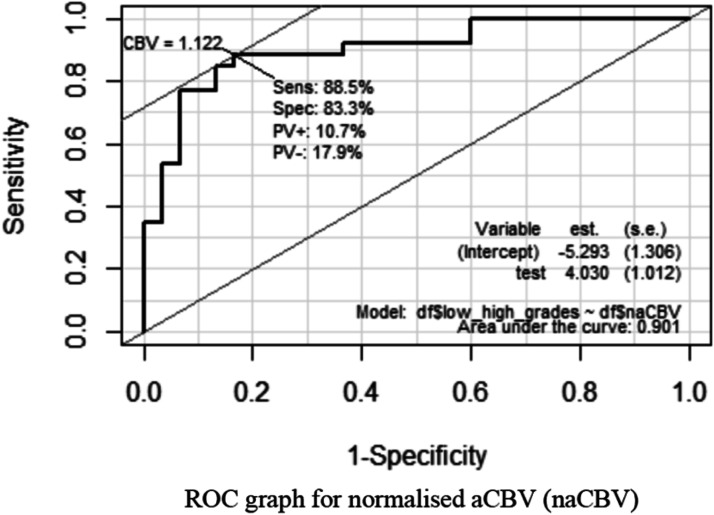
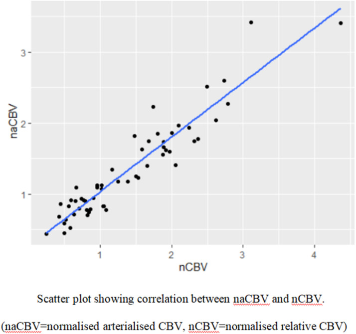
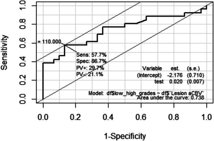
Results: In the study, ASL-derived normalized aCBV (naCBV) and T2*DSC-derived normalized CBV (nCBV) are showing very high correlation (Pearson's correlation coefficient value of 0.94) in grading glial neoplasms. naCBV and nCBF are also showing very high correlation (Pearson's correlation coefficient value of 0.876). The study also provides cutoff values for differentiating LGG from HGG for normalized aCBV(naCBV) of ASL, normalized CBV (nCBV), and normalized nCBF derived from T2* DCS as 1.12, 1.254, and 1.31, respectively. ASL-derived aCBV also shows better diagnostic accuracy than ASL-derived CBF.
Conclusion: This study is one of its kind to the best of our knowledge where multi-delay ASL perfusion-derived aCBV is used as a novel imaging biomarker for grading glial neoplasms, and it has shown high statistical correlation with T2* DSC-derived perfusion parameters.
Keywords: Perfusion magnetic resonance imaging; T2*dynamic susceptibility contrast; arterial spin labeling; cerebral blood volume; glioma.
Conflict of interest statement
Declaration of conflicting interestsThe author(s) declared no potential conflicts of interest with respect to the research, authorship, and/or publication of this article.
Figures
Similar articles
-
Comparison of ASL and DSC perfusion methods in the evaluation of response to treatment in patients with a history of treatment for malignant brain tumor.BMC Med Imaging. 2024 Mar 22;24(1):70. doi: 10.1186/s12880-024-01249-w. BMC Med Imaging. 2024. PMID: 38519901 Free PMC article.
-
Pediatric astrocytic tumor grading: comparison between arterial spin labeling and dynamic susceptibility contrast MRI perfusion.Neuroradiology. 2018 Apr;60(4):437-446. doi: 10.1007/s00234-018-1992-6. Epub 2018 Feb 16. Neuroradiology. 2018. PMID: 29453753
-
A radiomics-based comparative study on arterial spin labeling and dynamic susceptibility contrast perfusion-weighted imaging in gliomas.Sci Rep. 2020 Apr 9;10(1):6121. doi: 10.1038/s41598-020-62658-9. Sci Rep. 2020. PMID: 32273523 Free PMC article.
-
Differentiation Between High-Grade Glioma and Brain Metastasis Using Cerebral Perfusion-Related Parameters (Cerebral Blood Volume and Cerebral Blood Flow): A Systematic Review and Meta-Analysis of Perfusion-weighted MRI Techniques.J Magn Reson Imaging. 2025 Feb;61(2):758-768. doi: 10.1002/jmri.29473. Epub 2024 Jun 20. J Magn Reson Imaging. 2025. PMID: 38899965
-
Clinical utility of arterial spin labeling imaging in disorders of the nervous system.Neurosurg Focus. 2019 Dec 1;47(6):E5. doi: 10.3171/2019.9.FOCUS19567. Neurosurg Focus. 2019. PMID: 31786550 Review.
Cited by
-
Advances of MR imaging in glioma: what the neurosurgeon needs to know.Acta Neurochir (Wien). 2025 Jun 21;167(1):174. doi: 10.1007/s00701-025-06593-6. Acta Neurochir (Wien). 2025. PMID: 40542873 Free PMC article. Review.
References
-
- Golay X, Petersen ET. Arterial spin labeling: benefits and pitfalls of high magnetic field. Neuroimaging Clin 2006; 16: 259–268. - PubMed
-
- Kety SS, Schmidt CF. The determination of cerebral blood flow in man by the use of nitrous oxide in low concentrations. American Journal of Physiology-Legacy Content 1945; 143: 53–66.
-
- Sadowski EA, Bennett LK, Chan MR, et al. Nephrogenic systemic fibrosis: risk factors and incidence estimation. Radiology 2007; 243: 148–157. - PubMed
Publication types
MeSH terms
Substances
LinkOut - more resources
Full Text Sources
Medical
