. 2023 Aug;12(8):e12339.
doi: 10.1002/jev2.12339.

Depletion of soluble cytokines unlocks the immunomodulatory bioactivity of extracellular vesicles

Affiliations

Depletion of soluble cytokines unlocks the immunomodulatory bioactivity of extracellular vesicles

Quentin Roux et al. J Extracell Vesicles. 2023 Aug.

Abstract

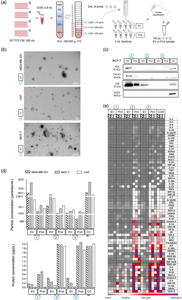
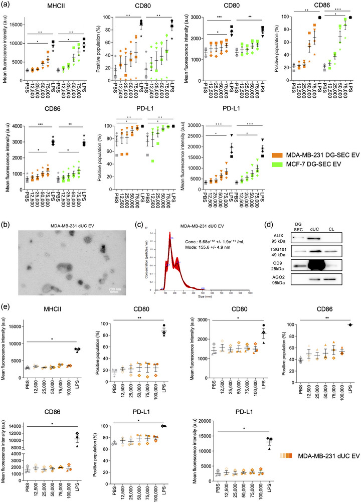
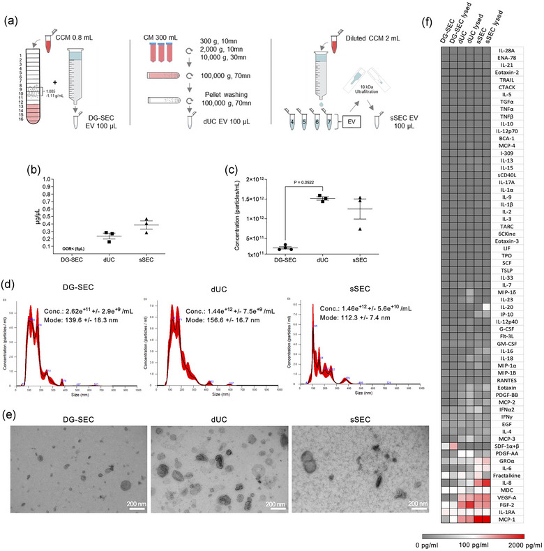
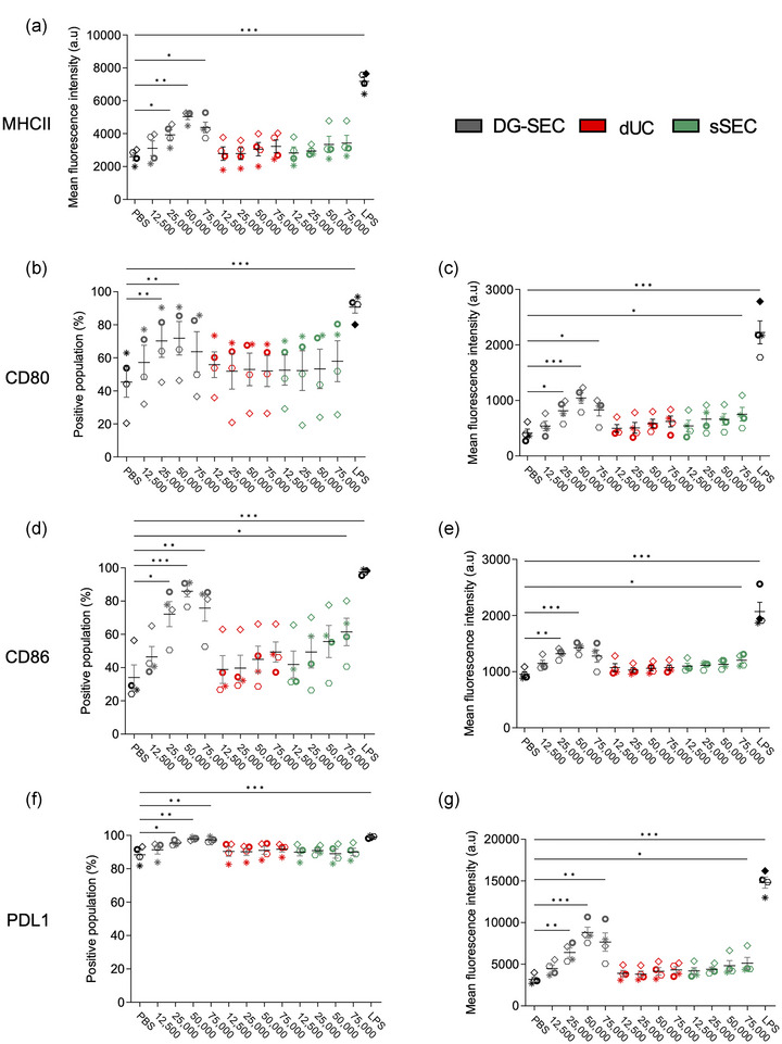
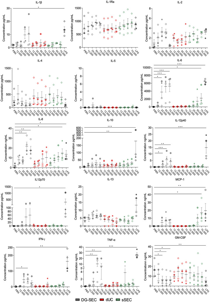
Despite an enormous interest in understanding the bioactivity of extracellular vesicles (EV) in physiology and disease for the development of therapeutic applications, the impact of EV preparation methods remains minimally explored. In this study, we implemented density gradient ultracentrifugation combined with size-exclusion chromatography (DG-SEC), differential ultracentrifugation (dUC) and/or stand-alone SEC (sSEC) to fractionate media conditioned by different cancer cells and/or cancer-associated fibroblasts (CAF). EV-enriched but protein-depleted versus EV-depleted but protein-enriched DG-SEC fractions, and EV-containing dUC and sSEC preparations were quality controlled for particle number, protein concentration, selected protein composition and ultrastructure, characterized for their cytokine content, and dose-dependently evaluated for monocyte-derived dendritic cell (MoDC) maturation by measuring surface marker expression and/or cytokine secretion. EV preparations obtained by DG-SEC from media conditioned by different cancer cell lines or CAF, were depleted from soluble immune suppressive cytokines such as VEGF-A and MCP-1 and potently stimulated MoDC maturation. In contrast, EV-containing dUC or sSEC preparations were not depleted from these soluble cytokines and were unable to mature MoDC. Subsequent processing of dUC EV preparations by SEC dose-dependently restored the immunomodulatory bioactivity. Overall, our results demonstrate that method-dependent off-target enrichment of soluble cytokines has implications for the study of EV immunomodulatory bioactivity and warrants careful consideration.

Keywords: corona; dendritic cells; exosomes; isolation; maturation; microvesicles; separation; vaccines.


Conflict of interest statement

The authors declare no conflicts of interest.

Figures

FIGURE 1
FIGURE 1
Bottom‐up DG‐SEC efficiently separates EV from soluble cytokines. (a) Schematic representation of the DG‐SEC protocol. CM: conditioned medium, CCM: concentrated CM, EV: EV‐enriched, Prot: protein‐enriched. (b, d, and e) Particle and protein characterization of CCM and pools (p)1, p2 and p3 EV and protein‐enriched samples generated as described in (a) from a representative MDA‐MB‐231, MCF‐7 and CAF harvesting (n = 1) (see Figures S1 c, d,g). (b) Transmission electron microscopy images from MDA‐MB‐231, MCF‐7 or CAF p1 EV‐enriched SEC fractions. Magnification × 30,000, scale bar: 200 nm. (c) Western blot analysis of MCF‐7 p1, p2 and p3 EV and protein‐enriched (Prot) SEC fractions. Thirty microlitres of samples generated by DG‐SEC (a) were denatured as described in the methods section and loaded in each lane. CL: cell lysate (15 μg). (d) Particle and protein concentrations measured in CCM and p1, p2 and p3 EV and Prot samples. Particle and protein concentration were measured by nanoparticle tracking analysis (see (d)) and Qubit protein assay respectively (n = 1). (e) Luminex® cytokine 65‐analytes profiling of CCM and p1, p2 and p3 EV and Prot samples characterized in (b) and (d) (n = 1).
FIGURE 2
FIGURE 2
EV separated by DG‐SEC induce a dose‐dependent increase in MoDC maturation markers. a. Flow cytometry analysis after 24 h treatment of 200,000 immature MoDC from healthy volunteers with DG‐SEC EV from MDA‐MB‐231 or MCF‐7 (n = 4 MoDC donors per cell line). Treatment is indicated as corresponding number of NTA‐measured particles per MoDC. LPS (100 ng/mL) was used as a positive control to induce MoDC maturation. PBS was used as a negative control. Data represent mean and standard error. Statistical analysis: Friedman test with Dunn's multiple comparisons test (*p < 0.05, **p < 0.01, ***p < 0.001). (b) Transmission electron microscopy images from MDA‐MB‐231 dUC EV. Magnification x30,000, scale bar: 200 nm. (c) NTA size distribution profile of MDA‐MB‐231 dUC EV represented as mean (black line) and standard error (red shaded area). Conc.: particle concentration. Particle concentration and mode are expressed as mean and standard error (n = 3 videos). (d) Western blot analysis of MDA‐MB‐231 EV DG‐SEC and dUC EV preparations. Thirty microliters of EV preparations were denatured as described in the methods section and loaded in each lane. CL: cell lysate (15 μg). (e) Flow cytometry analysis after 24 h treatment of 200,000 immature MoDC from healthy volunteers with dUC EV from MDA‐MB‐231 (n = 3 MoDC donors). LPS (100 ng/mL) was used as a positive control to induce MoDC maturation. PBS was used as a negative control. Data represent mean and standard error. Statistical analysis: Friedman test with Dunn's multiple comparisons test (*p < 0.05, **p < 0.01, ***p < 0.001).
FIGURE 3
FIGURE 3
Impact of separation method on breast CAF EV particle and protein content. (a) Schematic view of CAF EV separation using different methods. (b) Protein concentration measured by Qubit using 5 μL of each preparation (DG‐SEC [n = 4], dUC [n = 3], sSEC [n = 3]). (c) Particle concentration measured by NTA in DG‐SEC (n = 4), dUC (n = 3), sSEC (n = 3). Three videos of 30 s were acquired for each replicate. (b–d) Data represent mean and standard error. Statistical analysis: Kruskal Wallis test with Dunn's multiple comparisons test. (d) Representative NTA size distribution profile of DG‐SEC, dUC and sSEC EV represented as mean (black line) and standard error (red shaded area). Conc.: particle concentration. Particle concentration and mode are expressed as mean and standard error. (e) Transmission EM of DG‐SEC, dUC, and sSEC CAF EV, magnification × 30,000, scale bar 200 nm. (f) Cytokine profiling in one representative DG‐SEC, dUC and sSEC EV preparation lysed or not with 0.2% Triton X‐100. Signal from PBS 0.2% Triton X100 control was subtracted.
FIGURE 4
FIGURE 4
Impact of separation method on CAF EV potential to stimulate MoDC maturation markers expression. (a–g) Flow cytometry analysis after 24 h treatment of 200,000 immature MoDC from healthy volunteers with DG‐SEC, dUC or sSEC EV breast CAF (n = 4 MoDC donors). Treatment is indicated as corresponding number of NTA‐measured particles per MoDC. LPS (100 ng/mL) was used as a positive control to induce MoDC maturation. PBS was used as a negative control. Data represent mean and standard error. Statistical analysis: Friedman test with Dunn's multiple comparisons test (*p < 0.05, **p < 0.01, ***p < 0.001).
FIGURE 5
FIGURE 5
Impact of separation method on CAF EV‐induced cytokine release by MoDC. Luminex 15‐plex cytokine profiling in MoDC culture medium following treatment with breast CAF DG‐SEC, dUC and sSEC EV preparations (Figure 4). LPS (100 ng/mL) was used as a positive control to induce MoDC maturation. PBS was used as a negative control. Data represent mean and standard error. Statistical analysis: Friedman test with Dunn's multiple comparisons test (*p < 0.05, **p < 0.01, ***p < 0.001).

References

    1. Alfaro, C. , Suarez, N. , Gonzalez, A. , Solano, S. , Erro, L. , Dubrot, J. , Palazon, A. , Hervas‐Stubbs, S. , Gurpide, A. , Lopez‐Picazo, J. M. , Grande‐Pulido, E. , Melero, I. , & Perez‐Gracia, J. L. (2009). Influence of bevacizumab, sunitinib and sorafenib as single agents or in combination on the inhibitory effects of VEGF on human dendritic cell differentiation from monocytes. British Journal of Cancer, 100(7), 1111–1119. 10.1038/sj.bjc.6604965 - DOI - PMC - PubMed
    1. Andre, F. , Schartz, N. E. , Movassagh, M. , Flament, C. , Pautier, P. , Morice, P. , Pomel, C. , Lhomme, C. , Escudier, B. , Le Chevalier, T. , Tursz, T. , Amigorena, S. , Raposo, G. , Angevin, E. , & Zitvogel, L. (2002). Malignant effusions and immunogenic tumour‐derived exosomes. The Lancet, 360(9329), 295–305. 10.1016/s0140-6736(02)09552-1 - DOI - PubMed
    1. Anguille, S. , Smits, E. L. , Lion, E. , van Tendeloo, V. F. , & Berneman, Z. N. (2014). Clinical use of dendritic cells for cancer therapy. The Lancet Oncology, 15, e257–267. - PubMed
    1. Bonsergent, E. , Grisard, E. , Buchrieser, J. , Schwartz, O. , Théry, C. , & Lavieu, G. (2021). Quantitative characterization of extracellular vesicle uptake and content delivery within mammalian cells. Nature Communications, 12(1), 1864. 10.1038/s41467-021-22126-y - DOI - PMC - PubMed
    1. Cocozza, F. , Grisard, E. , Martin‐Jaular, L. , Mathieu, M. , & Théry, C. (2020). SnapShot: Extracellular vesicles. Cell, 182, 262–262.e1. - PubMed

Publication types

MeSH terms

LinkOut - more resources