. 2023 Sep 1;80(9):989-995.
doi: 10.1001/jamaneurol.2023.2523.

Immunoglobulin A Antibodies Against Myelin Oligodendrocyte Glycoprotein in a Subgroup of Patients With Central Nervous System Demyelination

Affiliations

Immunoglobulin A Antibodies Against Myelin Oligodendrocyte Glycoprotein in a Subgroup of Patients With Central Nervous System Demyelination

Ana Beatriz Ayroza Galvão Ribeiro Gomes et al. JAMA Neurol. .

Erratum in

  • Error in Figure 2B.
    [No authors listed] [No authors listed] JAMA Neurol. 2024 Jan 1;81(1):88. doi: 10.1001/jamaneurol.2023.4587. JAMA Neurol. 2024. PMID: 37955904 Free PMC article. No abstract available.

Abstract

Importance: Differential diagnosis of patients with seronegative demyelinating central nervous system (CNS) disease is challenging. In this regard, evidence suggests that immunoglobulin (Ig) A plays a role in the pathogenesis of different autoimmune diseases. Yet little is known about the presence and clinical relevance of IgA antibodies against myelin oligodendrocyte glycoprotein (MOG) in CNS demyelination.

Objective: To investigate the frequency of MOG-IgA and associated clinical features in patients with demyelinating CNS disease and healthy controls.

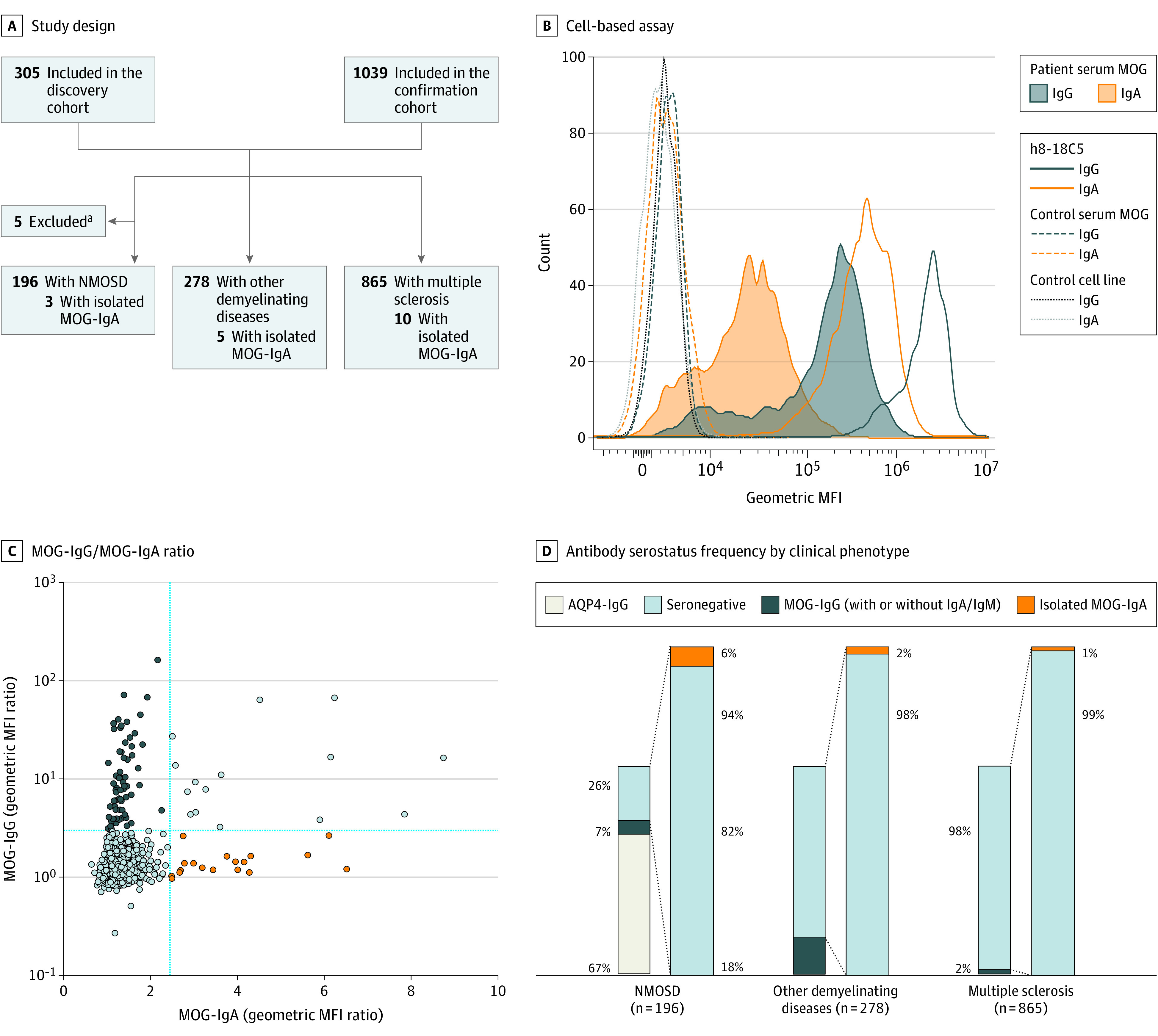
Design, setting, and participants: This longitudinal study comprised 1 discovery and 1 confirmation cohort derived from 5 centers. Participants included patients with suspected or confirmed demyelinating diseases and healthy controls. MOG-IgA, MOG-IgG, and MOG-IgM were measured in serum samples and cerebrospinal fluid (CSF) of patients, who were assessed from September 2012 to April 2022.

Main outcomes and measures: Frequency and clinical features of patients who were seropositive for MOG-IgA and double-seronegative for aquaporin 4 (AQP4) IgG and MOG-IgG.

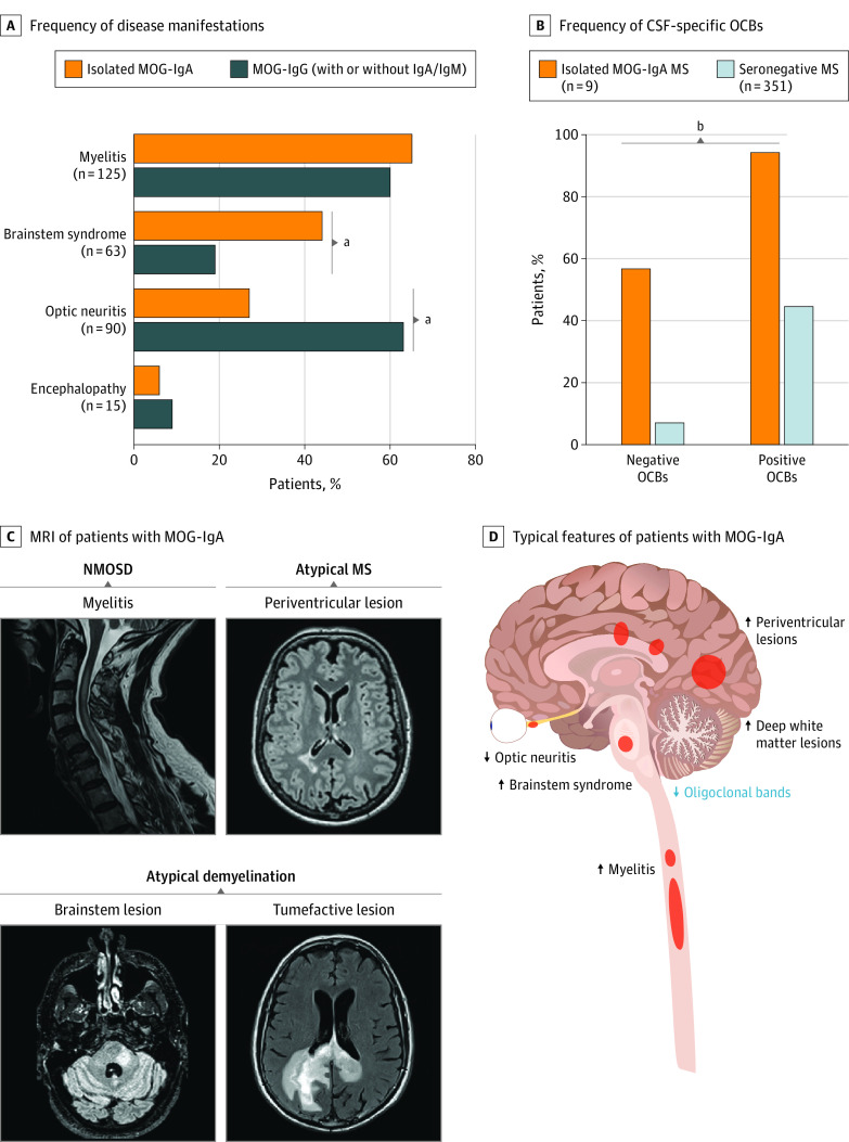
Results: After the exclusion of 5 participants with coexisting AQP4-IgG and MOG-IgA, MOG-IgG, and/or MOG-IgM, 1339 patients and 110 healthy controls were included; the median follow-up time was 39 months (range, 0-227 months). Of included patients with isolated MOG-IgA, 11 of 18 were female (61%), and the median age was 31.5 years (range, 3-76 years). Among patients double-seronegative for AQP4-IgG and MOG-IgG (1126/1339; 84%), isolated MOG-IgA was identified in 3 of 50 patients (6%) with neuromyelitis optica spectrum disorder, 5 of 228 patients (2%) with other CNS demyelinating diseases, and 10 of 848 patients (1%) with multiple sclerosis but in none of the healthy controls (0/110). The most common disease manifestation in patients seropositive for isolated MOG-IgA was myelitis (11/17 [65%]), followed by more frequent brainstem syndrome (7/16 [44%] vs 14/75 [19%], respectively; P = .048), and infrequent manifestation of optic neuritis (4/15 [27%] vs 46/73 [63%], respectively; P = .02) vs patients with MOG-IgG. Among patients fulfilling 2017 McDonald criteria for multiple sclerosis, MOG-IgA was associated with less frequent CSF-specific oligoclonal bands (4/9 [44%] vs 325/351 [93%], respectively; P < .001) vs patients with multiple sclerosis who were MOG-IgG/IgA seronegative. Further, most patients with isolated MOG-IgA presented clinical attacks after recent infection or vaccination (7/11 [64%]).

Conclusion and relevance: In this study, MOG-specific IgA was identified in a subgroup of patients who were double-seronegative for AQP4-/MOG-IgG, suggesting that MOG-IgA may be a novel diagnostic biomarker for patients with CNS demyelination.


Conflict of interest statement

Conflict of Interest Disclosures: Dr Ayroza Galvão Ribeiro Gomes reported grants from Roche during the conduct of the study. Dr de Moura Brasil Matos reported grants from Hoffmann La Roche outside the submitted work. Dr Pereira reported grants from CNPq–Conselho Nacional de Desenvolvimento Científico e Tecnológico (308172/2018-3) during the conduct of the study. Dr Ribeiro Monteiro reported grants from CNPq–Conselho Nacional de Desenvolvimento Científico e Tecnológico during the conduct of the study. Dr Schindler reported nonfinancial support from UCB Pharma outside the submitted work. Dr Chien reported grants from Novartis and Alexion during the conduct of the study and nonfinancial support as a member from the Canadian Institutes of Health Research Standing Committee on Science outside the submitted work. Dr Schwake reported speaker honoraria from Alexion and travel support from Novartis and UCB outside the submitted work. Dr Schneider reported research grant support from Novartis and speaker honoraria from Roche and Alexion outside the submitted work. Dr Aktas reported personal fees from Alexion, Almirall, Horizon, Novartis, and Roche outside the submitted work and serving as steering committee member and co-coordinator of the German Neuromyelitis Optica Study Group (NEMOS). Dr Fischer reported grants to their institution from Medtronic, Stryker, Rapid Medical, Penumbra, and Phenox; consultant fees paid to their institution from Medtronic, Stryker, and CSL Behring outside the submitted work; participation in an advisory board for Alexion/Portola, Boehringer Ingelheim, Biogen, and Acthera (fees paid to institution); member of a clinical event committee of the COATING study (Phenox); member of the data and safety monitoring committee of the TITAN, LATE_MT, and IN EXTREMIS trials; and vice-presidency of the Swiss Neurological Society. Dr Mehling reported grants from the Swiss National Science Foundation, Roche, and Merck and fees for advisory board activities paid to their institution from Merck, Roche, Novartis, and Biogen outside the submitted work. Dr Derfuss reported grants from Alexion, Novartis, and Roche and fees paid to their institution for membership in advisory boards, data and safety monitoring boards, and/or steering committees from Actelion, Biogen, Celgene, Sanofi, GeNeuro, Merck, MedDay, Roche, Alexion, and Novartis outside the submitted work. Dr Kappos reported consultant, speaking, advisory board, and/or steering committee fees from Bayer, Biogen, Bristol Myers Squibb, Celltrion, df-mp Molnia & Pohlman, Eli Lilly (Suisse), EMD Serono, F&U confirm, Genentech, GlaxoSmithKline, Janssen, Japan Tobacco, Merck (Schweiz), Minoryx Therapeutics, Novartis, Roche, Santhera Pharmaceuticals, Shionogi, and Wellmera, grants from the European Union, Innosuisse, and Swiss National Science Foundation; and product license fees from Neurostatus outside the submitted work. Dr Ayzenberg reported personal fees from Roche, Alexion, Merck, Horison, and Sanofi and grants from Diamed from outside the submitted work. Dr Ringelstein reported personal fees from Alexion, Horizon, Roche, and Biogen outside the submitted work. Dr Callegaro reported being on the board of BCTRIMS, the Brazilian commitment for treatment and research in multiple sclerosis and related diseases, and serving as coordinator of the Neuroimmunology Center, Hospital das Clínicas, São Paulo University. Dr Kuhle reported grants from the Swiss MS Society, Swiss National Science Foundation, Novartis, Biogen, Merck, Bristol Myers Squibb, and Roche outside the submitted work. Dr Papadopoulou reported grants from University of Basel, grants from University Hospital of Basel, and grants from Swiss Multiple Sclerosis Society during the conduct of the study; advisory board and/or speaking fees to their institution from AbbVie, Eli Lilly, Lundbeck, and TEVA and conference travel support from TEVA outside the submitted work. Dr Pröbstel reported advisory board and/or consultant fees from Roche, Biogen, and Novartis outside the submitted work. No other disclosures were reported.

Figures

Figure 1.
Figure 1.. Study Design and Frequency of Isolated Myelin Oligodendrocyte Glycoprotein (MOG) Immunoglobulin (Ig) A in Central Nervous System Demyelination
A, Flowchart of patients in the discovery and confirmation cohort who were screened for MOG-IgA, MOG-IgG, and MOG-IgM. Aquaporin 4 (AQP4) was tested as part of the routine clinical diagnosis. B, Representative IgG and IgA binding of humanized 8-18C5 (h8-18C5) monoclonal antibody, MOG-Ig seropositive patient serum, and MOG-Ig seronegative control sample to human MOG–transfected or control cells. C, Individual patients’ geometric mean fluorescence intensity (MFI) ratio based on up to 4 measurements as XY plot for MOG-IgG and MOG-IgA, all cohorts combined. D, Antibody serostatus frequency according to clinical phenotype. Seronegative indicates AQP4-/MOG-IgG double-seronegative. aFive patients with neuromyelitis optica spectrum disorder (NMOSD) were excluded from downstream analysis: 3 with AQP4-IgG/MOG-IgG, 1 with AQP4-IgG/MOG-IgA, and 1 with AQP4-IgG/MOG-IgG/MOG-IgM.
Figure 2.
Figure 2.. Clinical Characterization of Patients Seropositive for Myelin Oligodendrocyte Glycoprotein (MOG) Immunoglobulin (Ig) A
A, Frequency of disease manifestations for patients with isolated MOG-IgA and MOG-IgG. B, Frequency of positive and negative cerebrospinal fluid (CSF)–specific oligoclonal bands (OCBs) in MOG-IgA seropositive multiple sclerosis (MS) compared with seronegative MS. C, Magnetic resonance imaging (MRI) of patients with MOG-IgA highlighting the following disease phenotypes: neuromyelitis optica spectrum disorder (NMOSD, often presenting with myelitis), atypical MS (often presenting with periventricular lesions), and atypical demyelination (often associated with brainstem syndrome or with tumor-mimic/atypical demyelination). D, Clinical features frequently observed in isolated MOG-IgA seropositive central nervous system demyelination. Arrows indicate high and low frequencies. aFisher exact test, P < .05. bFisher exact test, P < .001.

References

    1. Lennon VA, Wingerchuk DM, Kryzer TJ, et al. . A serum autoantibody marker of neuromyelitis optica: distinction from multiple sclerosis. Lancet. 2004;364(9451):2106-2112. doi:10.1016/S0140-6736(04)17551-X - DOI - PubMed
    1. O’Connor KC, McLaughlin KA, De Jager PL, et al. . Self-antigen tetramers discriminate between myelin autoantibodies to native or denatured protein. Nat Med. 2007;13(2):211-217. doi:10.1038/nm1488 - DOI - PMC - PubMed
    1. Pröbstel AK, Dornmair K, Bittner R, et al. . Antibodies to MOG are transient in childhood acute disseminated encephalomyelitis. Neurology. 2011;77(6):580-588. doi:10.1212/WNL.0b013e318228c0b1 - DOI - PubMed
    1. Marignier R, Hacohen Y, Cobo-Calvo A, et al. . Myelin-oligodendrocyte glycoprotein antibody-associated disease. Lancet Neurol. 2021;20(9):762-772. doi:10.1016/S1474-4422(21)00218-0 - DOI - PubMed
    1. Pröbstel AK, Rudolf G, Dornmair K, et al. . Anti-MOG antibodies are present in a subgroup of patients with a neuromyelitis optica phenotype. J Neuroinflammation. 2015;12(46):46. doi:10.1186/s12974-015-0256-1 - DOI - PMC - PubMed

Publication types