

Detection rate of contrast-enhanced brain magnetic resonance imaging in patients with cognitive impairment
- PMID: 37549181
- PMCID: PMC10406288
- DOI: 10.1371/journal.pone.0289638
Detection rate of contrast-enhanced brain magnetic resonance imaging in patients with cognitive impairment
Abstract
Introduction: The number of brain MRI with contrast media performed in patients with cognitive impairment has increased without universal agreement. We aimed to evaluate the detection rate of contrast-enhanced brain MRI in patients with cognitive impairment.
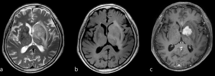
Materials and methods: This single-institution, retrospective study included 4,838 patients who attended outpatient clinics for cognitive impairment evaluation and underwent brain MRI with or without contrast enhancement from December 2015 to February 2020. Patients who tested positive for cognitive impairment were followed-up to confirm whether the result was true-positive and provide follow-up management. Detection rate was defined as the proportion of patients with true-positive results and was compared between groups with and without contrast enhancement. Individual matching in a 1:2 ratio according to age, sex, and year of test was performed.
Results: The overall detection rates of brain MRI with and without contrast media were 4.7% (57/1,203; 95% CI: 3.6%-6.1%) and 1.8% (65/3,635; 95% CI: 1.4%-2.3%), respectively (P<0.001); individual matching demonstrated similar results (4.7% and 1.9%). Among 1,203 patients with contrast media, 3.6% was only detectable with the aid of contrast media. The proportion of patients who underwent follow-up imaging or treatment for the detected lesions were significantly higher in the group with contrast media (2.0% and 0.6%, P < .001).
Conclusions: Detection rate of brain MRI for lesions only detectable with contrast media in patients with cognitive impairment was not high enough and further study is needed to identify whom would truly benefit with contrast media.
Copyright: © 2023 Joo et al. This is an open access article distributed under the terms of the Creative Commons Attribution License, which permits unrestricted use, distribution, and reproduction in any medium, provided the original author and source are credited.
Conflict of interest statement
The authors have declared that no competing interests exist.
Figures


References
-
- American Psychiatric Association. Diagnostic and Statistical Manual of Mental Disorders. 5th ed. Arlington, VA: American Psychiatric Association; 2013.
-
- Knopman DS, DeKosky ST, Cummings JL, Chui H, Corey-Bloom J, Relkin N, et al.. Practice parameter: diagnosis of dementia (an evidence-based review). Report of the Quality Standards Subcommittee of the American Academy of Neurology. Neurology. 2001;56(9): 1143–1153. doi: 10.1212/wnl.56.9.1143 - DOI - PubMed
Publication types
MeSH terms
Substances
LinkOut - more resources
Full Text Sources
Medical
