. 2023 Aug 15;120(33):e2300036120.
doi: 10.1073/pnas.2300036120. Epub 2023 Aug 7.

Silver electroceutical technology to treat sarcopenia

Affiliations

Silver electroceutical technology to treat sarcopenia

Min Young Kim et al. Proc Natl Acad Sci U S A. .

Erratum in

Abstract

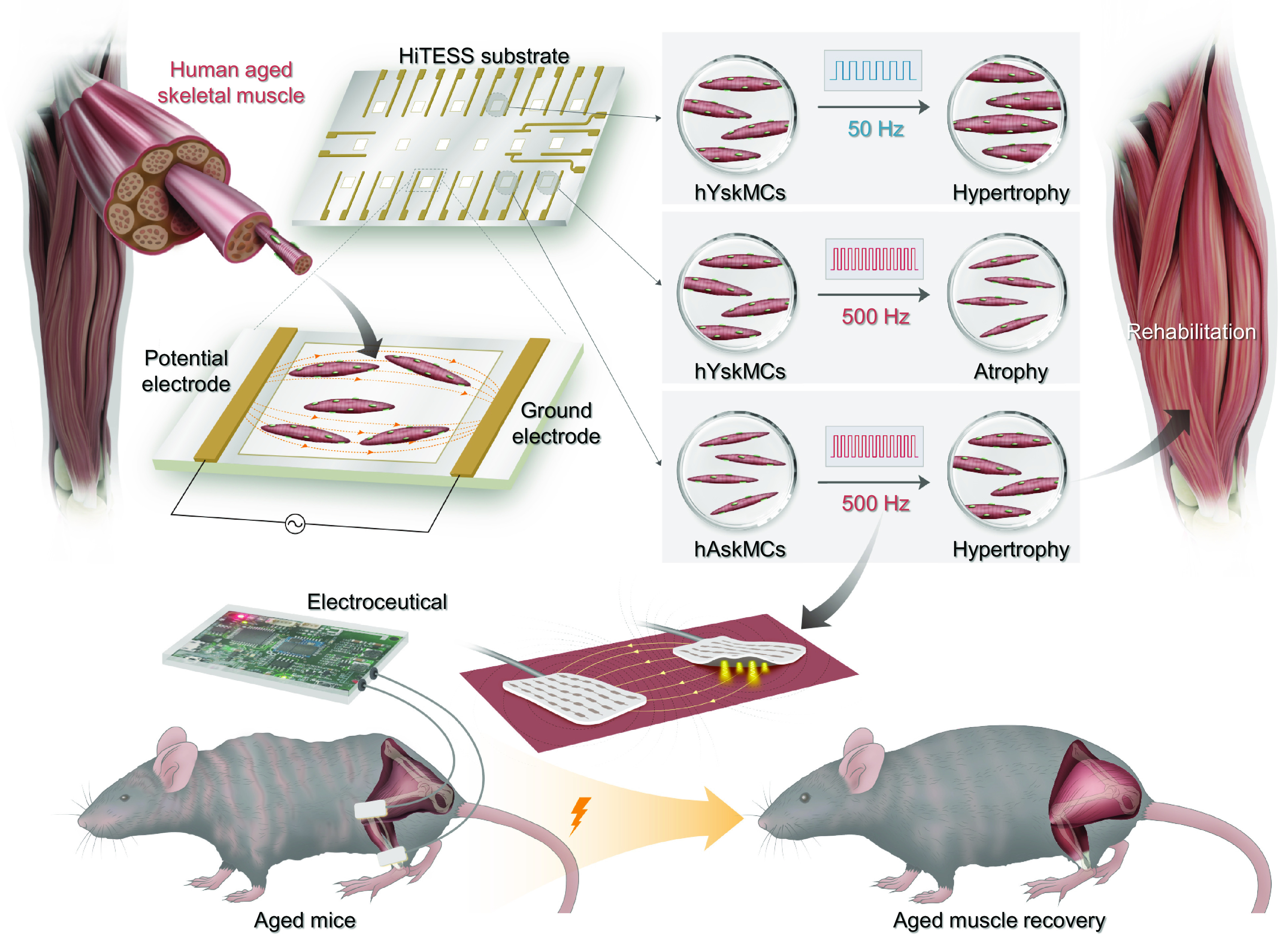
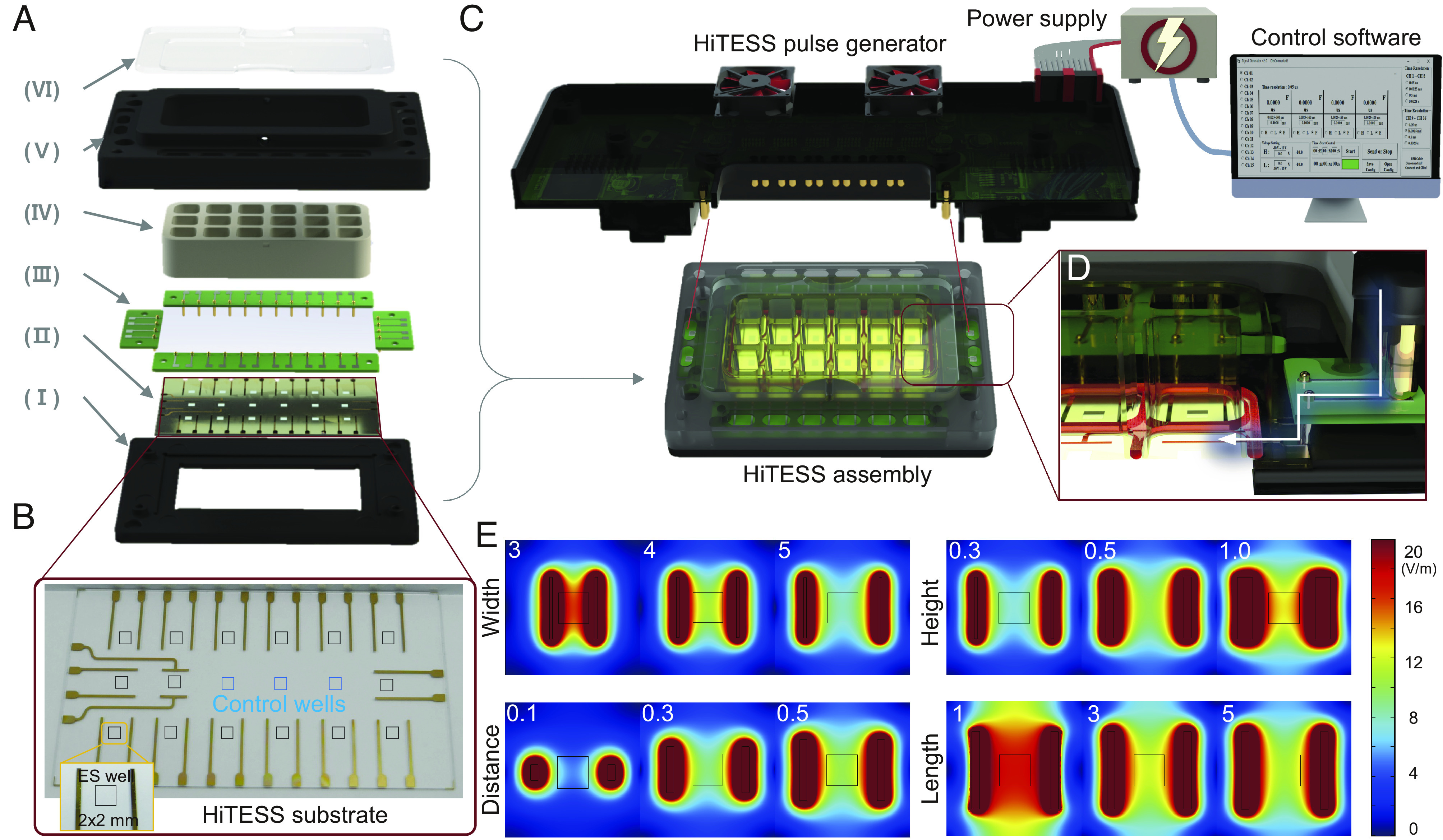
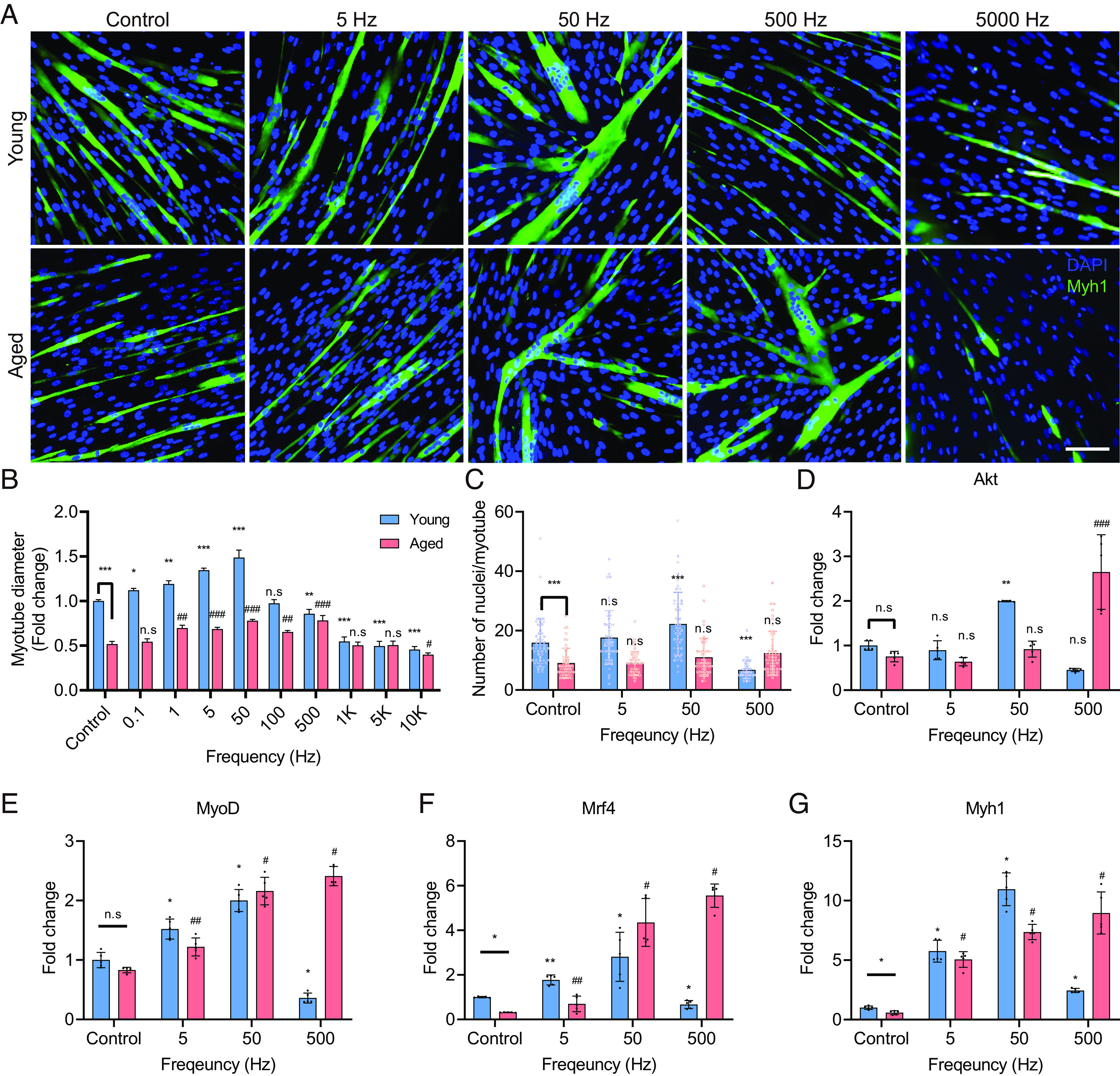
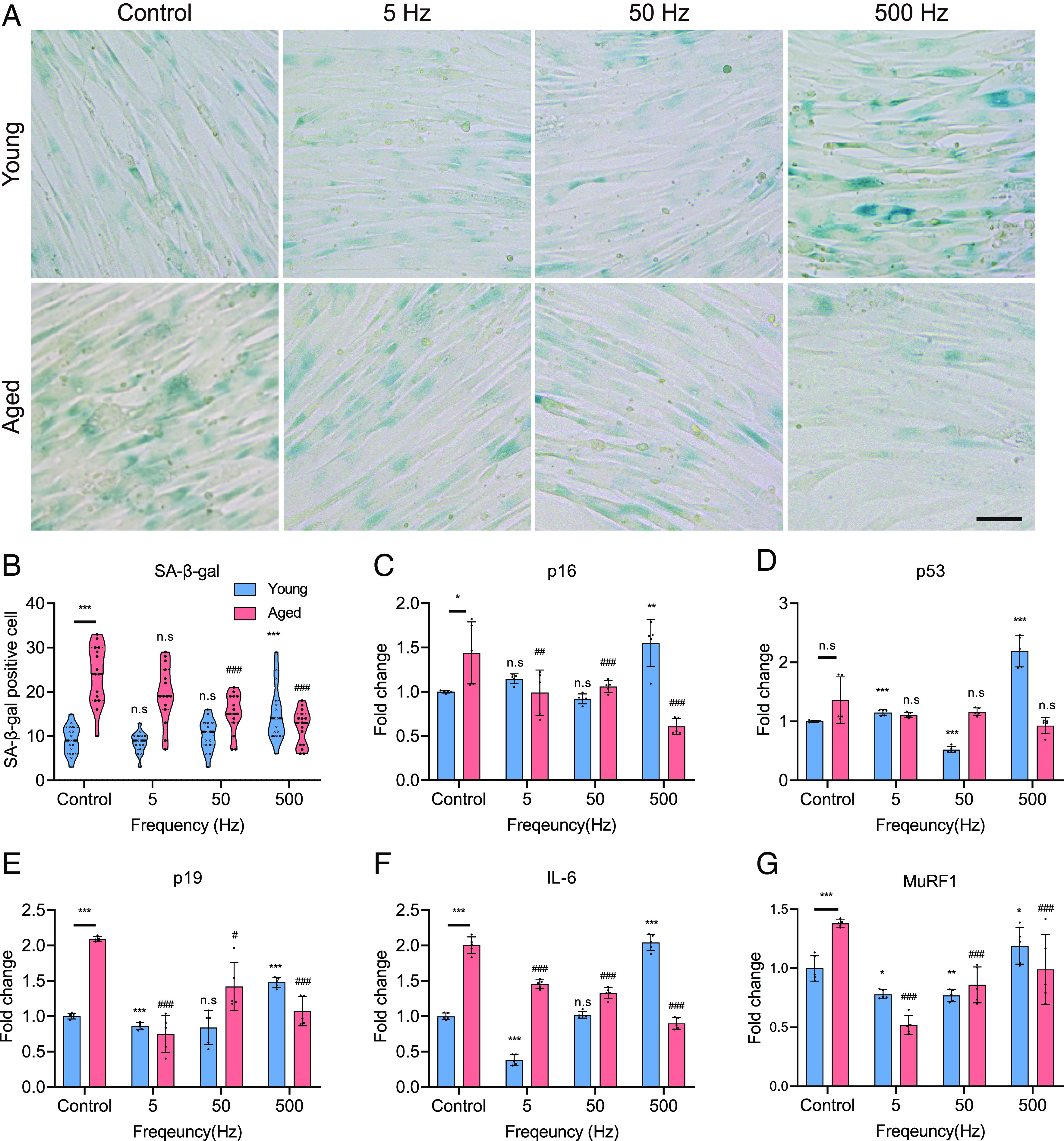
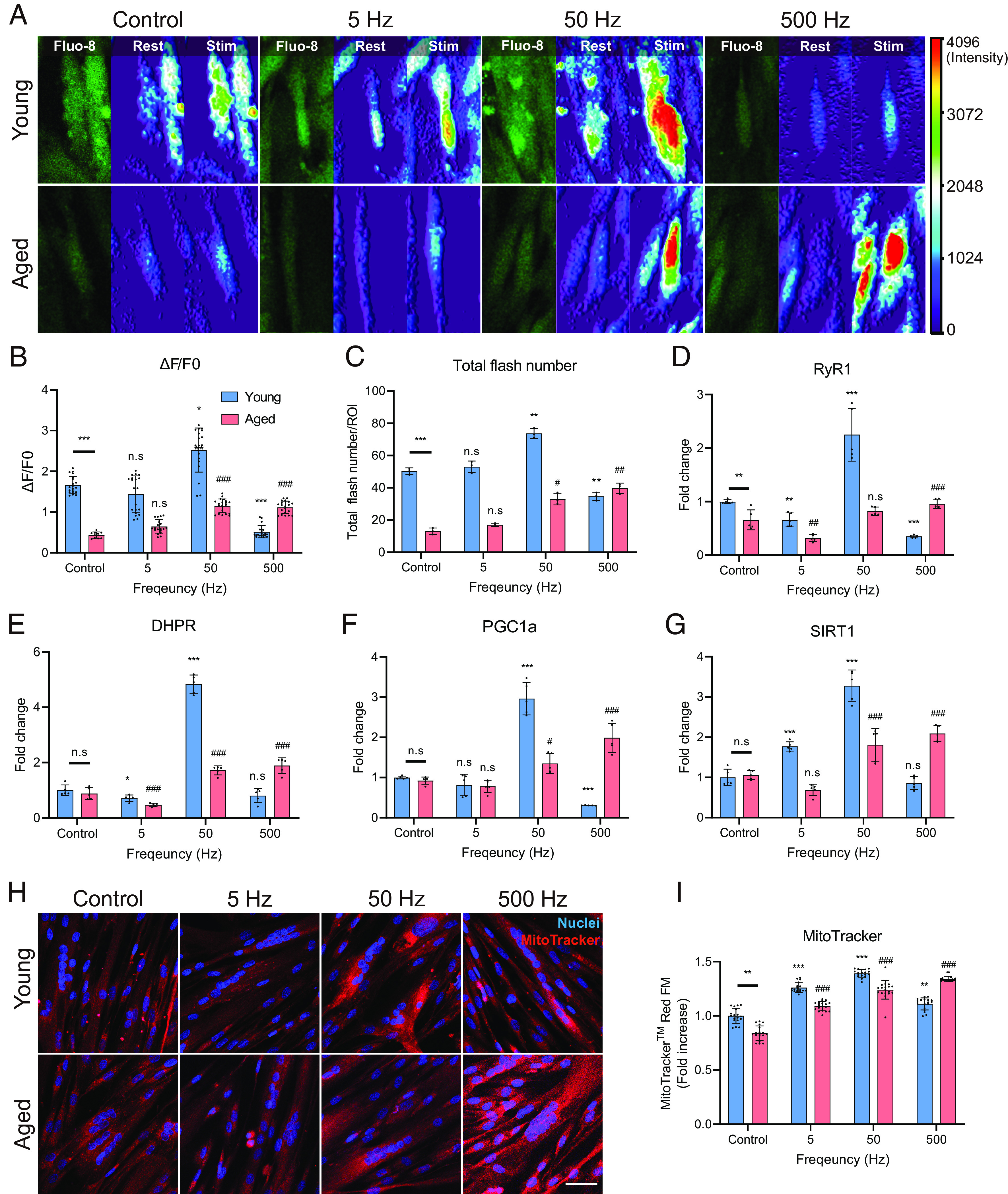
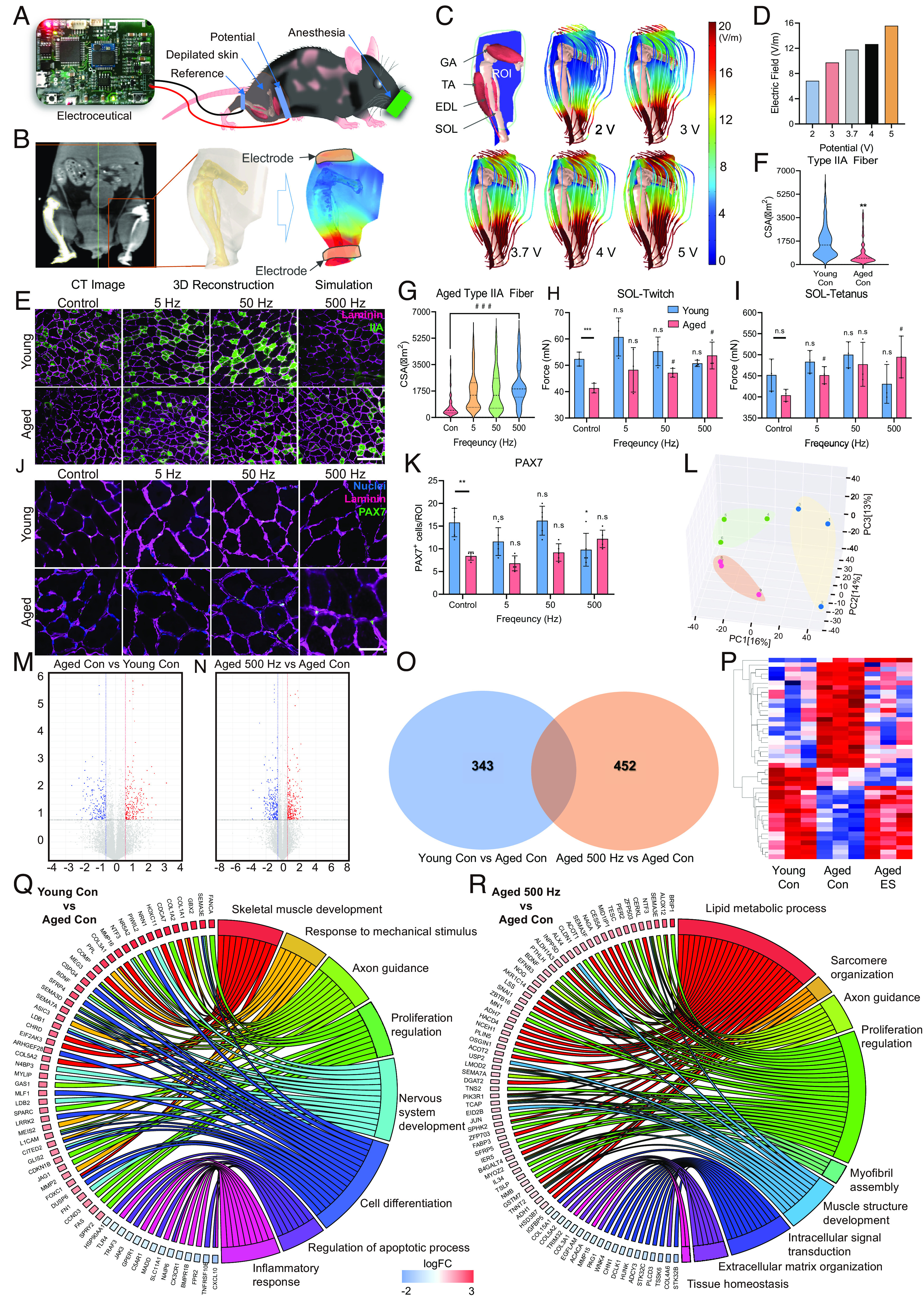
While the world is rapidly transforming into a superaging society, pharmaceutical approaches to treat sarcopenia have hitherto not been successful due to their insufficient efficacy and failure to specifically target skeletal muscle cells (skMCs). Although electrical stimulation (ES) is emerging as an alternative intervention, its efficacy toward treating sarcopenia remains unexplored. In this study, we demonstrate a silver electroceutical technology with the potential to treat sarcopenia. First, we developed a high-throughput ES screening platform that can simultaneously stimulate 15 independent conditions, while utilizing only a small number of human-derived primary aged/young skMCs (hAskMC/hYskMC). The in vitro screening showed that specific ES conditions induced hypertrophy and rejuvenation in hAskMCs, and the optimal ES frequency in hAskMCs was different from that in hYskMCs. When applied to aged mice in vivo, specific ES conditions improved the prevalence and thickness of Type IIA fibers, along with biomechanical attributes, toward a younger skMC phenotype. This study is expected to pave the way toward an electroceutical treatment for sarcopenia with minimal side effects and help realize personalized bioelectronic medicine.

Keywords: electroceutical; integrated electrical stimulation biochip; multiplex screening technology; personalized electric medicine; sarcopenia.


Conflict of interest statement

The authors declare no competing interest.

Figures

Fig. 1.
Fig. 1.
Schematic of the silver electroceutical to restore the aged skeletal muscle into a young-phenotypic muscle. Skeletal muscle responds differently depending on ES conditions, and identical ES conditions result in different cell fates depending on young and aged skeletal muscles.
Fig. 2.
Fig. 2.
Configuration of the HiTESS platform and simulation for electric field optimization. (A) Configuration of the high-throughput electrical stimulation screening (HiTESS) assembly. (I) Bottom holder, (II) HiTESS substrate, (III) connecting PCB, (IV) chamber with O-ring, (V) upper holder, and (VI) cover. (B) Plane view of the HiTESS substrate. The black (ES) and blue (no ES) boxes indicate the individual culture well. (C) HiTESS assembly connected with HiTESS pulse generator. The HiTESS pulse generator was controlled by custom-made control software. (D) HiTESS assembly and HiTESS pulse generator are connected through the contact between the connecting PCB and the pogo pins of the pulse generator. (E) Simulation results of E- fields based on distance, width, length, and medium height.
Fig. 3.
Fig. 3.
Specific ES conditions induce myotube differentiation. (A and B) Representative images (A) and quantification (B) of myotubes stained with Myh1 and nuclei, respectively. Optimal ES frequencies vary with age. (Scale bar, 100 μm.) (C) Quantification of the number of nuclei/myotube. The optimal ES frequency for hYskMC and hAskMC is 50 and 500 Hz, respectively. (DG) mRNA expression levels of Akt (D), MyoD (E), Mrf4 (F), and Myh1 (G). The gene expression profiles are up-regulated at the appropriate ES condition. (*P < 0.05; **P < 0.01; ***P < 0.001; compared to young control, #P < 0.05; ##P < 0.01; ###P < 0.001; compared to aged control).
Fig. 4.
Fig. 4.
Specific ES conditions reduce senescence-associated factors. (A) SA-β-Gal staining of hYskMC and hAskMC. Specific ES conditions that could recover the senescence-associated phenotype in the hAskMC and the optimal ES frequency for young and aged were different. (Scale bar, 50 μm.) (B) Quantification of SA-β-Gal-positive cells. (CG) mRNA expression levels of p16 (C), p53 (D), p19 (E), IL-6 (F), and MuRF1 (G). The gene expression data elucidated that the hAskMC can be restored to the hYskMC-like phenotype. (*P < 0.05; **P < 0.01; ***P < 0.001; compared to young control, #P < 0.05; ##P < 0.01; ###P < 0.001; compared to aged control).
Fig. 5.
Fig. 5.
Specific ES conditions lead to calcium signaling improvement and metabolic improvement in hAskMC. (AC) Results of calcium imaging (A) quantification result of ∆F/F0 (B) and quantification result of total flash number (C). The optimal ES condition enhanced the calcium flux in hYskMC and hAskMC. (D and E) mRNA expression level of RyR1 (D) and DHPR (E). These results showed that there was an improvement in the specific ES-mediated calcium signaling in hAskMC. (F and G) mRNA Expression level of PGC1a (F) and SIRT1 (G). (H and I) Representation (H) and quantification (I) of MitoTrackerTM Red FM staining of skMC. The specific ES condition enhances metabolic function in the hAskMC. (Scale bar, 100 μm.) (*P < 0.05; **P < 0.01; ***P < 0.001; compared to young control, #P < 0.05; ##P < 0.01; ###P < 0.001; compared to aged control).
Fig. 6.
Fig. 6.
Validation of the silver electroceutical in aged mice. (A) Schematics of the preclinical ES experiment. (B) E-field simulation process in a mouse leg based on 3D anatomy reconstructed by CT images. (C) Simulation results of E-field distribution in a mouse leg. (D) Quantification result of E-field value in the ROI of a mouse leg. (E) Representative CSA images of Type IIA muscle fibers in young and aged mice (Green; Type IIA fiber, Magenta; Laminin). (Scale bar, 200 μm.) (F) Quantification of myofiber CSA in young versus aged mice. (G) Quantification of myofiber CSA for ES conditions in aged mice. (H and I) Isometric twitch (H) and tetanus contraction (I) force measurement of soleus muscles depending on ES conditions in young and aged mice. (J and K) Representative images (Scale bar, 50 μm.) (J) and quantification (K) of the expression of PAX7 (Green) and muscle fibers (Magenta; Laminin). (L) Result of PCA. (M and N) Volcano plot of RNA sequencing transcriptome data displaying the pattern of gene expression values for young control (M) and aged 500 Hz mice (N) relative to aged control mice. (O) Venn diagram of differentially expressed genes found in aged con/young con and aged 500 Hz/young con. (P) Gene clustering heatmap of RNA sequencing using TA muscle from mice. (Q and R) Gene ontology analysis results for young control (Q) and aged 500 Hz mice (R) relative to aged control mice. (*P < 0.05; **P < 0.01; ***P < 0.001; compared to young control, #P < 0.05; ##P < 0.01; ###P < 0.001; compared to aged control).

References

    1. Walston J. D., Sarcopenia in older adults. Curr. Opin. Rheumatol. 24, 623–627 (2012). - PMC - PubMed
    1. Cruz-Jentoft A. J., Sayer A. A., Sarcopenia. Lancet 393, 2636–2646 (2019). - PubMed
    1. Marty E., Liu Y., Samuel A., Or O., Lane J., A review of sarcopenia: Enhancing awareness of an increasingly prevalent disease. Bone 105, 276–286 (2017). - PubMed
    1. Feike Y., Zhijie L., Wei C., Advances in research on pharmacotherapy of sarcopenia. Aging Med. (Milton) 4, 221–233 (2021). - PMC - PubMed
    1. Parise G., Snijders T., Myostatin inhibition for treatment of sarcopenia. Lancet Diabetes Endocrinol. 3, 917–918 (2015). - PubMed

Publication types

LinkOut - more resources