. 2023 Aug 15;120(33):e2307513120.
doi: 10.1073/pnas.2307513120. Epub 2023 Aug 7.

Uncoupling of Ca2+ sparks from BK channels in cerebral arteries underlies hypoperfusion in hypertension-induced vascular dementia

Affiliations

Uncoupling of Ca2+ sparks from BK channels in cerebral arteries underlies hypoperfusion in hypertension-induced vascular dementia

Jade L Taylor et al. Proc Natl Acad Sci U S A. .

Abstract

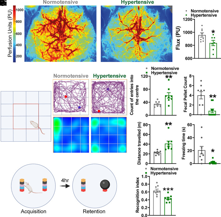
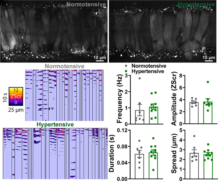
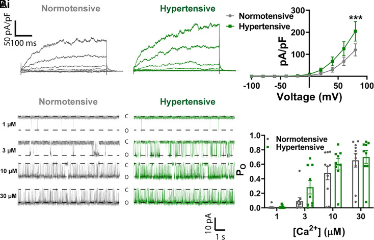
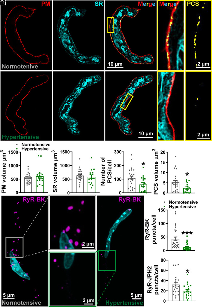
The deficit in cerebral blood flow (CBF) seen in patients with hypertension-induced vascular dementia is increasingly viewed as a therapeutic target for disease-modifying therapy. Progress is limited, however, due to uncertainty surrounding the mechanisms through which elevated blood pressure reduces CBF. To investigate this, we used the BPH/2 mouse, a polygenic model of hypertension. At 8 mo of age, hypertensive mice exhibited reduced CBF and cognitive impairment, mimicking the human presentation of vascular dementia. Small cerebral resistance arteries that run across the surface of the brain (pial arteries) showed enhanced pressure-induced constriction due to diminished activity of large-conductance Ca2+-activated K+ (BK) channels-key vasodilatory ion channels of cerebral vascular smooth muscle cells. Activation of BK channels by transient intracellular Ca2+ signals from the sarcoplasmic reticulum (SR), termed Ca2+ sparks, leads to hyperpolarization and vasodilation. Combining patch-clamp electrophysiology, high-speed confocal imaging, and proximity ligation assays, we demonstrated that this vasodilatory mechanism is uncoupled in hypertensive mice, an effect attributable to physical separation of the plasma membrane from the SR rather than altered properties of BK channels or Ca2+ sparks, which remained intact. This pathogenic mechanism is responsible for the observed increase in constriction and can now be targeted as a possible avenue for restoring healthy CBF in vascular dementia.

Keywords: calcium imaging; dementia; hypertension; ion channels.


Conflict of interest statement

The authors declare no competing interest.

Figures

Fig. 1.
Fig. 1.
Hypertensive mice display a vascular dementia phenotype. (A, i) Laser speckle images showing the average perfusion of the dorsal surface of the brain from normotensive (Left) and hypertensive (Right) mice over a 5-min period. (A, ii) Average median flux through a surface artery (N = 8 normotensive mice and 7 hypertensive mice; *P < 0.05, unpaired t test). (B, i) Schematic of the open field maze, with the center shown as a red square. (B, ii) Upper panels show the pathway traveled by a normotensive (Left) and hypertensive (Right) mouse, with the blue spot showing the starting position and the red spot showing the final position of the mouse. Lower panels show a heat map of the time spent in certain regions of the maze. Summary data showing the number of entries into the center of the maze (B, iii), total fecal pellet count (B, iv), distance traveled (B, v), and time freezing (B, vi) in the hypertensive mice compared to the normotensive mice (N = 9 normotensive mice and 8 hypertensive mice; *P = 0.05, **P = 0.01, unpaired t test). (C, i) Schematic of the novel object recognition test. (C, ii) Recognition index for hypertensive mice versus normotensive mice (N = 9 normotensive mice and 8 hypertensive mice; ***P < 0.001, unpaired t test).
Fig. 2.
Fig. 2.
BK channel function is impaired in hypertension. (A) Pressure myography traces showing the changes in arterial diameter in response to increasing intraluminal pressure (mmHg) in pial cerebral arteries from a normotensive mouse (A, i) and a hypertensive mouse (A, ii). Percent myogenic tone was determined by comparing active control of arterial diameter in the presence of Ca2+ (normotensive, gray; hypertensive, green) with passive diameter in Ca2+-free solutions (black trace). (A, iii) Summary data comparing myogenic tone in arteries isolated from hypertensive mice and normotensive mice (n = 14 arteries from 11 normotensive mice and 11 arteries from 9 hypertensive mice; ***P < 0.001, two-way ANOVA). (B) Pressure myography experiments showing the change in diameter in response to paxilline (1 μM) in pial arteries isolated from normotensive mice (B, i, gray) and hypertensive mice (B, ii, green). (B, iii) Summary data comparing constriction to paxilline in arteries from hypertensive mice and normotensive mice (n = 5 arteries from 5 normotensive mice and 5 arteries from 5 hypertensive mice; *P < 0.05, unpaired t test). (C, i) Perforated patch electrophysiological recordings of STOCs in isolated SMCs from pial arteries from normotensive (gray) and hypertensive (green) mice at holding potentials of −50 mV (Upper), −30 mV (Middle), and −10 mV (Lower). Summary data showing STOC frequency (C, ii) and amplitude (C, iii) in cells isolated from normotensive and hypertensive mouse pial arteries (n = 13 cells from 6 normotensive mice and 7 cells from 4 hypertensive mice; *P < 0.05, ***P < 0.001, two-way ANOVA).
Fig. 3.
Fig. 3.
Ca2+ spark events are comparable between normotensive and hypertensive vessels. (A) Cerebral pial arteries from normotensive and hypertensive mice loaded with Fluo-4-AM, pressurized to 60 mmHg, and imaged using a spinning-disk confocal microscope. (A, ii) ST maps generated from recordings processed using Volumetry. Events were converted to pixels and given a ZScr to allow categorization into sparks and waves based on duration (length) and spatial spread (width). Summary data showing spark frequency, amplitude, duration, and spread (B) in normotensive (gray) and hypertensive (green) mice (n = 7 arteries from 7 normotensive mice and 10 arteries from 10 hypertensive mice; unpaired t test).
Fig. 4.
Fig. 4.
No decrease in functional BK subunit expression in hypertension. (A, i) Whole-cell ruptured-patch electrophysiological recordings of paxilline-sensitive currents obtained between −100 and 100 mV in SMCs isolated from pial arteries from normotensive (gray) and hypertensive (green) mice. (A, ii) Summary data comparing BK current density in cells isolated from hypertensive mice and normotensive mice (n = 14 arteries from 5 normotensive mice and 11 arteries from 4 hypertensive mice; *P < 0.05, **P < 0.01, two-way ANOVA). (B, i) Single-channel inside-out BK channel recordings at −40 mV in exercised patches from SMCs isolated from normotensive (gray) and hypertensive (green) pial arteries. Openings were evoked by exposing patches to 1 μM (Upper), 3 μM (Upper-Middle), 10 μM (Lower Middle), and 30 μM (Lower) free Ca2+. (B, ii) Summary data comparing the open probability (PO) between groups at different Ca2+ concentrations (n = 10 cells from 4 normotensive mice and 9 cells from 5 hypertensive mice; two-way ANOVA).
Fig. 5.
Fig. 5.
Spatial uncoupling between Ca2+ sparks and BK channels in hypertension. (A) Live-cell deconvolved confocal imaging of SMCs isolated from normotensive (A, i) and hypertensive (A, ii) pial arteries. The PM (red) and SR (cyan) of VSMCs were labeled with specific membrane dyes, with sites of colocalization (yellow) deemed to be PCS. The yellow box is expanded to the right to show the interaction between the two membranes in both merged and PCS only. 3D-reconstructed cells showing the PM volume (A, iii) and SR volume (A, iv) and number of PCS (A, v) and mean PCS volume (A, vi) per cell (n = 25 cells from 4 normotensive mice and 16 cells from 4 hypertensive mice; *P < 0.05, unpaired t test). (B) Maximum projection images of proximity ligation assay experiments in isolated SMCs from normotensive (B, i) and hypertensive (B, ii) pial arteries, labeled with primary antibodies against RyRs and BK channels. Positive association (<40 nm apart) of the two channels results in fluorescent puncta (magenta). (B, iii) Summary data showing the number of RyR-BK puncta per cell in SMCs isolated from normotensive and hypertensive mouse pial arteries (n = 29 cells from 6 normotensive mice and 24 cells from 4 hypertensive mice; ***P < 0.001, unpaired t test). (B, iv) Summary data showing RyR–junctophilin (JPH2) puncta per cell in SMCs isolated from hypertensive and normotensive pial arteries (n = 21 cells from 4 normotensive mice and 20 cells from 4 hypertensive mice; *P < 0.05, unpaired t test).

References

    1. Wardlaw J. M., Smith C., Dichgans M., Mechanisms of sporadic cerebral small vessel disease: Insights from neuroimaging. Lancet Neurol. 12, 483–497 (2013). - PMC - PubMed
    1. Graham N. L., Emery T., Hodges J. R., Distinctive cognitive profiles in Alzheimer’s disease and subcortical vascular dementia. J. Neurol. Neurosurg. Psychiatry 75, 61–71 (2004). - PMC - PubMed
    1. Lohner V., Brookes R. L., Hollocks M. J., Morris R. G., Markus H. S., Apathy, but not depression, is associated with executive dysfunction in cerebral small vessel disease. PLoS One 12, e0176943 (2017). - PMC - PubMed
    1. Moretti R., Cavressi M., Tomietto P., Gait and apathy as relevant symptoms of subcortical vascular dementia. Am. J. Alzheimers Dis. Other Demen. 30, 390–399 (2015). - PMC - PubMed
    1. Williamson J. D., et al. , Effect of intensive vs standard blood pressure control on probable dementia: A randomized clinical trial. JAMA 321, 553–561 (2019). - PMC - PubMed

Publication types

Substances