. 2023 Sep 7:140:90-99.
doi: 10.1016/j.placenta.2023.07.297. Epub 2023 Jul 31.

Early pregnancy imaging predicts ischemic placental disease

Affiliations

Early pregnancy imaging predicts ischemic placental disease

Brian Lee et al. Placenta. .

Abstract

Introduction: To characterize early-gestation changes in placental structure, perfusion, and oxygenation in the context of ischemic placental disease (IPD) as a composite outcome and in individual sub-groups.

Methods: In a single-center prospective cohort study, 199 women were recruited from antenatal clinics between February 2017 and February 2019. Maternal magnetic resonance imaging (MRI) studies of the placenta were temporally conducted at two timepoints: 14-16 weeks gestational age (GA) and 19-24 weeks GA. The pregnancy was monitored via four additional study visits, including at delivery. Placental volume, perfusion, and oxygenation were assessed at both MRI timepoints. The primary outcome was defined as pregnancy complicated by IPD, with group assignment confirmed after delivery.

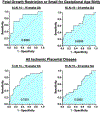
Results: In early gestation, mothers with IPD who subsequently developed fetal growth restriction (FGR) and/or delivered small-for gestational age (SGA) infants showed significantly decreased MRI indices of placental volume, perfusion, and oxygenation compared to controls. The prediction of FGR or SGA by multiple logistic regression using placental volume, perfusion, and oxygenation revealed receiver operator characteristic curves with areas under the curve of 0.81 (Positive predictive value (PPV) = 0.84, negative predictive value (NPV) = 0.75) at 14-16 weeks GA and 0.66 (PPV = 0.78, NPV = 0.60) at 19-24 weeks GA.

Discussion: MRI indices showing decreased placental volume, perfusion and oxygenation in early pregnancy were associated with subsequent onset of IPD, with the greatest deviation evident in subjects with FGR and/or SGA. These early-gestation MRI changes may be predictive of the subsequent development of FGR and/or SGA.

Trial registration: ClinicalTrials.gov NCT02786420.

Keywords: Ischemic placental disease; Oxygenation; Perfusion; Placental blood flow.


Conflict of interest statement

Declaration of competing interest None of the authors have any conflict of interest.

Figures

Figure 1.
Figure 1.
Subject recruitment flowchart D&C = dilatation and curettage; MRI = magnetic resonance imaging; GA = gestational age; IPD refers to the ischemic placental disease group patients
Figure 2.
Figure 2.
Representative MRI Images of placental perfusion and oxygenation showing the clear visual difference in placental perfusion and oxygenation as obtained by MRI. Placental perfusion of a control patient at 16 0/7 weeks gestation (A, top left) has more areas of higher placental perfusion, as well as clear areas of high placental perfusion, when compared to a patient with ischemic placental disease manifesting as fetal growth restriction at 16 3/7 weeks gestation (FGR, C, bottom left), which shows low perfusion throughout the placenta. Placental oxygenation of the same control patient (B, top right), is increased, as evidenced by lower values of R2*, when compared to the same patient with FGR (D, bottom right). Higher R2* values indicate an increased amount of deoxygenated hemoglobin.
Figure 3.
Figure 3.
Receiver operating curves for the prediction of fetal growth restriction or small for gestational age delivery, and ischemic placental disease overall. Numbers within the curves are the area under the curve: A) AUC=0.81 (p < .01), B) AUC=0.66 (p = 0.04), C) AUC=0.7231 (p<0.01), D) AUC= 0.593 (p=0.22).
Figure 4:
Figure 4:
Placental volume, perfusion, and oxygenation at 14 – 16 weeks gestational age (GA) and 19 – 24 weeks GA. IPD: Ischemic placental disease group; FGR+SGA: fetal growth restriction and small for gestational age subgroup. FGR+SGA+PE: Fetal growth restriction, small for gestational age, and preeclampsia subgroup. * = p < 0.05.

References

    1. Ananth CV. Ischemic placental disease: a unifying concept for preeclampsia, intrauterine growth restriction, and placental abruption. Semin Perinatol. 2014;38(3):131–2. doi: 10.1053/j.semperi.2014.03.001. - DOI - PubMed
    1. Ananth CV, Peltier MR, Chavez MR, Kirby RS, Getahun D, Vintzileos AM. Recurrence of ischemic placental disease. Obstet Gynecol. 2007;110(1):128–33. Epub 2007/07/03. doi: 10.1097/01.AOG.0000266983.77458.71. - DOI - PubMed
    1. Burton GJ, Jauniaux E. Pathophysiology of placental-derived fetal growth restriction. Am J Obstet Gynecol. 2018;218(2S):S745–S61. Epub 2018/02/10. doi: 10.1016/j.ajog.2017.11.577. - DOI - PubMed
    1. Ananth CV, Vintzileos AM. Maternal-fetal conditions necessitating a medical intervention resulting in preterm birth. Am J Obstet Gynecol. 2006;195(6):1557–63. Epub 20061002. doi: 10.1016/j.ajog.2006.05.021. - DOI - PubMed
    1. Murray E, Fernandes M, Fazel M, Kennedy SH, Villar J, Stein A. Differential effect of intrauterine growth restriction on childhood neurodevelopment: a systematic review. BJOG. 2015;122(8):1062–72. Epub 2015/05/21. doi: 10.1111/1471-0528.13435. - DOI - PubMed

Publication types

Associated data